Hemolytic anemias 1 of 2 Ali Al Khader


- Slides: 21

Hemolytic anemias (1 of 2) Ali Al Khader, M. D. Faculty of Medicine Al-Balqa’ Applied University Email: ali. alkhader@bau. edu. jo

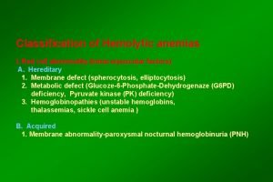
Overview • = anemias due to accelerated red cell destruction ……may be due to intrinsic (intracorpuscular) red cell defects…usually inherited or extrinsic (extracorpuscular) factors…usually acquired Elsevier. Kumar et al. Robbins basic pathology 10 th, , , modified

Overview, cont’d • Erythropoietin release from the kidney growth of erythroid elements + increased release of reticulocytes from the bone marrow …so: erythroid hyperplasia and reticulocytosis are hallmarks of all hemolytic anemias…may be also: extramedullary hematopoiesis

A more clinical way to classify hemolytic anemias • Extravascular hemolysis …destruction of RBCs by phagocytes (mainly in spleen) or • Intravascular hemolysis…within the circulation *Direct damage, examples: -mechanical forces (e. g. , defective heart valve) -fixation of complement -bacterial toxins -heat

Findings that distinguish intravascular hemolysis from extravascular hemolysis • Hemoglobinemia, hemoglobinuria, and hemosiderinuria…intravascular hemolysis • Loss of iron…intravascular hemolysis …by contrast, in extravascular hemolysis: iron recycling by phagocytes is very efficient • No splenomegaly…intravascular hemolysis **Both extra- and intravascular pathways of hemolysis cause decreased serum levels of haptoglobin and both cause jaundice and pigment cholelithiasis

Hereditary Spherocytosis • Nondeformable cells…sequestration and destruction in the spleen • Usually: autosomal dominant …a more severe, autosomal recessive form in minority of patients Elsevier. Kumar et al. Robbins basic pathology 10 th, , , modified

Hereditary Spherocytosis, cont’d • Splenomegaly is more common and prominent than in any other form of hemolytic anemia • Beneficial effect of splenectomy…partial splenectomy is better, Why? • Morphology: dark red and lack central pallor • Cholelithiasis. . in 40% to 50% of patients • Anemia • Jaundice • RBCs show osmotic fragility when placed in hypotonic salt solutions

Hereditary Spherocytosis, cont’d • Stable clinical course…may be punctuated by aplastic crises …the most severe of which are triggered by parvovirus B 19 infection …marked tropism for erythroblasts …until the immune response controls the infection (usually in 10– 14 days), the marrow may be virtually devoid of red cell progenitors …blood transfusions may be needed to support patients until the infection is cleared

Glucose-6 -Phosphate Dehydrogenase Deficiency • Normally: reduced glutathione (GSH) inactivates endogenous and exogenous oxidants • The abnormality here affects the enzyme responsible for the synthesis of GSH…G 6 PD (on X chromosome) • Many variants of G 6 PD and few of them are associated with the disease • Of the variants: G 6 PD A-… 10% of black males in USA …normal activity but decreased half-life …the problem appears in older cells

G 6 PD deficiency, cont’d • Transient episodes of intravascular hemolysis caused by exposure to an environmental factor (usually infectious agents or drugs) • Examples of drugs: antimalarials (e. g. , primaquine), sulfonamides, nitrofurantoin, phenacetin, aspirin (in large doses), and vitamin K derivatives …infections are more common to cause the episodes • Oxidized hemoglobin denatures and accumulates as “Heinz bodies” these will damage the RBC membrane…intravascular hemolysis

Heinz bodies and bite cells Elsevier. Kumar et al. Robbins basic pathology 10 th, , , modified…(Courtesy of Dr. Robert W. Mc. Kenna, Department of Pathology, University of Texas Southwestern Medical School, Dallas, Texas. ) These will be trapped in th spleen…so we have also extravascular hemolysis

G 6 PD deficiency, cont’d • Hemolysis typically develops 2 or 3 days after drug exposure • The red cells of affected males are uniformly deficient and vulnerable to oxidant injury • Random inactivation of one X chromosome in heterozygous females creates two populations of red cells, one normal and the other G 6 PDdeficient • Most carrier females are unaffected except for those with a large proportion of deficient red cells (a chance situation known as unfavorable lyonization)

G 6 PD deficiency, cont’d • In the case of the G 6 PD A− variant, it is mainly older red cells that are susceptible to lysis …Because the marrow compensates for the anemia by increasing its production of new red cells with adequate levels of G 6 PD, the hemolysis abates even if the drug exposure continues • G 6 PD Mediterranean variant: …more severe deficiency and more severe hemolysis

Paroxysmal Nocturnal Hemoglobinuria (PNH) • Acquired mutations in PIGA, a gene required for the synthesis of phosphatidylinositol glycan (PIG), which serves as a membrane anchor for many proteins • X-linked…normal cells have only one active PIGA gene, a mutation of which is sufficient to cause PIGA deficiency • Mutations in PNH occur in an early hematopoietic progenitor that is capable of giving rise to red cells, leukocytes, and platelets …the resulting progeny lack the ability to make “PIG-tailed” proteins, including several that limit the activity of complement…so? ? ?

PNH, cont’d • RBCs in this case are more sensitive than the affected WBCs • Nocturnal hemolysis…because complement fixation is more with low p. H • Most patients: only anemia and iron deficiency • The most feared complication of PNH is thrombosis …often occurs within abdominal vessels such as the portal vein and the hepatic vein …Eculizimab, an antibody that binds C 5 and inhibits the assembly of the C 5 b–C 9 membrane attack complex…effective …doesn’t affect early phases of complement fixation…C 3 b deposition will cause some degree of extravascular hemolysis …all treated patients must be vaccinated against meningococci

Immunohemolytic Anemia • Antibodies bind to RBC membrane • Diagnosis depends on the detection of antibodies and/or complement on red cells …using Coombs test: 1 -Direct Coombs test 2 -Indirect Coombs test

Immunohemolytic Anemia, cont’d Ig. G or rarely Ig. A Phagocytosis in the spleen …phagocytes cause changes in the membrane of RBCs resembling spherocytosis Most patients: chronic mild anemia & splenomegaly…do not need treatment More than 60% of the cases Low affinity Ig. M. . bind at <30 degrees…distal body parts Elsevier. Kumar et al. Robbins basic pathology 10 th, , , modified Ig. M fixes complement but early steps not membrane attack complex (because of the low temperature)…opsonization by C 3 b and resultant phagocytosis Ig. M also crosslinks red cells and causes them to clump (agglutinate). Sludging of blood in capillaries because of agglutination often produces Raynaud phenomenon

Hemolytic Anemia Resulting From Mechanical Trauma to Red Cells • Defective cardiac valve prostheses or • Activity involving repeated physical pounding of one or more body parts (e. g. , marathon racing, karate chopping, bongo drumming) modified

Microangiopathic hemolytic anemia…another hemolytic anemia resulting from mechanical trauma to red cells …occurs in pathologic states in which small vessels become partially obstructed or narrowed by lesions that predispose passing red cells to mechanical damage …The most frequent of these conditions is disseminated intravascular coagulation (DIC) …also: • Malignant hypertension • Systemic lupus erythematosus • Thrombotic thrombocytopenic purpura (TTP) • Hemolytic uremic syndrome (HUS) • Disseminated cancer

Microangiopathic hemolytic anemia, cont’d • Mechanical fragmentation of red cells = schistocytosis …leads to the appearance of characteristic “burr cells, ” “helmet cells, ” and “triangle cells” in peripheral blood smears Elsevier. Kumar et al. Robbins basic pathology 10 th, , , modified Check https: //www. pinterest. com/pin/372532200421659963/ for references

Thank You
 Ali al khader
 Ali al khader
 Ali al khader
 Ali al khader
 Ali al khader
 Microbes
 Ali al khader
 Jamil khader
 Pus formation
 Jamil khader
 Ciclo da bilirrubina direta e indireta
 Tipos de anemia
 Clasificacion morfologica de la anemia
 Anemia valores oms
 Clasificación morfológica de las anemias
 Anemias hemolíticas
 Mil eritrocito monterrey
 Classificacao das anemias
 Anemias premedulares
 Megaloblast vs normoblast
 Robert koch institute
 Extrinsic hemolytic anemia


