HYPOPHYSIS PITUITARY GLAND DEFINITION The pituitary gland is
 DEFINITION: - The pituitary gland is reddish-grey, small ovalshaped structure, situated")

 Adenohypophysis(ant. pituitary) (a) pars distalis or anterior (b) pars tuberalis (c)")


: There are different kind of cell secreting different types of trophic hormones")

 : -Secreted by acidophil cells. CHEMISTRY: -mol.")


 Hormonal factors. Oestrogen and other ovarion hormones have got")
 THYROID STIMULATING HORMONES (TSH) CHEMISTRY : - it is a glycoprotein with molecular")
 Thyroxin level in the blood: - the rate of secretion of thyrotrophin is")

 ADRENOCORTICOTROPHIC HORMONE (ACTH) Secreted by basophil cells. CHEMISTRY : - polypeptide in nature")


 The basophil cells secrete gonadotrophins which control the growth and activity")


 Hypothalamus ,")


 in the young: -Gigantism 2.")


. This is same as frohich’s syndrome(children type) plus (a) polydactylism,")
 (1) In children. progeria apperance of")
 Chemistry: -it is")
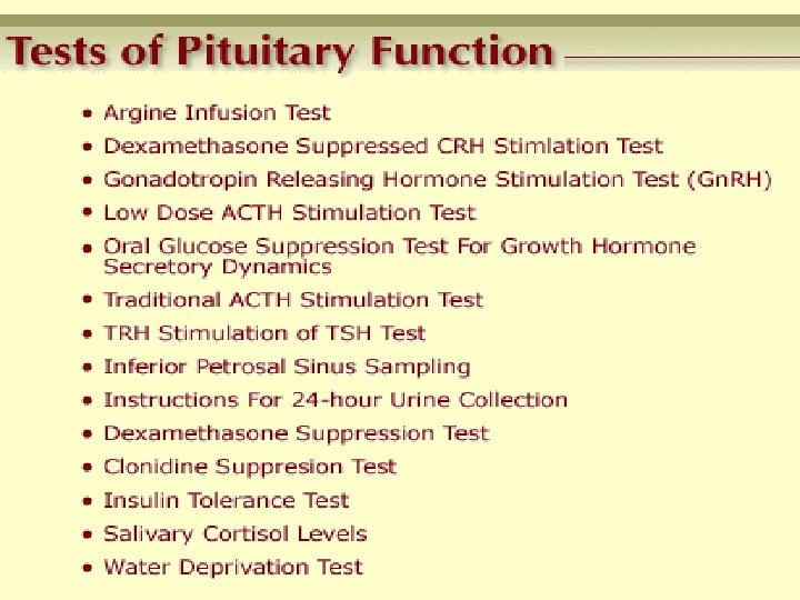
 1. 2. 3. 4. 5. Water deprivation")
 CHEMISTRY- it is octapeptide molecular weight of oxytosin is about 1000 oxytocin shares")


- Slides: 55
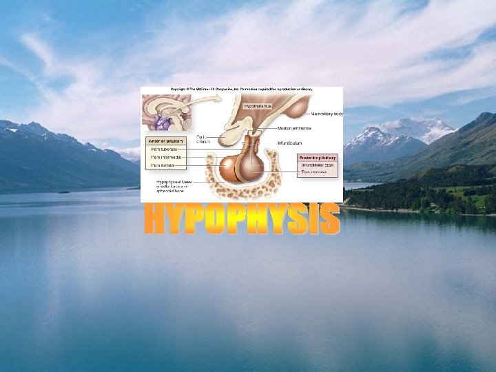
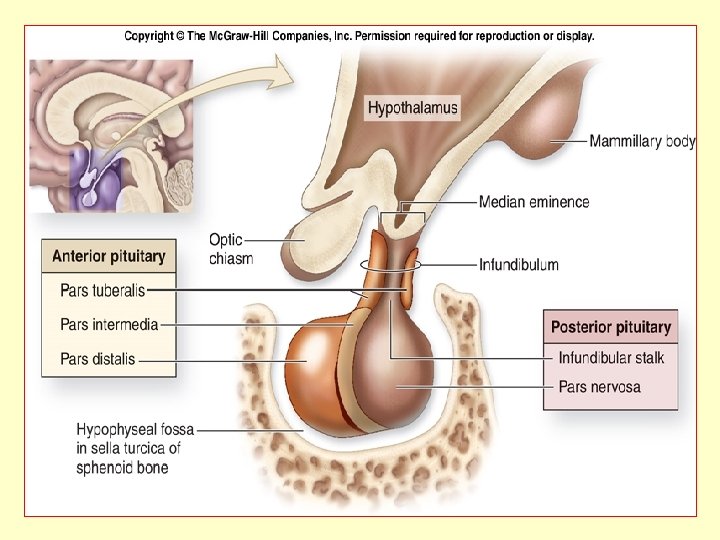
HYPOPHYSIS (PITUITARY GLAND) DEFINITION: - The pituitary gland is reddish-grey, small ovalshaped structure, situated at the base of brain in the sella turcica of sphinoid bone. WORD MEANING : The pituitary(L. pituita=phlegm ) which collected wastes from the brain and these lubricated the nasal and pharyngeal passages. When the true function of the gland was established the term hypophysis( hypo+Gr. Phyein=to grow) was substituted
ANATOMY It situated in the sella turcica of the sphenoid bone. It is attached to the floor of third ventricle by the stalk. average Weight in adults- 0. 5 to 0. 6 g. the weight varries with age , sex and physiologic state. It is somewhat larger in females, weighing from 0. 6 to 0. 7 g. It increases in size through the fourth decade of life, and during pregnancy. Anatomically it consist of two parts but microscopically it consist of 6 parts
DIVISION: - (1) Adenohypophysis(ant. pituitary) (a) pars distalis or anterior (b) pars tuberalis (c) pars intermedia (2) Neurohypophysis (post. pituitary) (a) pars nervosa (b) median eminence (c) infundibulum(neural stalk)
DEVELOPMENT Pituitary gland is originate from the ectoderm. it develops from two out growths. A glandular diverticuluam-rathkes pouch-grows upwards from the primitive buccal cavity and meets with a similar neural out growth from the floor of third ventricle. The stalk of rathke’s pouch disapperas but that nural out growth persists and forms infundibulum. Ant. Wall of rathke’s pouch develops the pars distails from the top arises the pars tuberalis, from the post. wall develops the pars intermedia. The pars intermedia invest the pars nervosa to some extent. The original cavity of rathke’s pouch forms the interglandular cleft.
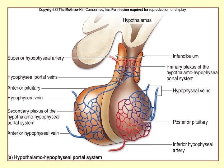
BLOOD SUPPLY The pituitary gland gets blood supply from the hypophysial arteries which originating from the internal carotid artery and circle of willis. the superior hypophyseal arteries supply the anterior lobe and the infirior hypophyseal arteries supply the posterior lobe. NERVE SUPPLY Ant. Lobe-there is definite evidence of a nervous control of ant. Lobe but only a few fibres either from the hypothalamohypophyseal tract of nerve fibers or from carotid plexus of cervical sympathatic have been traced to it. They are probably vasomotor nerves. Post. Lobe – two tracts of non-medullated nerve fibers arises from the hypothalamus and supply the pars nervosa.
ADENOHYPOPHYSIS Histology- it includes pars destalis 75% , pars tuberalis , and intermedia 25%. The present idea is that the different pituitary harmones are liberated by seprate cell types. so possibly six kinds of cells secrete six seprate harmones. studies employing histochemical, electronmicroscopic techniques have made such classifications possible CHROMOPHOBES (chief cells-50%) These cells are not stained with either basic dyes or acid dyes and lack typical secretory granules. chromophobe cells in general have got less cytoplasm than chromophils. the earlier hypothesis is that the chromophobes are non-functional and have recently been doubted.
CHROMOPHIL Cells are so named as they have got affinity toward dyes. Early staining methods show only twotypes of chromophils : Acidophil (eosinphil or oxyphil, 35%) – recent studies claim that acidophil cell secretes both growth hormone. (a) Somatotropic cells: - these cells are oval and the cytoplasm is packed with dense, round granules- 350 -400 nm in size, stained with orange G of an azan stain. tumors of an acidophil cells are associated with acromegaly & gigantism. (b) Lactotropic cells: - these types of cells stained with erythropsin or acid fuschin. No. of these cells varies in relation to the reproductive cycle– increasing significantly during pregnency &lactation. these cells are belived to secrete prolactin.
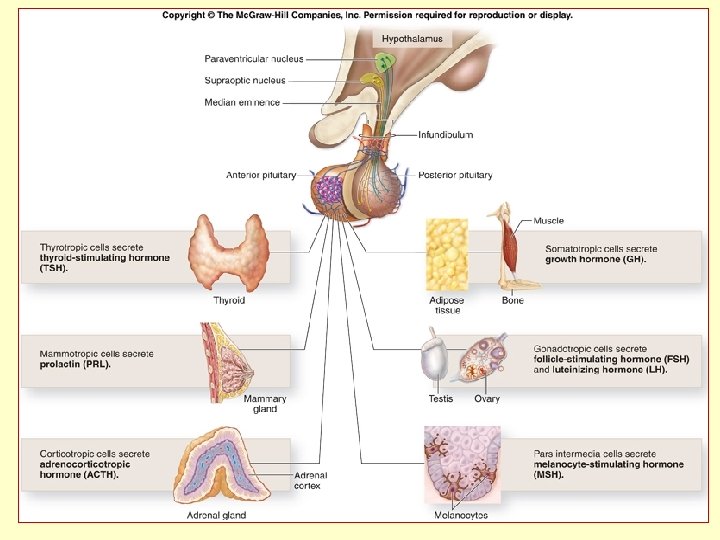
Basophils(mucoid cell-15%): There are different kind of cell secreting different types of trophic hormones like – (a) Thyrotropic cells: - secrete TSH. (b) Gonadotropic cells: - secrete GTH which are 2 types 1. FSH. 2. LH or ICSH. (c) Somatotropic cells: - secrete GH. (d) Corticotropic cells: - secrete ACTH and LH.
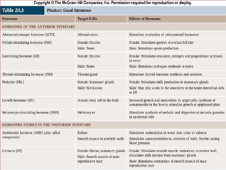
FUNCTIONS OF PARS DISTALIS OF PITUITARY Anterior pituitary is the master gland of the endocrine system because it produces protien tropic hormone which affect the other ductless gland. Following hormones are believed to be secreted : a. Growth hormone or somatotropic hormone(GH or STH). b. Adrenocorticotropic hormone(ACTH). c. Thyrotropic hormone(TSH). d. Gonadotropic hormone: 1. FSH 2. LH or ICSH 3. MH or LTH.
1. GROWTH HORMONE or SOMATOTROPIC HORMONE(GHor. STH) : -Secreted by acidophil cells. CHEMISTRY: -mol. Weight is about 21000 -48000. it is not effect- ive by oral administration as it is inactivated by pepsin &trypsin. ACTION OF GROWTH HORMONE: Skeletal Growth: -stimulates the multiplication of the epiphyseal cortilage and thus increases the length of cartilage bone. in hypophysectomised animals a membrane appears at the epiphyseal line which inhibit the growth of the cortilage cells. Regulates general body growth: - It increases the body growth due to the direct effect in the tissue. 1. Muscles – stimulates the growth of muscles. In gigantism & acromegaly there is an increased growth of muscles.
2. Visceras- in hypophysectomised animal the liver cells and reduced in size with impared functions. 3. It stimulates the growth of thymus. 4. Increases secretion of milk during lactation. 5. Nervous system is not affected by STH. Metabolism – a. on protein metabolism: - it increases the neuclic acid and protien synthesis, decreases nitrogen excrition in the urine and the nitrogen thus retained helps in the synthesis of tissue protein. STH is a protein anabolic hormone and prevents the catabolism of amino acids. b. on fat metabolism: - it increases the oxidation of fat and decreases the catabolism of amino acid causing utilisation of amino acid in the protein synthesis. c. on carbohydrates metabolism: - it stimulates carbohydrates storage. Administration of GH produces hyper - glycemia and glycosurea, GH is diabetogenic especially in man
Administration of ACTH produces similar effect as induced by GH. STH and ACTH exert antagonistic action to those of insulin. d. Ion or mineral metabolism: - it increases intestinal absorption of calcium as well as its excretion. In addition to Ca, Na, K, Mg, phosphate and chloride are retained. e. the growth hormone also has lactogenic effects. REGULATION OF GH SECRETION: 1. Hypothalamus: - secretion of growth hormone by pituitary acidophil cells are regulated by the hypothalamic hormone, the GHRF. 2. Other factors : - Certain factors may affect the GH secretion, these are –stimuli which increase GH secretion are- Hypoglycemia, Fasting Exercise Sleep: -Deep sleep stimulate growth. The young allowed to sleep always stimulate growth.
STRESS : Different stressful stimuli affect the secretion of GH. Surgical stress emotional stress etc. darkness inhibits growth, crowding stimulates growthes slowly, noise sometimes inhibits growth. HARMONS : (a) Thyroid hormone stimulates growth through the secretion of GH. (b) Androgen stimulate growth , but on prolongad treatment stops growth by closing the epiphysis. (c) ACTH infusion can stimulate HGH release during starvation FEEDBACK CONTROL: - Growth hormone secretion also controlled by the feedback inhibition process which is controlled by the GHIH releasing of the hypothalamus.
INHIBITORY FACTORS OF GROWTH (a) Hormonal factors. Oestrogen and other ovarion hormones have got influence on growth as because females are shorter than the males. It is suggested that ovarion hormones retard growth through inhibition of GH secretion. (b) Adrenal corticoids. Corticoids interfere with the growth process. Administration of corticoids causes dwarfism. The corticoids inhibit the stimulating effect of GH on epiphysis.
(2) THYROID STIMULATING HORMONES (TSH) CHEMISTRY : - it is a glycoprotein with molecular weight of about 25000. It is not effective by oral administration as it is inactivated by proteolytic enzymes. CONTROL OF TSH SECRETION: - thyrotrophin secretion is under the control of (1) hypothalamus (2) thyroxin level in the blood. (1) Hypothalamus: - stimulation of hypothalamus by fine electrodes causes release of a tripeptide humoral factor known as thyrotrophin releasing factor (TRF) which circulates through the blood stream (hypophyseal portal system) and helps in the liberation of thyrotrophin from the anterior pituitary.
(2) Thyroxin level in the blood: - the rate of secretion of thyrotrophin is controlled by the thyroxin content of the blood. High thyroxin content in the blood inhibits a low thyroxin content in the blood stimulates secretions. If the low thyroxin content persists for a prolonged period there will be continuous secretion of a large amount of thyrotrophin which causes thyroid hyperplasia.
FUNCTIONS OF TSH: The main role of TSH is to regulate the synthesis and production of thyroid hormone from the thyroid gland. These peripheral hormones play a basic job in our metabolism. In fact, they are important in catabolism, the mechanism by which our body burns calories and produces heat. Thyroid hormones are also very important in regulating other physiological actions: o brain development during fetal period, o heart function, o emotional reactions such as anxiety, nervousness or happiness Finally, they can interfere in the reproductive system by altering menstrual regularity and libido, or in the skeletal system by regulating bone mass.
(3) ADRENOCORTICOTROPHIC HORMONE (ACTH) Secreted by basophil cells. CHEMISTRY : - polypeptide in nature consisting of 39 amino acides residues. Molecular weight about 4500. It has been isolated in a α and β forms. Both these forms contain a large number of aminoacid and have got straight chain linkage. Unlike other pituitary hormones. ACTION: The hypophysis can regulate some aspects of the stress. Generally, stress can be defined as an emotional and physiological response to a disagreeable situation.
The pituitary is important in some severe and acute stress situations, during which it produces a hormone called ACTH stimulates the adrenal cortex, where it regulates the synthesis and production of cortisol. During severe trauma, burns, illness, fever, hypotension, physical exercises, surgery and hypoglycemia, cortisol production increases. Cortisol supports the body's adaptation to the stress condition, by increasing glucose use, the heart activity or electrolyte exchanges. Finally, ACTH also exerts a less important function in the adrenal cortex: steroid production. In 1975 scientists identified another peptide: endorphin, which acts in experimental animals as a natural pain reliever in times of stress.
Endorphin and ACTH are made as parts of a single large protein, which subsequently splits. This may be the body's mechanism for coordinating the physiological activities of two stress-induced hormones. The same large pro-hormone that contains ACTH and endorphin also contains short peptides called melanocytestimulating hormones. These substances are analogous to the hormone that regulates pigmentation in fish and amphibians, but in humans they have no known function. CONTROL OF ACTH SECRETION: ACTH secretion is under the of (1) hypothalamus (2) steroid feedback mechanism. (3) short feedback mechanism. (4) stress control mechanism.
4. GONADOTROPHIC HORMONES(GTH) The basophil cells secrete gonadotrophins which control the growth and activity of the gonad and indirectly all other process connected with it. the anterior pituitry controls the gonads is proved by the following facts. 1. Hypophysectomy, in young, retards growth and activity of the gonads. 2. Hypopituitarism in children (dwarfism) retards sexual devlopment and in adults leads the sexual degeneration (impotency, amenorrhoea etc. ). There are two gonadotrophins (1) FSH (2) LH or LTH or ICSH. (3) LTH(prolactin)
CHEMISTRY: The GTH are glycoprotein in nature and the molecular weight of about 30000. -: FUNCTIONS OF GTH: Function of FSH: - 1. In females: -a. increases the no. & size of Graffian follicals and prepares them for ovulation. After the menopause production rate of FSHis increased approximately 15 -fold. 2. In males: - stimulates spermatoogenesis. In old age in the male (as testicular function wanes) the production of FSH increases. Function of LH: 1. In females: The adenohypophysis produces two hormones, called luteinizing hormone (LH) and follicle stimulating hormone (FSH) which are required for development and maintenance of the sexual characteristics.
LH and FSH begin to be secreted at puberty in a pulsatile fashion. In women, FSH favors the development of one ovarian follicle at each menstrual cycle, and the production of estradiol by the granulosa cells. LH mediates ovulation (departure of the oocyte from the ovarian follicle) and the production of progesterone by the corpus luteum.
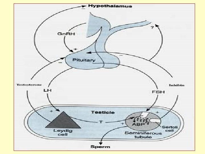
2. In males: - In men, LH stimulates production of testosterone by the Leydig cells of the testis. Testosterone is the main androgen hormone and it is responsible for male sexual development (male hairs, muscles, voice changes at puberty). In this general scenario, it is easy to understand how big is the role of pituitary in sexual life: by gonadotropins, this gland regulates the entire fertile period of woman and the fertility of men. LH stimulates the production of androgen-binding protein (ABP) by the Sertoli cells of the testis. This protein allows high local, near the sperm, concentrations of testosterone, on which the full maturation of the spermatozoa depends.
CONTROL OF GONADOTROPHIN SECRETION Gonadotrophin secretion is under the control of (1) Hypothalamus , and (2) sex hormones. 1 Hypothalamus : - hypotalamus nuclei are known to secrete specific releasing factors for the release of specific gonadotrophic hormones. luteinising hormone-releaseing, factor(LRF) for LH (ICSH) and Follicle-stimulateing hormone-releasinfg factor (FSH-RF) for FSH are secretecd from hypothalamus when they are necessry 2 Sex hormone : - injection of oestrogen or androgen for long periods, cause degeneration of anterior pituitary. secretion of gonadotrophin is inhibited causing atrophy of gonads
This shows that conc. Of sex hormons in blood regulates secretions of gonadotrophins. A high conc. Inhibits , where as a low concentration stimulates secretion. (c) LUTEOTROPHIC HORMONES (LTH) or MAMMOTROPHIC HORMONE (MH) : it is secreted during pregnancy and lactation in women by acidophil cells. It is peptied hormones, molecular weight is about 25000 and 180 to 190 amino acids are found. ACTION OF PROLACTIN : The pituitary gland regulates lactation by producing the hormone prolactin. This hormone is responsible for the maintenance of lactation, after the mammary gland has been prepared for milk production by estrogens. During the entire post-partum period prolactin is increased
because breast suckling stimulates prolactin production. The high prolactin value is also the reason why women are infertile during this period. In fact, this hormone can inhibit the production of gonadotropins and block follicle maturation in the ovary. EVIDENCES : - 1. Hypophysectomy in lectating animals prevents the onset of lactations (e. g. simmonds disease) 2. Injection of prolactin stimulate milk secretion in the fully develops mammary glands 3. Synthetic ergot alkaloids, which diminish prolactin secretion probably inhibit the release of performed hormone granules from the lactotrophs this shows that the ant. Pituitary produces a hormone(LTH) which controls the activity of mammary glands. The physiological role of LTH in the male is not known. ’
CONTROL OF PROLACTIN SECRETION: - 1. HORMONES: - The sex hormones and the placental gonado trophins inhibit prolactin secretion. although breast are fully devloped, yet lactation does not occur during pregnency. It starts after parturition and expulsion of placenta. 2. NERVOUS SYSTEM: - Suckling of the baby generates afferent impulses which reflexly stimulate prolactin secretion through htpothalamus. It is believed that suckling inhibits the median eminence factor (PIF) and thus prolactin secretion from the anterior pituitary is increased.
1. DYSFUNCTION OF ACIDOPHIL CELLS: a. Hyperactivity 1. ) in the young: -Gigantism 2. ) in the adult : -Acromegaly b. Hypoactivity – a. in the young-dwarfism, three types : 1. ) lorain-levy type 2. ) brissaud type 3. ) mixed type. b. in the adult- acromicria (very rare)
GIGANTISM Caused by hyperactivity of the acidophil cells, in the young. 1. Skeleton- avery tall (7 -8 feet) minimum 6. 8 feet 2. Muscles and visceras-proportionally large 3. Metabolism- a. hyperglycaemia, reduced sugar tolerance and may be , glycosurea(due to over secretion of STH and ACTH) b. BMR- raised c. SWEATING-increased(to help hit lost) 4. Changes in other glands a. thyroid- enlarged and hyperactive, hence may cause additional graves disease. b. adrienal cortex-hyper active and enlarged c. thymus- enlarged d. gonads-at first hyper active than may a trophy(over work and exhaustion)
G I G A N T I S M
ACROMEGALY Caused by hyperactivity of the acidophil cells, in adults 1. Skeletona. overgrowth of two jaws. b. hand feet- enlarged. c. Bowing of the spine (kyphosis) due to above mentioned skeletal causes a, b, c, the subject assumes gorilla like appearance. 2. Subcutaneous tissues- of the hand , feet, nose, scalp and skin increase in amount producing deep furrows. 3. Metabolism-a. hyperglycaemia, reduced sugar tolerance and may be , glycosurea(due to over secretion of STH and ACTH) b. BMR- raised c. SWEATING-increased(to help hit lost) 4. Viscera- are all enlarged such as heart, lungs, lever, spleen etc.
5. Other endocrines: a. thyroid- enlarged and hyperactive, hence may cause additional graves disease. b. adrienal cortex-hyper active and enlarged c. thymus- enlarged d. gonads-at first hyper active than may a trophy(over work and exhaustion) Other manifestation: In males- diminished libido and sometimes impotence. In females- devlopement of sterility, menstrual disturbences, failure of breast devlopment.
DWRFISM Caused by hypoactivity of the acidophil cells, in the young. Three types – 1. lorain-levy type(infantism): a. stunded growth. b. sex organ & secondary sex character do not grow c. intelligence- normal and proportionally to age. d. metabolism- normal. 2. Brissaud type- (fat boy of dikens) same as lorain-levy type, plus excess deposition of fat in the body. 3. Mixed type: -
DYSFUNCTION OFBASOPHIL CELLS CUSHING’S DISEASE Definition: - Cushing syndrome is a disease that occurs when your body produces too much of the hormone cortisol. It may also occur if you use take too much cortisol or other steroid hormones Causes : - Cushing syndrome is a condition that results from an excess of cortisol, a hormone produced by the adrenal glands. The most common cause of Cushing syndrome is Cushing's disease, caused by excessive production of the hormone ACTH by the pituitary gland. ACTH stimulates the adrenal glands to produce cortisol.
Cushing syndrome can be caused by a tumor of the pituitary gland, a tumor of the adrenal gland, a tumor somewhere other than the pituitary or adrenal glands (ectopic Cushing syndrome), or by long-term use of corticosteroids (drugs commonly used to treat conditions such as rheumatoid arthritis and asthma). Risk factors for Cushing syndrome are adrenal or pituitary tumors, long-term therapy with corticosteroids, and being female.
D W A R F I S M
DYSFUNCTION OF THE CHROMOPHOBE CELLS Usually caused by atumour of the inactive chromophobe cells. 1. In children; A; frochich’s syndroe (adiposogential dystrophy). (a) stunted growth(dwarfism). (b) idiotic. (c)sexual infantilism. (d) generalised obesity
LAURENCE-MOON-BIEDL SYNDROME (often familial). This is same as frohich’s syndrome(children type) plus (a) polydactylism, and (b) retinitis pigmentosa. 2. In adults- frochich’s syndrome-(adult type). A. in males (a) mental disposition and apperance resemble females (b) degeneration of sex (c) hand feet are small and tapering (d)skin of face and body is smooth and hairless. B. in female- (a) extreme adiposity (b) degeneration of sex.
TOTAL DISFUNCTION OF PARS DISTALIS Hyperactivity (degenrative changes) (1) In children. progeria apperance of early senility-often seen in pituitary dwarfs. (2) In adults . Simmonds disease Due to degeneration of anteriar lobe of pituitary 1. Cachexia-Loss of weight 2. Sexual degeneration –amenorrhoea , sterility, impotency 3. Asthenia –extreme weakness 4. Anorexia- loss of appetite 5. Low BMR 6. Mental deterioration 7. Hypoglycaemia ( 8) anaemia
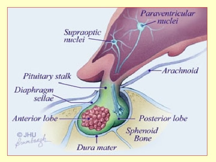
NEUROHYPOPHYSIS SYNTHESIS, TRANSPORT AND STORAGE OF NEUROHYPO- PHYSEAL HORMONES 1. VASOPRESSIN(PITRESSIN) Chemistry: -it is octapeptide. Vasopressin has molecular weight of about 1100. Vasopressin(ADH) is released in response to stress, and also in response to dehydration. Vasopressin has got antidiuretic propertyand increases facultative reabsorption of H 2 O and thus reduces the volume of urine formed. EVIDENCES: -disease of the post pituitary, hypothalamus or injury to the nerve tracts of gland produces a condition in which large amount of very dilute urine is produced. This condition is called diabetes insipidus.
CONTROL OF THE SECRETION OF VASOPRESSIN (ADH) 1. 2. 3. 4. 5. Water deprivation Negative pressure breathing Positive pressure breathing Haemorrhage and other changes in the blood volumue Hypoxia
OXYTOCIN(PITOCIN) CHEMISTRY- it is octapeptide molecular weight of oxytosin is about 1000 oxytocin shares some of the activities of ADH or vice versa and thus oxytocin is minimally antidiuretic. ACTION OF OXYTOCIN; 1. Uterus – the action of oxytocin on uterus depends upon the species and pretreatment of the animal with oestrogen. 2. Milk ejection –it causes contraction of the myoepithilial cells around the mammery alveoli. 3. Metabolism –it produces hyperglycemia. 4. Sperm transport –oxytocin facilitates the transport of sperm in the female genital tract. 5. Water metabolism –oxytocin given alarge doses and in abundant fluid volume may cause water intoxication due to antidiuretic effect of oxytocin on the nephron.
DISORDERS OF THE FUNCTION OF THE POSTERIOR PITUITARY Destruction of the neurohypophysis causes dibetes insipidus (insipid =taste less). this is a condition in which large quantities of urine of very low sp. Gr. – 1. 002 to 1. 006 and low cloride content are excreted. but further development of lesion may destroy the pars distalis with amelioration of the diabetes.
SOME IMPORTANT QUESTIONS: 1. Which endocrine Glands are regulated by anterior pituitary hormones? 2. Describe Hypophysis with its hormone. 3. Anterior pituitary is called the master gland of endocrine system. Why? 4. Write short notes on: - 1. Cushing’s syndrome. 2. Gigantism. 3. Acromagaly. 4. Dwarfism. 5. Describe hormones of post. pituitary. 6. Functions of ACTH. 7. Describe trophic hormones. 8. Describe regulation of Growth hormone secretion.
REFERED BOOKS: 1. Dr. C. C. Chatterjee 2. Gerard J Tortora 3. A K Jain. 4. Dr. Choudhary