PITUITARY GLAND THYROID GLAND HYPOPHYSIS CEREBRI PITUITARY GLAND


PITUITARY GLAND THYROID GLAND
")
HYPOPHYSIS CEREBRI ( PITUITARY GLAND )
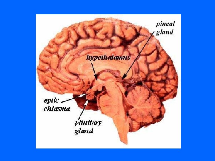

PITUITARY GLAND
 ADENOHYPOPHYSIS CEREBRI: 1 - Pars Distalis (pars anterior) 2 - Pars Tuberalis")
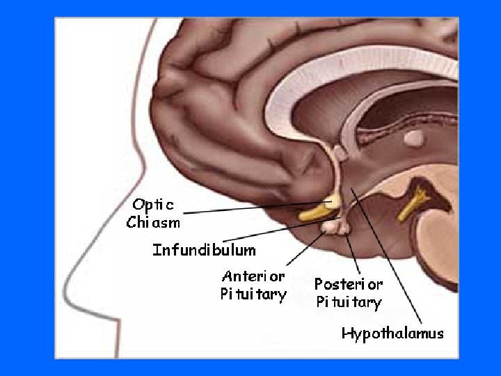
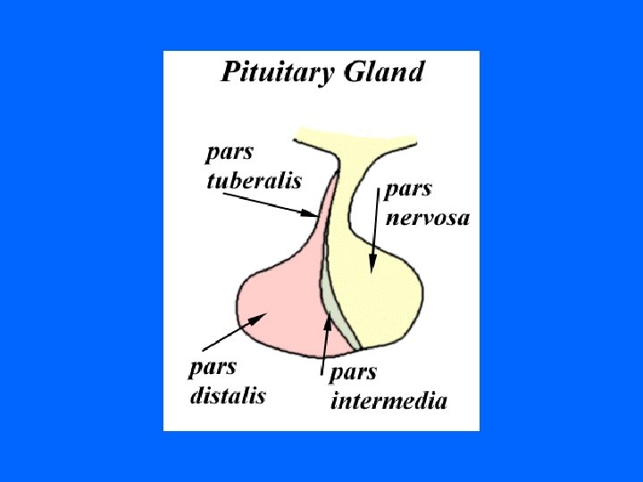
COMPONENTS (A) ADENOHYPOPHYSIS CEREBRI: 1 - Pars Distalis (pars anterior) 2 - Pars Tuberalis 3 - Pars Intermedia (B) NEUROHYPOPHYSIS CEREBRI: 1 - Median eminence 2 - Infundibulum: Neural (Infundibular) Stalk 3 - Pars Nervosa

DEVELOPMENT
 ADENOHYPOPHYSIS: Arises as outpocketing (evagination) of ectoderm from the roof of the")
DEVELOPMENT (A) ADENOHYPOPHYSIS: Arises as outpocketing (evagination) of ectoderm from the roof of the primitive mouth of embryo → Rathke’s Pouch → Separates from oral cavity. (B) NEUROHYPOPHYSIS: Arise as a down growth from the floor of diencephalon, without detaching from the brain.

PITUITARY GLAND

PITUITARY GLAND

NEUROHYPOPHYSIS xxxxxx XXXXX

PARS NERVOSA

PARS NERVOSA

PARS NERVOSA

PARS NERVOSA

PARS NERVOSA
 PARS NERVOSA CONTENTS: 1 - Unmyelinated axons of secretory neurons situated in")
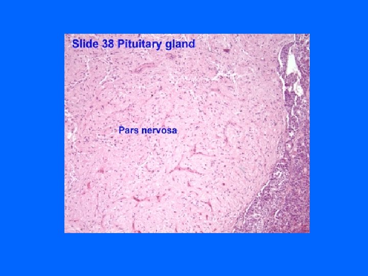
NEUROHYPOPHYSIS (A) PARS NERVOSA CONTENTS: 1 - Unmyelinated axons of secretory neurons situated in supraoptic & paraventricular nuclei (i. e. Axons of hypothalamhypophyseal tract). 2 - Herring’s Bodies: contain neurosecretory granules. 3 - Fenestrated blood capillaries. 4 - Pituicytes: branched glial-like cells. N. B. No secretory cells in pars nervosa.

HERRING BODIES - Are blue-black stained distentions of the axons in p. nervosa. - Representing accumulation of neurosecretory granules at axon termini and along the length of the axons in p. nervosa.

PITUICYTES Are glial-like cells in p. nervosa. Structure: Have numerous cytoplasmic processes with gap junctions. Functions: 1 - Support the axons of the p. nervosa. 2 - May have a trophic function.
 2 -")
Function of p. nervosa: Storage & release of: 1 - Vasopressin (ADH) 2 - Oxytocin

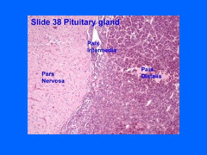
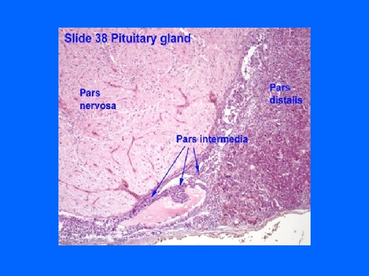
P. DISTALIS, P. INTERMEDIA & P. NERVOSA
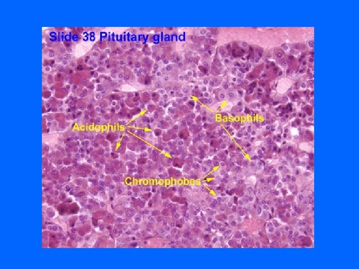

PARS DISTALIS Blue arrow: acidophils Red arrow: basophils Yellow arrow: chromophobes

Blue arrow: acidophils Red arrow: basophils Yellow arrow: chromophobes

PARS DISTALIS

PARS DISTALIS

GH CELLS

PROLACTIN CELLS

ACTH CELLS

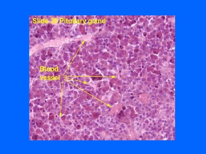
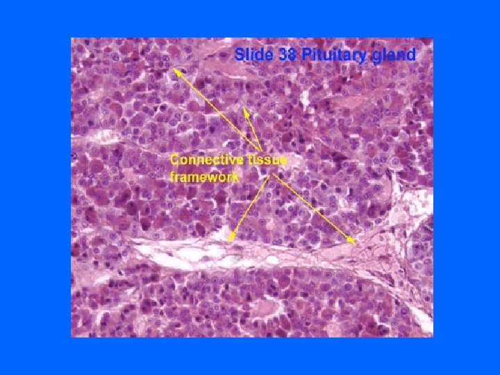
PARS DISTALIS Stroma: 1 - Fibrous capsule. 2 - Scant C. T. located mainly around aa &vv. (hypophyseal arteries & portal veins). 3 - Reticular fibers: around the cords of cells & “ the sinusoidal capillaries.

PARS DISTALIS Parenchyma: 1 - Cords of parenchymal cells. 2 - Sinusoidal capillaries: With fenestrated endothelium.
 Chromophils: a- Acidophils: 1 - Somatotrophs (Somatotropic")
PARS DISTALIS Types of parenchymal cells (1) Chromophils: a- Acidophils: 1 - Somatotrophs (Somatotropic cells). 2 - Mammotrophs (Mammotropic cells). b- Basophils: 1 - Thyrotrophs (Thyrotropic cells)(TSH) 2 - Gonadotrophs (Gonadotropic cells) (FSH, LH) 3 - Corticotrophs (ACTH, Lipotropic H) 4 - Melanotropes ? ? ? ?
 Chromophobes: may represent: 1 - stem cells. 2 -")
Types of parenchymal cells (2) Chromophobes: may represent: 1 - stem cells. 2 - degranulated chromophils. 3 - degenerated cells. (3) Folliculostellate cells: Structure: have long processes with gap j. Function: Not clear ( are non-secretory) ? ? ? (may be: 1 -supporting, 2 -phagocytic).

PARS TUBERALIS Structure: Cuboidal basophilic cells. Function: Possibly contain FSH & LH.
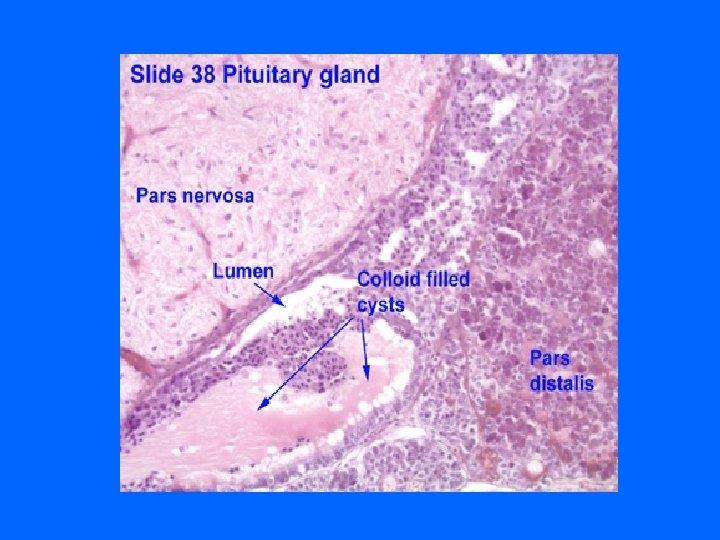
 L/M: 1 -Numerous colloid-containing cysts (Rathke’s cysts): Are lined with")
PARS INTERMEDIA (ZONA INTERMEDIA) L/M: 1 -Numerous colloid-containing cysts (Rathke’s cysts): Are lined with cuboidal cells. Are remnants of Rathke’s pouch. 2 -Cords of weakly basophilic cells. 3 - Network of blood capillaries. Function: Secrete proopiomelanocortin→ Gives: α-MSH, Corticotropin, ß-lipotropin & ß-endorphin. N. B. α-MSH in human stimulates prolactin release.

BLOOD SUPPLY
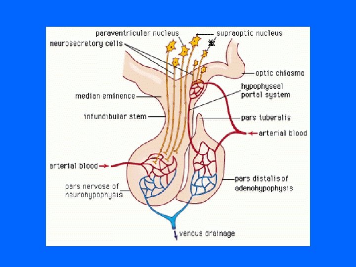
Sup. Hypoph. Arteries (Rt & Lt): To median eminence & Neural stalk")
BLOOD SUPPLY (1)Sup. Hypoph. Arteries (Rt & Lt): To median eminence & Neural stalk → 1 ry capillary plexus of fenestrated capillaries → Hypophyseal portal Veins (or venules) → 2 ry capillary plexus of capillaries in adenohyp [ Hypophyseal Portal System ] [“ “ Circulation ] It carries neurohormones from median eminence to adenohypophysis. (2) Inf. Hypoph. Arteries (Rt & Lt): Mainly to pars nervosa, They are Not participating in hypophyseal portal circulation. !!!!!!!!!!!!!!!!!!!

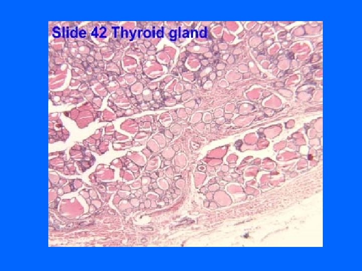
THYROID GLAND

THYROID GLAND


THYROID GLAND

THYROID GLAND

THYROID GLAND

THYROID GLAND

THYROID GLAND

THYROID GLAND
")
THYROID GLAND (E/M)

PARAFOLLICULAR CELL

STROMA OF THYROID GLAND 1 - Capsule: dense irregular collagenous C. T. 2 - Septa (Interlobular septa): “ “. 3 - Reticular fibers: Thin C. T. , composed mostly of reticular fibers with rich capillary plexus surrounds each thyroid follicle.

PARENCHYMA OF THYROID GLAND THYROID FOLLICLES: Are the structural and functional units of the thyroid gland.

THYROID FOLLICLES L/M: 1 - Simple cuboidal epithelium: a- Follicular cells. b- Parafollicular cells. 2 - Colloid: central colloid-filled lumen. N. B. Each follicle is surrounded by thin basal lamina.
 CELLS L/M: Simple cuboidal cells May be squamous or low columnar. Round")
FOLLICULAR (PRINCIPAL) CELLS L/M: Simple cuboidal cells May be squamous or low columnar. Round nucleus with prominent nucleoli. Basophilic cytoplasm.
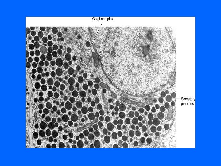
 CELLS E/M: - Mitochondria. - RER - Supranuclear Golgi Complex. - Numerous")
FOLLICULAR (PRINCIPAL) CELLS E/M: - Mitochondria. - RER - Supranuclear Golgi Complex. - Numerous apically-located lysosomes. - Numerous dispersed small vesicles: contain newly formed thyroglobulin. - Numerous apical short microvilli.
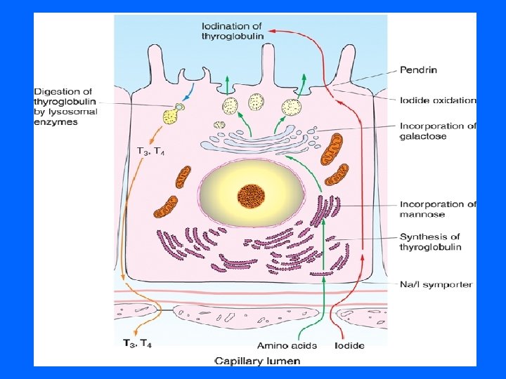
 CELLS Function: Synthesis of thyroid hormones (T 4 & T 3). 1")
FOLLICULAR (PRINCIPAL) CELLS Function: Synthesis of thyroid hormones (T 4 & T 3). 1 - Synthesis of thyroglobulin (glycoprotein). 2 - Trapping of circulating iodide. 3 - Oxidation of iodide into activated iodide (by lysosomal peroxidase). 4 - Iodination of tyrosine of thyroglobulin in the colloid at the colloid-follicular cell interface. 5 - Endocytosis of hormone-bound thyroglobulin→ Cleavage of T 4 & T 3 by lysosomal proteases.
 (C CELLS) L/M: - Pale stained cells. - Are found")
PARAFOLLICULAR CELLS (CLEAR CELLS) (C CELLS) L/M: - Pale stained cells. - Are found singly or in clusters in between the follicular cells. - Do not reach the lumen of the follicle. - Are larger than follicular cells (2 -3 times). - Only 0. 1% of the epithelial cells. - Have round nucleus
 (C CELLS) E/M: - Mitochondria. - RER (moderate). - Well-developed")
PARAFOLLICULAR CELLS (CLEAR CELLS) (C CELLS) E/M: - Mitochondria. - RER (moderate). - Well-developed Golgi. - Small dense secretory granules located mainly in the basal cytoplasm. Function: Secrete calcitonin.
- Slides: 68