Histology is the microscopic study of the structure


 that form continuous sheets. Desmosomes hold")
 arrangement (layers) i) simple (one layer of cells) ii) stratified")

 salivary glands and pancreas")


 Adipose tissue b) Areolar tissue c) Dense fibrous tissue-")


 that")

 • • • SUPPORTS & PROTECTS MOST RIGID CONNECTIVE TISSUE Hardness")


")


 -allows fluid rich in proteins & other")


- Slides: 55
Histology is the microscopic study of the structure and function of cells, tissues, and organs The four basic types of tissues are: 1) epithelial tissue 2) connective tissue 3) muscle tissue 4) nerve tissue
Epithelial tissue Lining, covering & glandular tissue of body n Lining and covering epithelium covers external and internal surfaces of the body n Glandular epithelium forms various glands in the body. Epithelium functionsabsorption, protection, filtration & secretion
Epithelial tissue -Fit closely together ( excluding glandular) that form continuous sheets. Desmosomes hold neighboring cells together. -Always one free (unattached) surface called the apical surface. -The lower surface rests on a basement membrane. -Avascular -Rely on diffusion from capillaries below. -Regenerate themselves easily if well nourished.
CLASSIFICATION Based on: 1) arrangement (layers) i) simple (one layer of cells) ii) stratified (> than one cell layer) 2) basis of cell size i) squamous ii) cuboidal iii) columnar
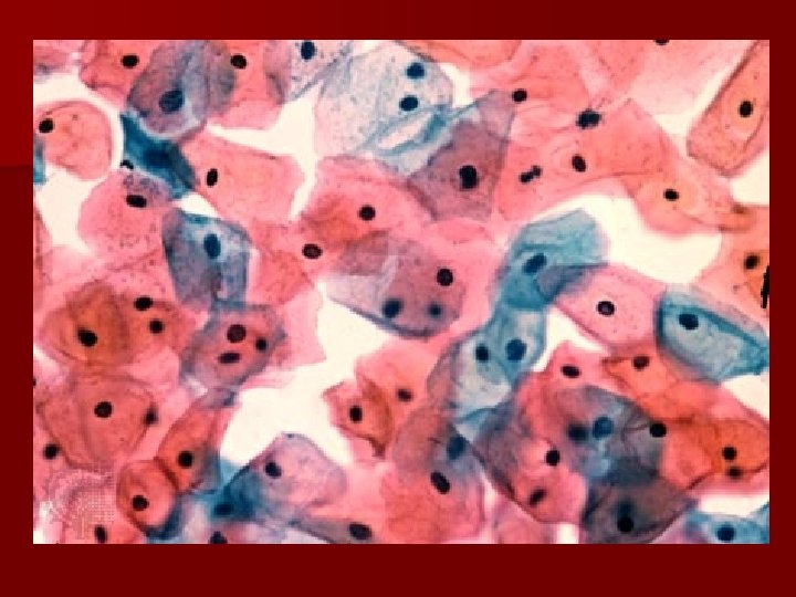
Squamous epithelial n Thin coverings and flat n Irregular shaped n Line heart, blood and lymphatic alveoli & body cavities vessels,
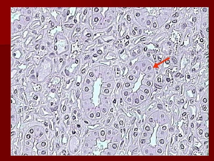
Cuboidal epithelium n Common in glands and their ducts ex) salivary glands and pancreas Covers surface of ovaries, kidney tubules
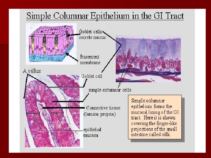
n Involved Columnar epithelium in secretion and uptake of materials n Elongated with nucleus near bottom n Digestive tract, parts of respiratory tract and glands
Glandular epithelium n Specialized to secrete materials such as digestive juices, hormones, milk, perspiration and wax. n Can be columnar or cuboidal in shape Endocrine and exocrine glands
Connective tissue is found throughout the body. Its functions range from filling spaces, connecting, supporting, and protecting other tissues to fighting disease. Major cell types 1) Fibroblasts- produce fibers Collagen fibers- ligaments & tendons Elastic fibers (elastin) 2) Macrophages- phagocytic WBC 3) Mast cell- produce histamine & heparin
CONNECTIVE TISSUE Types include: a) Adipose tissue b) Areolar tissue c) Dense fibrous tissue- tendons, ligaments, fascia d) Supportive tissue- bone & cartilage e) Vascular tissue- blood & lymph
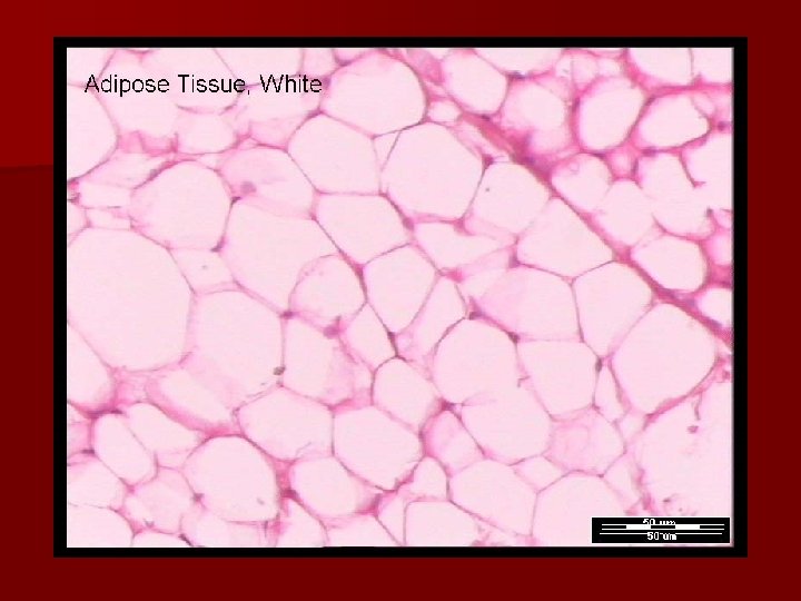
Adipose tissue n Type of loose connective tissue n Composed of saclike adipose cells n Stores lipids (fat) n Acts as filler tissue n Cushions and supports n Insulates body
Areolar tissue Also a type of LOOSE connective tissue n Most widely distributed connective tissue n Fluid matrix containing all types of cells & fibers: collagen, elastin, mast cells and various WBCs n When a body region is inflamed the loose areolar tissue soaks up excess fluid and swell = EDEMA n Supports nerve cells, blood vessels
Dense connective tissue n Dense fibrous tissue n Ligaments- strong flexible band (cords) that hold bones together at joints. ** n Tendons are white bands that connects skeletal muscle to bone n Fascia- fibrous sheet that wrap around muscle bundles to hold in place n Poor blood supply- heal slowly
Supportive connective tissue BONE & CARTILAGE
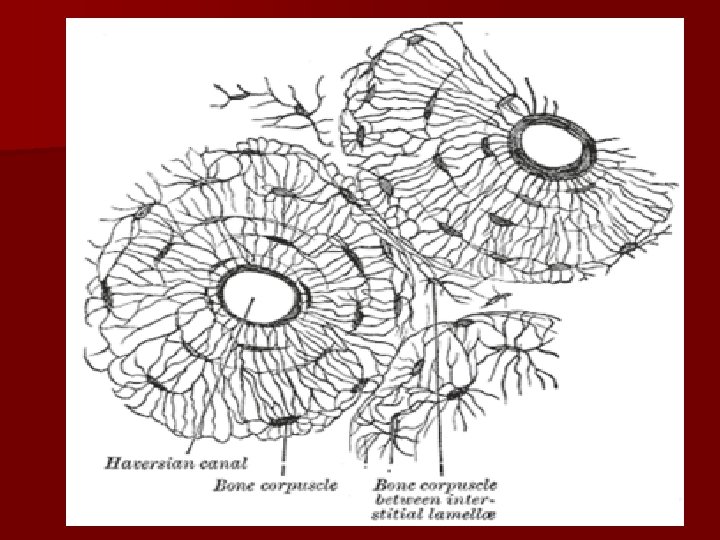
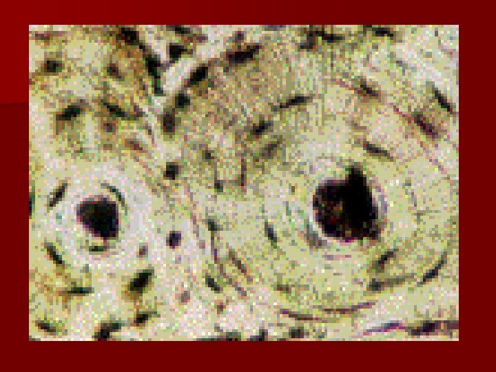
BONE (OSSEOUS TISSUE) • • • SUPPORTS & PROTECTS MOST RIGID CONNECTIVE TISSUE Hardness due to mineral salts COMPOSED OF BONE CELLS IN CAVITIES and surrounded by collagen fibers. Haversian canals have blood vessels which allows exchange of materials between blood and bone cells.
Cartilage n Rigid connective tissue n Provides support, frameworks, attachments, protects underlying tissues, and forms structural models for many developing bones n Cartilage cells are chondrocytes Three types: hyaline cartilage, fibrocartilage and elastic cartilage
Hyaline cartilage n most common ends of bones at joints. soft part of nose bronchi & bronchiole tubes costal cartilage
Fibrocartilage n Very tough tissue that contain many cartilagenous fibers n Form pads (discs) n Knee meniscus n Pubic symphysis
Elastic cartilage n More flexible that hyaline due to larger numbers of elastic fibers in extrcellular matrix. n External ears n Part of larynx
Vascular connective tissue Blood & Lymph
Blood n Composed of cells suspended in a fluid extracellular matrix called plasma n The cells: RBCs, WBCs and platelets n Carries nutrients, wastes, respiratory gases, plus other materials.
n Defend body n clotting n Carry oxygen
Lymph n Transports tissue fluid, proteins, fats from tissues to circulatory system. n Fluid that leaks out of vessels. n Lymphatic vessels carry lymph n Filtered by lymph nodes.
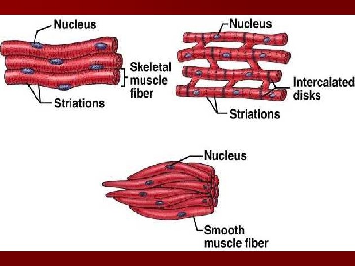
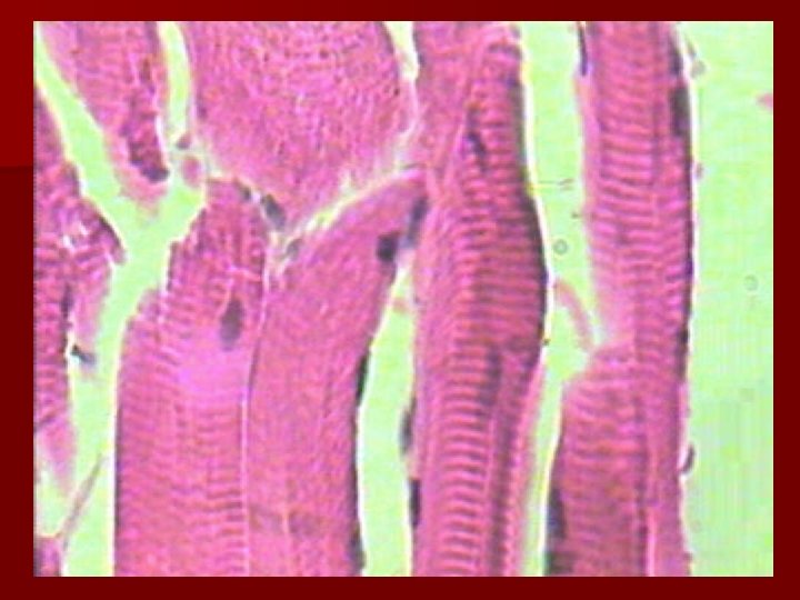
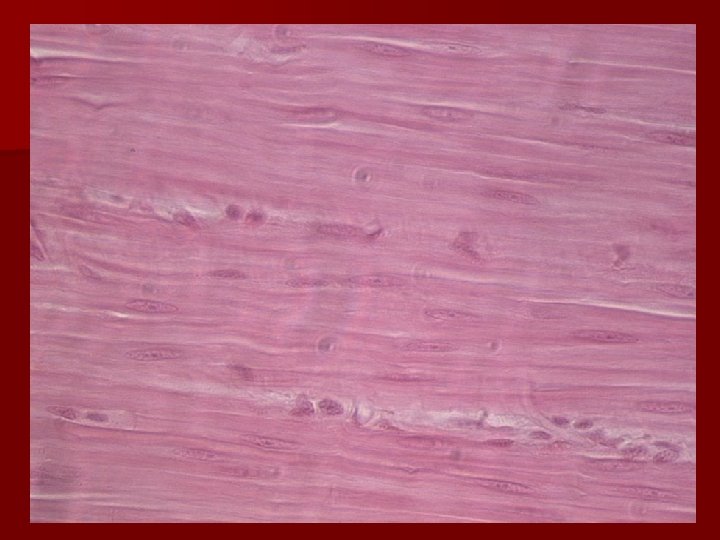
Muscle tissue • Functions to move and support body parts • Forms the walls of the heart and blood vessels Three subtypes of muscle tissue 1) striated (skeletal) muscle 2) Smooth (visceral) muscle 3) cardiac muscle
n Cardiac muscle found only in the HEART n Skeletal muscle is VOLUNTARY and striated. n Smooth muscle is INVOLUNTARY. Not striated and in organs & blood vessels.
Its time for a quiz !!!!
Nerve tissue Composed of specialized cells called nerve cells or NEURONS and supporting cells. Neurons receive stimuli and conduct impulses to and from all parts of the body. The supportive cells, which have a gluelike consistency, bind and support neurons
The Neuron TERMINAL BRANCHES DENDRITES CELL BODY AXON MYELIN SHEATH
After injury: n Capillaries become permeable (hemostasis) -allows fluid rich in proteins & other substance to seep into injured area. -clot forms & stops blood loss, walls off area and prevents bacteria and other harmful substance from spreading to other tissue. n Granulation tissue forms new capillaries, that bleed freely to form a scab. Contain phagocytes and fibroblasts n Surface epithelium regenerates under scab
n Hemostasis n Inflammatory n Remodeling phase
Tissue repair n 2 mechanism Regeneration: replacement of destroyed tissue by same kind of cells Fibrosis: repair by dense (fibrous) connective tissue = formation of scar tissue Which occurs depends on -type of tissue damaged -severity of the injury Generally speaking clean cuts heal more successively than ragged tears
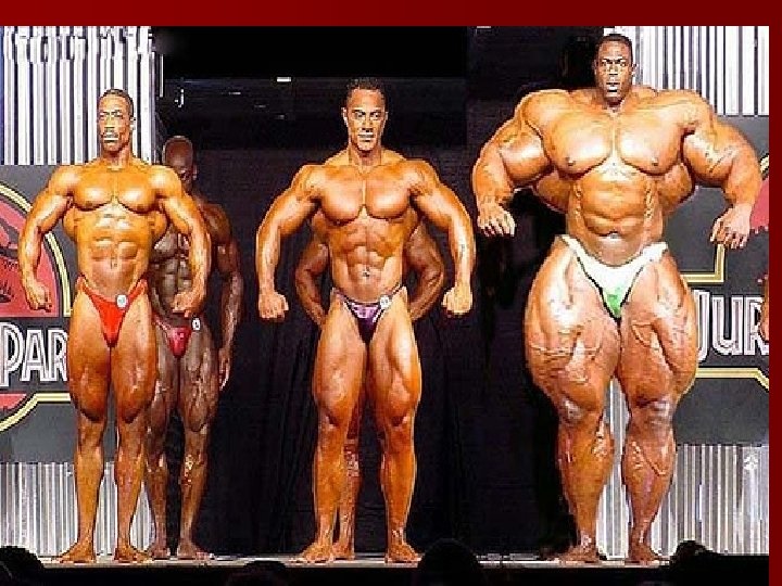
Disorders of cell structure n Atrophy- decrease in size n Hypertrophy- increase in size n Hyperplasia- increase in number n Metaplasia- change into other type of cell n Dysplasia- change in size, shape and organization (usually progresses to neoplasia. n Neoplasia- change is cell structure with an uncontrolled growth pattern
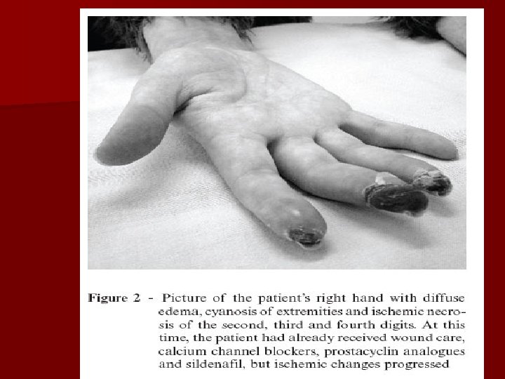
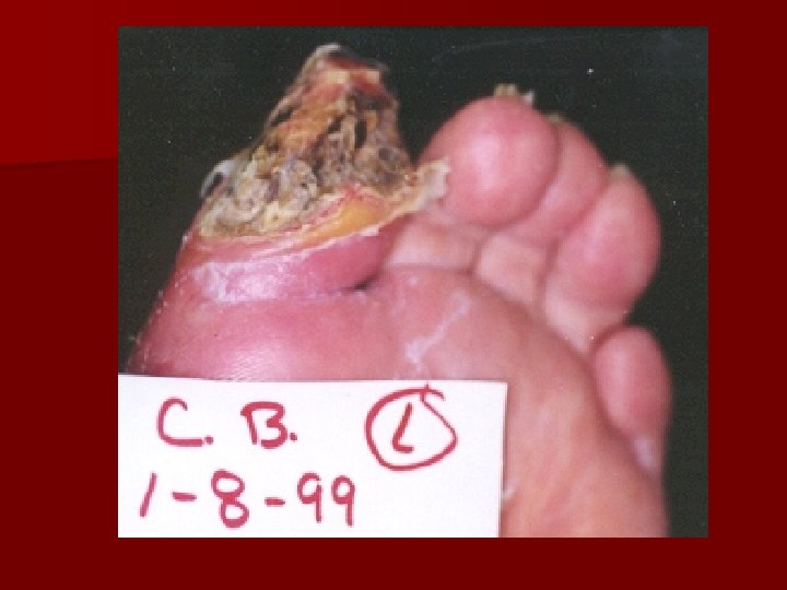
Trauma or injury can affect structure of a cell. n Hypoxia- decreased blood flow n Anoxia- lack of oxygen Bacterial toxins or viruses can also result death. Congenital defects (birth defects) alter structure.
decrease in size
increase in size
increase in size
increase in number
change into other type of cell
change in size, shape and organization
change is cell structure with uncontrolled growth pattern