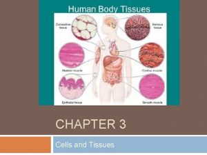
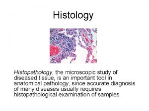
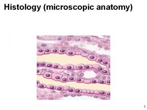
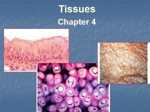
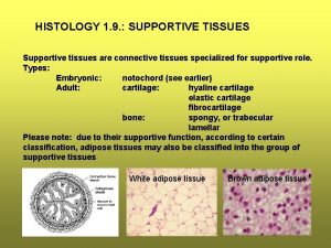
Histology Study of microscopic anatomy Study of tissues


- Slides: 42

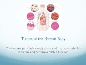
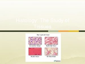
Histology • Study of microscopic anatomy • Study of tissues • Four primary tissue classes – – epithelial tissue connective tissue muscular tissue nervous tissue

I. Epithelial Tissue A. Characteristics 1. Cellularity – how the cells are arranged, composed of mainly closely packed cells with little or no extra cellular matrix.

2. Special contacts– cell components that tightly join each cell together. • Tight junctions – act as a “zipper” to hold plasma or cell membrane together. • Desmosomes – “rivet” like structures to hold plasma membrane together at certain spots. Tight junction desmosome

3. Polarity – cells differ at the free surface or or apical surface (top surface) than at basement membrane or basale (basal) layer (bottom layer) 4. Basement membrane – the border between the epithelium and connective tissue (CT). Made up of a supportive sheet of lamina membrane, made up of glycoproteins connected to reticular tissue– helps resist tearing and stretching of tissue, used in diffusion of needed nutrients to epithelial tissue

5. Innervated – it is supplied with nerve fibers 6. Avascular – NOT vascular – it has no blood vessels, all nourishment and gas exchange come from the underlying connective tissue. 7. Regeneration – it has a high mitotic division rate (it undergoes mitosis rapidly).

II. Types of Epithelial Tissue Membranous epithelilum Simple squamous simple cuboidal simple columnar simple ciliated columnar pseudostratified Stratified squamous (keratinized and non keratinized) stratified cuboidal stratified columnar transitional Glandular epithelium- special type of epithelium used to make glands in the body exocrine and endocrine glands will be discussed in later chapters

Simple Versus Stratified Epithelia • Simple epithelium – contains one layer of cells – named by shape of cells • Stratified epithelium – contains more than one layer – named by shape of apical cells

A. Simple Squamous Epithelium • Single row of flat cells • Allows rapid diffusion of substances; secretes serous fluid • Found in alveoli, endothelium, & serosa

B. Simple Cuboidal Epithelium • Single row of cube-shaped cells, often with microvilli • Absorption & secretion; produces mucus • most kidney tubules

C. Simple Columnar Epithelium Goblet cell Microvilli • • Single row of tall, narrow cells, some contain goblet cells Ciliated type- simple ciliated columnar Absorption & secretion; secretion of mucus Inner lining of GI tract, uterus, kidney & uterine tubes

D. Pseudostratified Epithelium Cilia Goblet cell Basal cell • Single row of cells not all of which reach the free surface – nuclei of basal cells give layer a stratified look • Secretes and propels respiratory mucus • Found in respiratory system (trachea)

E. Keratinized Stratified Squamous • Multilayered epithelium covered with layer of compact, dead squamous cells packed with protein keratin • Retards water loss & prevents penetration of organisms • Forms epidermal layer of skin

F. Nonkeratinized Stratified Squamous Epithelial layer • Multilayered, lacks surface layer of dead cells • protection • Found on tongue, esophagus & adult vagina

G. Stratified Cuboidal Epithelium • Two or more layers of cells; surface cells square • Secretion and protection • Found sweat gland ducts; ovarian follicles & seminiferous tubules

H. Stratified Columnar • Structure: many layers thick, top layer is column-shaped cells • Location: overall are rare but are usually found in the male urethra • Function: overall protection, absorption, secretion

I. Transitional Epithelium • Multilayered epithelium with rounded surface cells that flatten when the tissue is stretched • Stretches to allow filling of urinary tract, change shape • Found in urinary tract -- kidney, ureter, bladder

SIMPLE SQUAMOUS

SIMPLE CUBOIDAL

SIMPLE COLUMNAR

PSEUDOSTRATIFIED

STRATIFIED SQUAMOUS KERATINIZED

TRANSTIONAL

TRANSITIONAL (STRETCHED)

III. Connective Tissue Characteristics A. Found everywhere in the body – it is the most abundant tissue B. Chief subclasses – loose, dense, cartilage, other. C. Have a common origin – all connective tissue comes from mesenchyme (embryonic connective tissue) D. Degrees of vascularity – cartilage is avascular dense has poor vascularity, loose has high vascularity. E. Extra cellular matrix – connective tissue is composed of generally a non-living extra cellular matrix that separates cells in the tissue. Matrix is in the form of gel, liquid, solid, gel-sol. It gives connective tissue the ability to bear weight and withstand tension F. Made up of cells(cytes, blasts, clasts), fibers( reticular, elastic, collagen), and ground substance

Cellscytes- mature cell blasts- building cells clasts- repair or remodeling cells Common cells osteoblasts fibroblasts- most common chrondroblasts osteocytes osteoclasts fibrocytes chrondrocytes adipocytes erythrocytes leukocytes Also mast cells and macrophages used in immunity

• Fibers • Collagen fibers- “white fibers” provide strength • Elastic fibers- “yellow fibers” stretchy • Reticular fibers “fuzzy net” finely branched collagen fibers • Ground substance- unstructured material that fills spaces between cells and contains fibers. Varies from fluid to gel/sol.

IV. Types of Connective Tissue Loose areolar adipose reticular Dense dense regular dense irregular Cartilage hyaline cartilage elastic cartilage fibrocartilage Other bone (osseous) blood

A. Areolar Tissue • Type of loose tissue • Makes up soft packaging around organs

B. Reticular Tissue • Type of loose tissue • Lymphoid organs, ex. spleen

C. Adipose Tissue Types of loose tissue In subcutaneous layer of skin (hypodermis)

D. Dense Regular Connective Tissue • Type of dense tissue • Makes up tendons and ligaments

E. Dense Irregular Connective Tissue • Type of dense tissue • Makes up dermis of skin

F. Hyaline Cartilage • Type of cartilage tissue • Over ends of bones at movable joints; fetal skeleton

G. Elastic Cartilage • Type of cartilage tissue • External ear and epiglottis

H. Fibrocartilage • Type of cartilage tissue • Pubic symphysis & intervertebral discs

I. Bone Tissue (compact bone) • Type of “other” connective tissue • Found in bony skeleton

J. Blood • Type of “other” connective tissue • Found in heart and blood vessels

V. Muscle Tissue • Muscle Tissue (3 types) – gives the ability to contract • Classified by voluntary or involuntary or striated or smooth • Skeletal- voluntary and striated(banded) • Cardiac- involuntary and striated(banded) • Smooth- involuntary and smooth

Skeletal Muscle • Long, cylindrical, unbranched cells with striations and multiple nuclei • Skeletal muscles

Cardiac Muscle • Short branched cells with striations and intercalated discs; one central nuclei per cell • Found in the heart

Smooth Muscle • Short fusiform cells; nonstriated with only one central nucleus • Sheets of muscle in walls of hollow organs, iris of eye, smooth muscle in dermis

VI. Nerve Tissue • Large neurons with long cell processes surrounded by much smaller neuroglial cells lacking dendrites and axons • Conduct impulses • Found in brain, spinal cord, nerves
 Body tissue
 Four major tissue types
 Body tissues chapter 3 cells and tissues
 Eisonophil
 Body tissues chapter 3 cells and tissues
 Where is bile secreted
 Art labeling activity figure 23.5
 Microscopic anatomy of compact bone
 Microscopic anatomy of skeletal muscles
 Microscopic anatomy of skeletal muscle figure 6-2
 Microscopic anatomy of skeletal muscle figure 6-2
 Microscopic anatomy example
 Histopathology is a subdiscipline of microscopic anatomy.
 Microscopic anatomy of skeletal muscle
 Microscopic anatomy of the stomach
 Supportive connective tissue
 Study of diseased tissue
 Collagen location in body
 Anatomy histology slides
 Anatomy and physiology revealed
 Pancreas anatomy and physiology
 Liver histology
 Pseudostratified ciliated columnar epithelium
 Stool exam normal values
 Microscopic organism definition
 Gse test
 Reasonable care model of responsibility
 Ne2t/m
 Ohm's law microscopic form
 Heat is simply another word for
 Virtual urinalysis lab answers
 Microscopic view of current
 Glioblastoma multiforme
 Solid liquid gas particles
 Optical discs store items by using microscopic
 Microscopic examination of metals
 Preparat natif
 Father of bloodstain identification
 Microscopic algae
 Diagram microscopic structure of bone
 Evaluasi simplisia
 Shaft of the bone
 Direct microscopic count

