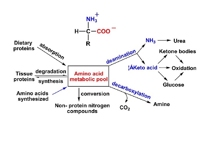
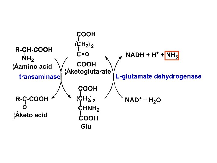
Four types of AA deamination transamination oxidative deamination


 Formation of urea (2) Formation of Gln (3) Excrete in urine")


 jaundice • Hepatocellular (hepatic) jaundice • Obstructive (posthepatic) jaundice")


 is a analog of hypoxanthine.")

 is a analog of thymine.")

 is a analog of Gln.")
 and Methotrexate (MTX)")
 inhibits the synthesis of d. CDP.")
- Slides: 42
Four types of AA deamination: transamination oxidative deamination union deamination non-oxidative deamination
Source and outlet of NH 3 1. Sources: ⑴ Endogenous sources: ① Deamination of AAs--main source ② Other nitrogen containing compounds ③ Kidney secretion (Gln) NH 3+H+ NH 4
⑵ Exogenous sources: ① Putrefaction in the intestine. ② Degradation of urea in the intestine NH 3+H+ NH 4
2. Outlets: (1) Formation of urea (2) Formation of Gln (3) Excrete in urine (4) Synthesis of AA
Summary
One carbon units are carried by FH 4
Transmethylation and Met cycle
PBG Heme ALA dehydratase ALA synthase ferrochelatase Gly succinyl Co. A Linear tetrapyrrol Protoporphyrin Ⅸ Protoporphyrinogen Ⅸ CPGⅢ UPGⅢ
coenzyme -pyridoxal phosphate ① ALA synthase ② decarboxylase ③ transaminase
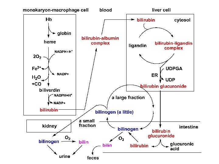
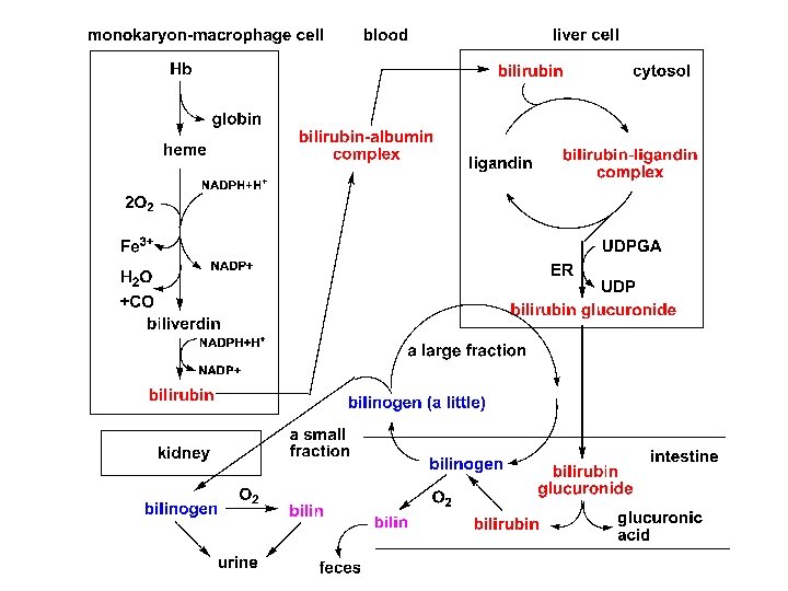
1. Formation and transport of bilirubin *The source of bilirubin The compounds involving iron prophyrin in the body are hemoglobin, myoglobin, cytochrome, peroxidase, and catalase, etc.
• Formation of bilirubin
hydrophobic
structure of bilirubin diglucuronide Carbosyl group Hydroxyl group
Jaundice • Hemolytic (prehepatic) jaundice • Hepatocellular (hepatic) jaundice • Obstructive (posthepatic) jaundice
Laboratory results in patients with jaundice normal Hemolytic jaundice < 1 mg/dl > 1 mg/dl Hepatocellular jaundice Obstructive jaundice Serum bilirubin total direct ↑ 0~ 0. 8 mg/dl indirect <1 > 1 mg/dl ↑↑ ↑ – ++ > 1 mg/dl ↑↑ Urine bile pigments urobilirubin – ++ urobilinogen A few ↑ uncertainty ↓ urobilin A few ↑ uncertainty ↓ Color of feces normal dark Simple or normal Clay color
two pathways of nucleotides De novo synthesis: precursors: amino acids, ribose-5 phosphate, CO 2, and one-carbon units. Salvage pathways: recycle the free bases or nucleosides.
Purine De novo synthesis a. Purines are synthesized using 5 phosphoribose as the starting material step by step. b. PRPP is active donor of R-5 -P. c. AMP and GMP are synthesized further at the base of IMP.
Pyrimidine De novo synthesis • The pyrimidine ring is first synthesized, then combines with PRPP. • UMP is first synthesized, then UMP is used for synthesizing other pyrimidine nucleotides.
Element sources of purine bases
Element source of pyrimidine
Regulation of de novo synthesis of purine nucleotides
Summary of purine biosynthesis IMP Deoxyribonucleotide
Summary of pyrimidine biosynthesis UMP
Catabolism of Purine Nucleotides
• Elevated concentration of uric acid cause gout. • Crystals of sodium urate deposited in the joints and the kidney tubules.
Allopurinol – a suicide inhibitor used to treat Gout
Antimetabolites of nucleotides • structural analogs of purine, amino acids and folic acid. They can interfere, inhibit or block synthesis pathway of purine nucleotides and further block synthesis of RNA, DNA, and proteins.
Competitive inhibition
inhibitor • structural similarities with substrates. • compete for the active sites with S.
Feed back inhibition
1. 1 Purine analogs • 6 -Mercaptopurine (6 -MP) is a analog of hypoxanthine.
• 6 -MP nucleotide is a analog of IMP de novo synthesis - amidotransferase - 6 -MP IMP 6 -MP nucleotide - AMP and GMP - HGPRT - salvage pathway
1. 2 Pyrimidine analogs • 5 -fluorouracil (5 -FU) is a analog of thymine.
d. UMP 5 -FU d. TMP 5 -Fd. UMP 5 -FUTP Synthesis of RNA Destroy structure of RNA
2. Amino acid analogs • Azaserine (AS) is a analog of Gln.
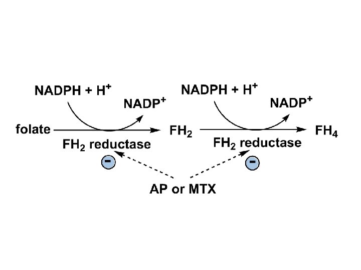
3. Folic acid analogs • Aminopterin (AP) and Methotrexate (MTX)
4. Nucleoside analogs • Arabinosyl cytosine (ara-c) inhibits the synthesis of d. CDP.