ELECTRICAL THERAPIES Z JAVADY NEJAD MD Tehran university


, 2:")


 is typically defined as")


 is consistently associated with a greater")

that")


 with")


 The recommended initial biphasic energy dose for cardioversion")

 VT with pulses is treated with synchronized cardioversion. Unstable polymorphic")


 impacts multiple key links in the chain of survival")


 reveals VF/VT,")


.")


 tachycardias (QRS< 0. 12 second), in order")


 with a pulse responds well to monophasic")
 will")

 Evaluation. The rhythm is considered to be")


 can also be used to")


 should not be attempted")


 VT Polymorphic (irregular) VT requires immediate defibrillation with the same")


 isomer is a potent beta-adrenergic agonist, whereas")

, inotropic (eg, dobutamine), agents should")


- Slides: 148
ELECTRICAL THERAPIES Z, JAVADY NEJAD, MD Tehran university of medical sciences
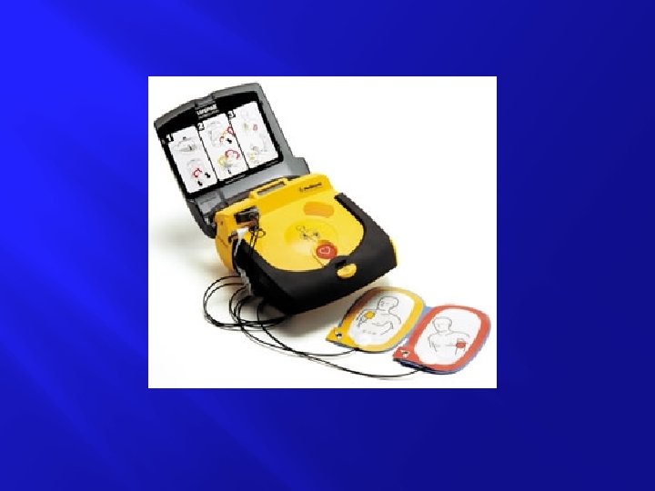
Automated External Defibrillators, Defibrillation, Cardioversion, and Pacing
Defibrillation Plus CPR: A Critical Combination Early defibrillation is critical to survival from sudden cardiac arrest (SCA) for several reasons
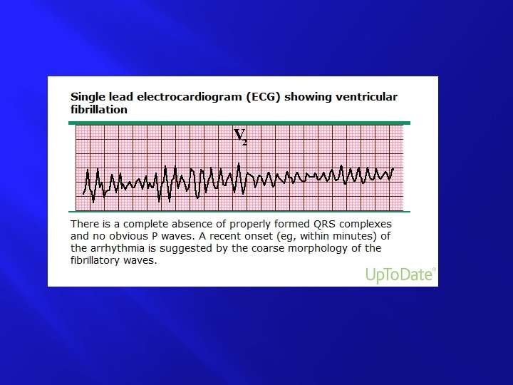
1: the most frequent initial rhythm in out-of-hospital witnessed SCA is ventricular fibrillation(VF), 2: the treatment for ventricular fibrillation is defibrillation, 3: the probability of successful defibrillation diminishes rapidly over time, 4: and VF tends to deteriorate to asystole over time
If bystanders provide immediate CPR, many adults in VF can survive with intact neurologic function, especially if defibrillation is performed within 5 to 10 minutes after SCA. CPR prolongs VF, delays the onset of asystole, and extends the window of time during which defibrillation can occur. Basic CPR alone, however, is unlikely to terminate VF and restore a perfusing rhythm.
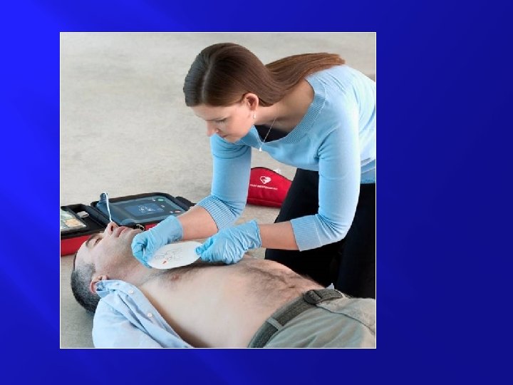
New Recommendations to Integrate CPR and AED Use To treat VF SCA, rescuers must be able to rapidly integrate CPR with use of the AED To give the victim the best chance of survival, 3 actions must occur within the first moments of a cardiac arrest 1: activation of the emergency medical services (EMS) system, 2 : provision of CPR, and 3: operation of an AED. When 2 or more rescuers are present, activation of EMS and initiation of CPR can occur simultaneously.
Two critical questions about integration of CPR with defibrillation The first question concerned whether CPR should be provided before defibrillation is attempted. The second question concerned the number of shocks to be delivered in a sequence before the rescuer resumes CPR.
Shock First Versus CPR First When any rescuer witnesses an out-of-hospital arrest and an AED is immediately available on-site, the rescuer should start CPR and use the AED as soon as possible. Healthcare providers who treat cardiac arrest in hospitals and other facilities with AEDs onsite should provide immediate CPR and should use the AED/defibrillator as soon as it is available. These recommendations are designed to support early CPR and early defibrillation, particularly when an AED is available within moments of the onset of SCA.
When VF is present for more than a few minutes, the myocardium is depleted of oxygen and metabolic substrates. A brief period of chest compressions can deliver oxygen and energy substrates, increasing the likelihood that a shock may terminate VF (defibrillation) and a perfusing rhythm will return (ie, ROSC).
1 -Shock Protocol Versus 3 -Shock Sequence Studies suggested significant survival benefit with the single shock defibrillation protocol compared with 3 -stacked-shock protocols. If 1 shock fails to eliminate VF, the incremental benefit of another shock is low, and resumption of CPR is likely to confer a greater value than another shock.
Defibrillation Waveforms and Energy Levels The term defibrillation (shock success) is typically defined as termination of VF for at least 5 seconds following the Shock. VF frequently recurs after successful shocks, but this recurrence should not be equated with shock failure. Shock success using the typical definition of defibrillation should not be confused with resuscitation outcomes such as restoration of a perfusing rhythm (ROSC), survival to hospital admission, or survival to hospital discharge.
Modern defibrillators are classified according to 2 types of waveforms: monophasic and biphasic. Monophasic waveform defibrillators were introduced first, but biphasic waveforms are used in almost all AEDs and manual defibrillators sold today. Energy levels vary by type of device and manufacturer.
Monophasic Waveform Defibrillators Monophasic waveforms deliver current of one polarity (ie, direction of current flow). Monophasic waveforms can befurther categorized by the rate at which the current pulse decreases to zero. The monophasic damped sinusoidal waveform (MDS) returns to zero gradually, whereas the monophasic truncated exponential waveform (MTE) current returns abruptly (is truncated) to zero current flow.
no specific waveform characteristic (either monophasic or biphasic) is consistently associated with a greater incidence of ROSC or higher survival to hospital discharge rates after cardiac arrest.
� � Biphasic Waveform Defibrillators Data from both out-of-hospital and in-hospital studies indicate that lower-energy biphasic waveform shocks have equivalent or higher success for termination of VF than either MDS or MTE monophasic waveform.
Defibrillation With Implanted Cardioverter Defibrillator If the patient has an implantable cardioverter defibrillator (ICD)that is delivering shocks (ie, the patient’s muscles contract in a manner similar to that observed during external defibrillation), allow 30 to 60 seconds for the ICD to complete the treatment cycle before attaching an AED. Occasionally, the analysis and shock cycles of automatic ICDs and AEDs will conflict. There is the potential for pacemaker or ICD malfunction after defibrillation when the pads are in close proximity to the device.
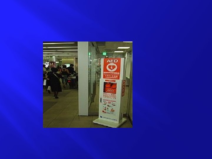
In-Hospital Use of AEDs � � � Defibrillation may be delayed when patients develop SCA in unmonitored hospital beds and in outpatient and diagnostic facilities. In such areas, several minutes may elapse before centralized response teams arrive with the defibrillator, attach it, and deliver shocks. Despite limited evidence, AEDs may be considered for the hospital setting as a way to facilitate early defibrillation (a goal of 3 minutes from collapse), especially in areas where staff have no rhythm recognition skills or defibrillators are used infrequently (Class IIb, LOE C).
� Fire v v v Hazard Several case reports have described fires ignited by sparks from poorly applied defibrillator paddles in the presence of an oxygen-enriched atmosphere. It may be reasonable for rescuers to take precautions to minimize sparking during attempted defibrillation; try to avoid defibrillation in an oxygen-enriched atmosphere (Class IIb, LOE C).
� � Synchronized Cardioversion Synchronized cardioversion is shock delivery that is timed (synchronized) with the QRS complex. This synchronization avoids shock delivery during the relative refractory portion of the cardiac cycle, when a shock could produce VF. VF
� � Synchronized cardioversion is recommended to treat supraventricular tachycardia due to reentry, atrial fibrillation, atrial flutter , and atrial tachycardia. Synchronized cardioversion is also recommended to treat monomorphic VT with pulses. Cardioversion is not effective for treatment of junctional tachycardia or multifocal atrial tachycardia.
� Synchronized cardioversion must not be used for treatment of VF as the device may not sense a QRS wave and thus a shock may not be delivered. Synchronized cardioversion should also not be used for pulseless VT or polymorphic (irregular VT). These rhythms require delivery of high-energy unsynchronized shocks (ie, defibrillation doses).
� � Supraventricular Tachycardias (Reentry Rhythms) The recommended initial biphasic energy dose for cardioversion of adult atrial fibrillation is 120 to 200 J (Class IIa, LOEA). If the initial shock fails, providers should increase the dose in a stepwise fashion. Cardioversion of adult atrial flutter and other supraventricular tachycardias generally requires less energy; an initial energy of 50 J to 100 J is often sufficient.
� � Ventricular Tachycardia The energy dose and timing of shocks for treatment of VT with pulses are determined by the patient’s condition and the morphological characteristics of the VT. Pulseless VT is treated as VF.
� Unstable monomorphic (regular) VT with pulses is treated with synchronized cardioversion. Unstable polymorphic (irregular) VT with or without pulses is treated as VF using unsynchronized high energy shocks (ie, defibrillation doses).
After shock delivery, the healthcare provider should be prepared to provide immediate CPR (beginning with chest compressions) and follow the ACLS Cardiac Arrest Algorithm if pulseless arrest develops.
Pacing is not recommended for patients in asystolic cardiac arrest.
� � It is reasonable for healthcare providers to be prepared to initiate pacing in patients with bradycardia who do not respond to atropine (or second-line drugs if these do not delay definitive management) (Class IIa, LOE B). Immediate pacing might be considered if the patient is severely symptomatic (Class. IIb, LOE C). If the patient does not respond to drugs or transcutaneous pacing, transvenous pacing is probably indicated (Class IIa, LOE C).
� � � Maintaining Devices in a State of Readiness User checklists have been developed to reduce equipment malfunction and operator errors. It is recommended to maintain devices in a state of readiness (Class I, LOE C).
�Adult Advanced Cardiovascular Life Support
Advanced cardiovascular life support (ACLS) impacts multiple key links in the chain of survival that include interventions to prevent cardiac arrest, treat cardiac arrest, and improve outcomes of patients who achieve return of spontaneous circulation (ROSC) after cardiac arrest. ACLS interventions aimed at preventing cardiac arrest include airway management, ventilation support, and treatment of bradyarrhythmias and tachyarrhythmias.
Cardiac arrest algorithms are simplified and redesigned to emphasize the importance of high-quality CPR (including chest compressions of adequate rate and depth, allowing complete chest recoil after each compression, minimizing interruptions in chest compressions and avoiding excessive ventilation).
� � Atropine is no longer recommended for routine use in the management of pulseless electrical activity (PEA)/asystole. There is an increased emphasis on physiologic monitoring to optimize CPR quality and detect ROSC. Chronotropic drug infusions are recommended as an alternative pacing in symptomatic and unstable bradycardia. Adenosine is recommended as a safe and potentially effective therapy in the initial management of stable undifferentiated regular monomorphic wide-complex tachycardia.
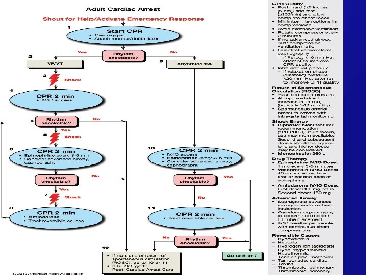
Management of Cardiac Arrest � Cardiac arrest can be caused by 4 rhythms: ventricular fibrillation(VF), pulseless ventricular tachycardia (VT), pulseless electric activity (PEA), and asystole.
� Periodic pauses in CPR should be as brief as possible and only as necessary to assess rhythm, shock VF/VT, perform a pulse check when an organized rhythm is detected, or place an advanced airway. In the absence of an advanced airway, a synchronized compression–ventilation ratio of 30: 2 is recommended at a compression rate of at least 100 per minute.
� � After placement of a supraglottic airway or an endotracheal tube, the provider performing chest compressions should deliver at least 100 compressions per minute continuously without pauses for ventilation. The provider delivering ventilations should give 1 breath every 6 to 8 seconds (8 to 10 breaths per minute) and should be particularly careful to avoid delivering an excessive number of ventilations.
Treatable Causes of Cardiac Arrest: The H’s and T’s � � � H’s Hypoxia Hypovolemia Hydrogen ion -acidosis Hypo-/hyperkalemia Hypothermia T’s Toxins Tamponade Tension pneumothorax Thrombosis,
� Paddles and electrode pads should be placed on the exposed chest in an anterior-lateral position. Acceptable alternative positions are anterior-posterior, anterior-left infrascapular, and anterior-right infrascapular. Rhythm checks should be brief, and if an organized rhythm is observed, a pulse check should be performed. If there is any doubt about the presence of a pulse, chest compressions should be resumed immediately.
� � � Rhythm-Based Management of Cardiac Arrest In most cases of witnessed and unwitnessed cardiac arrest the first provider should start CPR with chest compressions and the second provider should get or turn on the defibrillator, place the adhesive pads or paddles, and check the rhythm.
VF/Pulseless VT When a rhythm check by an automated external defibrillator (AED) reveals VF/VT, the AED will typically prompt to charge, “clear” the victim for shock delivery, and then deliver a shock, all of which should be performed as quickly as possible. CPR should be resumed immediately after shock delivery (without a rhythm or pulse check and beginning with chest compressions) and continue for 2 minutes before the next rhythm check
� � Drug Therapy in VF/Pulseless VT When VF/pulseless VT persists after at least 1 shock and a 2 -minute CPR period, a v a s o p r e s s o r can be given with the primary goal of increasing myocardial blood flow during CPR and achieving ROSC.
Amiodarone is the first-line antiarrhythmic agent given during cardiac arrest because it has been clinically demonstrated to improve the rate of ROSC and hospital admission in adults with refractory VF/pulseless VT.
� PEA/Asystole � When a rhythm check by an AED reveals a nonshockable � rhythm, CPR should be resumed immediately, beginning with chest compressions, and should continue for 2 minutes before the rhythm check is repeated. When a rhythm check using a manual defibrillator or cardiac monitor reveals an organized rhythm, a pulse check is performed.
� � Drug Therapy for PEA/Asystole A vasopressor can be given as soon as feasible with the primary goal of increasing myocardial and cerebral blood flow during CPR and achieving ROSC (Class IIb, LOE A).
Monitoring During CPR � � Physiologic Parameters In humans cardiac arrest is the most critically ill condition, yet it is typically monitored by rhythm assessment using selected electocardiographic (ECG) leads and pulse checks as the only physiologic parameters to guide therapy.
v v v v Pulse Clinicians frequently try to palpate arterial pulses during chest compressions to assess the effectiveness of compressions. No studies have shown the validity or clinical utility of checking pulses during ongoing CPR. Because there are no valves in the inferior vena cava, retrograde blood flow into the venous system may produce femoral vein pulsations. Thus, palpation of a pulse in the femoral triangle may indicate venous rather than arterial blood flow. Carotid pulsations during CPR do not indicate the efficacy of myocardial or cerebral perfusion during CPR. Palpation of a pulse when chest compressions are paused is a reliable indicator of ROSC but is potentially less sensitive than other physiologic measures discussed below.
� � End-Tidal CO 2 End-tidal CO 2 is the concentration of carbon dioxide in exhaled air at the end of expiration. It is typically expressed as a partial pressure in mm Hg (PETCO 2). Because CO 2 is a trace gas in atmospheric air, CO 2 detected by capnography in exhaled air is produced in the body and delivered to the lungs by circulating blood. Under normal conditions PETCO 2 is in the range of 35 to 40 mm Hg.
� � Pulse Oximetry During cardiac arrest, pulse oximetry typically does not provide a reliable signal because pulsatile blood flow is inadequate in peripheral tissue beds.
� � Arterial Blood Gases Arterial blood gas monitoring during CPR is not a reliable indicator of the severity of tissue hypoxemia, hypercarbia (and therefore adequacy of ventilation during CPR), or tissue acidosis. Routine measurement of arterial blood gases during CPR has uncertain value (Class IIb, LOE C).
� � Echocardiography No studies specifically examine the impact of echocardiography on patient outcomes in cardiac arrest. However, a number of studies suggest that transthoracic and transesophageal echocardiography have potential utility in diagnosing treatable causes of cardiac arrest such as cardiac tamponade, pulmonary embolism, ischemia, ….
Medications for Arrest Rhythms � � The primary goal of pharmacologic therapy during cardiac arrest is to facilitate restoration and maintenance of a perfusing spontaneous rhythm. Toward this goal, ACLS drug therapy during CPR is often associated with increased rates of ROSC and hospital admission but not increased rates of long-term survival with good neurologic outcome.
� � � Vasopressors To date no placebo-controlled trials have shown that administration of any vasopressor agent at any stage during management of VF, pulseless VT, PEA, or asystole increases the rate of neurologically intact survival to hospital discharge. There is evidence, however, that the use of vasopressor agents is associated with an increased rate of ROSC.
� � Epinephrine hydrochloride produces beneficial effects in patients during cardiac arrest, primarily because of its a-adrenergic receptorstimulating (ie, vasoconstrictor) properties.
� It is reasonable to consider administering a 1 mg dose of IV/IO epinephrine every 3 to 5 minutes during adult cardiac arrest (Class IIb, LOE A). If IV/IO access is delayed or cannot be established, epinephrine may be given endotracheally at a dose of 2 to 2. 5 mg.
� � � Vasopressin is a nonadrenergic peripheral vasoconstrictor that also causes coronary and renal vasoconstriction. Because the effects of vasopressin have not been shown to differ from those of epinephrine in cardiac arrest, 1 dose of vasopressin 40 units IV/IO may replace either the first or second dose of epinephrine in the treatment of cardiac arrest (Class IIb, LOE A).
Antiarrhythmics There is no evidence that any antiarrhythmic drug given routinely during human cardiac arrest increases survival to hospital discharge.
� � � Amiodarone IV amiodarone affects sodium, potassium, and calcium channels and has a- and b -adrenergic blocking properties. It can be considered for treatment of VF or pulseless VT unresponsive to shock delivery, CPR, and a vasopressor.
� � Lidocaine There is inadequate evidence to recommend the use of lidocaine in patients who have refractory VT/VF, defined as VT/VF not terminated by defibrillation or that continues to recur after defibrillation during out-of-hospital cardiac arrest or inhospital cardiac arrest.
� � Lidocaine may be considered if amiodarone is not available (Class IIb, LOE B).
Magnesium Sulfate � Routine administration of magnesium sulfate in cardiac arrest is not recommended (Class III, LOE A) unless torsades de pointes is present.
�INTERVENTIONS NOT RECOMMENDED FOR ROUTINE USE �DURING CARDIAC ARREST
� Atropine � Available evidence suggests that routine use of atropine during PEA or asystole is unlikely to have a therapeutic benefit (Class Iib, LOE B). For this reason atropine has been removed from the cardiac arrest algorithm.
Electric pacing is not recommended for routine use in cardiac arrest (Class III, LOE B).
Management of Symptomatic Bradycardia and Tachycardia � � Bradycardia is defined as a heart rate of< 60 beats per minute. However, when bradycardia is the cause of symptoms, the rate is generally <50 beats per minute.
� Atropine remains the first-line drug for acute symptomatic bradycardia (Class IIa, LOE B). Atropine sulfate reverses cholinergic-mediated decreases in heart rate and should be considered a temporizing measure while awaiting a transcutaneous or transvenous pacemaker for patients with symptomatic sinus bradycardia, conduction block at the level of the AV node, or sinus arrest.
It is reasonable for healthcare providers to initiate TCP in unstable patients who do not respond to atropine (Class IIa, LOE B). Immediate pacing might be considered in unstable patients with high-degree AV block when IV access is not available (Class IIb, LOE C). If the patient does not respond to drugs or TCP, transvenous pacing is probably indicated (Class IIa, LOE C)
� Alternative � � Drugs to Consider Although not first-line agents for treatment of symptomatic bradycardia, dopamine, epinephrine, and isoproterenol are alternatives when a bradyarrhythmia is unresponsive to or inappropriate for treatment with atropine, or as a temporizing measure while awaiting the availability of a pacemaker.
� Dopamine hydrochloride is a catecholamine with both a- and b-adrenergic actions. At lower doses dopamine has a more selective effect on inotropy and heart rate; at higher doses (10 mcg/kg per minute), it also has vasoconstrictive effects. Dopamine infusion may be used for patients with symptomatic bradycardia, particularly if associated with hypotension, in whom atropine may be inappropriate or after atropine fails (Class IIb, LOE B).
� Epinephrine is a catecholamine with a – and b-adrenergic actions. Epinephrine infusion may be used for patients with symptomatic bradycardia, particularly if associated with hypotension, for whom atropine may be inappropriate or after atropine fails (Class IIb, LOE B).
� � Isoproterenol is a b-adrenergic agent with –b 1 and -b 2 effects, resulting in an increase in heart rate and vasodilation. The recommended adult dose is 2 to 10 mcg/ min by IV infusion, titrated according to heart rate and rhythm response.
Classification of Tachyarrhythmias � � Tachycardias can be classified in several ways, based on the appearance of the QRS complex, heart rate, and regularity. ACLS professionals should be able to recognize and differentiate between sinus tachycardia, narrow-complex supraventricular tachycardia (SVT), and widecomplex tachycardia. Because ACLS providers may be unable to distinguish between supraventricular and ventricular rhythms, they should be aware that most wide-complex (broadcomplex)tachycardias are ventricular in origin.
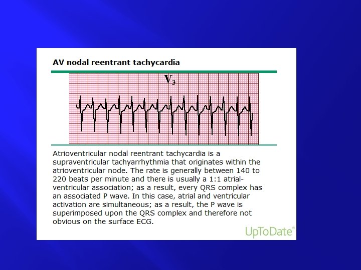
� � � � � Narrow–QRS-complex (SVT) tachycardias (QRS< 0. 12 second), in order of frequency ● Sinus tachycardia ● Atrial fibrillation ● Atrial flutter ● AV nodal reentry ● Accessory pathway–mediated tachycardia ● Atrial tachycardia (including automatic and reentry forms) ● Multifocal atrial tachycardia (MAT) ● Junctional tachycardia (rare in adults) Wide–QRS-complex tachycardias (QRS ≥ 0. 12 second) ● Ventricular tachycardia (VT) and ventricular fibrillation (VF) ● SVT with aberrancy ● Pre-excited tachycardias (Wolff-Parkinson-White [WPW] syndrome) ● Ventricular paced rhythms
� � Initial Evaluation and Treatment of Tachyarrhythmias Tachycardia is defined as an arrhythmia with a rate of> 100 beats per minute, although, as with defining bradycardia, the rate of a tachycardia takes on clinical significance at its greater extremes and is more likely attributable to an arrhythmia rate of ≥ 150 beats per minute.
� If oxygenation is inadequate or the patient shows signs of increased work of breathing, provide supplementary oxygen. Attach a monitor to the patient, evaluate blood pressure, and establish IV access. If available, obtain a 12 -lead ECG to better define the rhythm, but this should not delay immediate cardioversion if the patient is unstable. While initiating treatment, evaluate the patient’s clinical status and identify potential reversible causes of the tachycardia.
� � � Waveform and Energy The recommended initial biphasic energy dose for cardioversion of atrial fibrillation is 120 to 200 J (Class IIa, LOE A). Cardioversion of atrial flutter and other SVTs generally requires less energy; an initial energy of 50 J to 100 J is often sufficient.
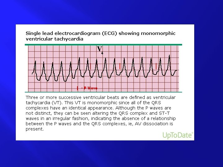
� Monomorphic VT (regular form and rate) with a pulse responds well to monophasic or biphasic waveform cardioversion (synchronized) shocks at initial energies of 100 J. If there is no response to the first shock, it may be reasonable to increase the dose in a stepwise fashion.
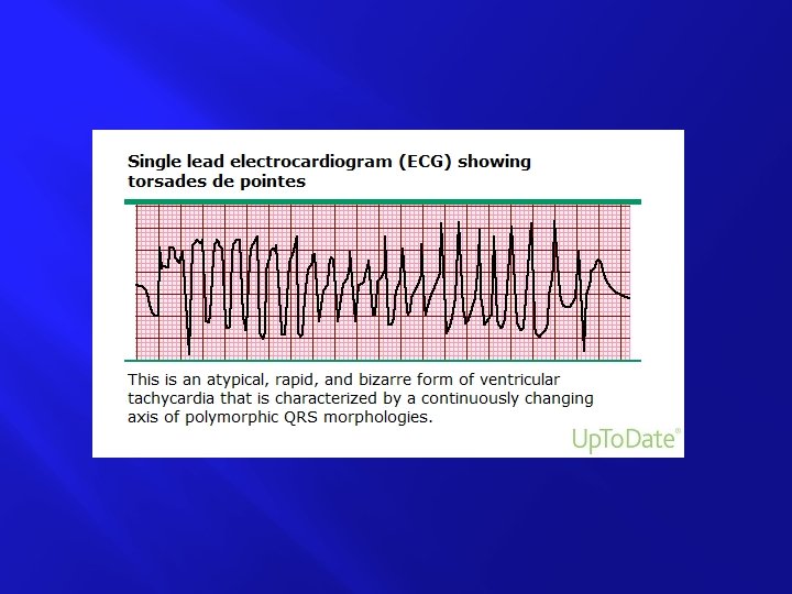
� � Arrhythmias with a polymorphic QRS appearance (such as torsades de pointes) will usually not permit synchronization. Thus, if a patient has polymorphic VT, treat the rhythm as VF and deliver high-energy unsynchronized shocks (ie, defibrillation doses). If there is any doubt whether monomorphic or polymorphic VT is present in the unstable patient, do not delay shock delivery to perform detailed rhythm analysis: provide high-energy unsynchronized shocks (ie, defibrillation doses).
� � � Regular Narrow-Complex Tachycardia Sinus Tachycardia : Sinus tachycardia is common and usually results from a physiologic stimulus, such as fever, anemia, or hypotension/shock. Sinus tachycardia is defined as a heart rate> 100 beats per minute.
� � � Supraventricular Tachycardia (Reentry SVT) Evaluation. The rhythm is considered to be of supraventricular origin if the QRS complex is narrow (<120 milliseconds or< 0. 12 second) or if the QRS complex is wide (broad) and preexisting bundle branch block or ratedependent aberrancy is known to be present.
� Therapy Vagal Maneuvers. Vagal maneuvers and adenosine are the preferred initial therapeutic choices for the termination of stable PSVT.
� � � � Adenosine. If PSVT does not respond to vagal maneuvers, give 6 mg of IV adenosine as a rapid IV push through a large(eg, antecubital) vein followed by a 20 m. L saline flush (Class. I, LOE B). If the rhythm does not convert within 1 to 2 minutes, give a 12 mg rapid IV push using the method above. Because of the possibility of initiating atrial fibrillation with rapid ventricular rates in a patient with WPW, a defibrillator should be available when adenosine is administered to any patient in whom WPW is a consideration.
� After conversion, monitor the patient for recurrence and treat any recurrence of PSVT with adenosine or a longer acting AV nodal blocking agent (eg, diltiazem or b-blocker). If adenosine or vagal maneuvers disclose another form of SVT (such as atrial fibrillation or flutter), treatment with a longer-acting AV nodal blocking agent should be considered to afford more lasting control of ventricular rate.
� Calcium Channel Blockers and B-Blockers. If adenosine or vagal maneuvers fail to convert PSVT or recurs after such treatment, or these treatments disclose a different form of SVT (such as atrial fibrillation or flutter), it is reasonable to use longer-acting AV nodal blocking agents, such as the nondihydropyridine calcium channel blockers (verapamil and diltiazem) (Class IIa, LOE B) or b -blockers (Class IIa, LOE C).
For verapamil, give a 2. 5 mg to 5 mg IV bolus over 2 minutes (over 3 minutes in older patients). If there is no therapeutic response and no drug-induced adverse event, repeated doses of 5 mg to 10 mg may be administered every 15 to 30 minutes to a total dose of 20 mg. An alternative dosing regimen is to give a 5 mg bolus every 15 minutes to a total dose of 30 mg. Verapamil should be given only to patients with narrow complex reentry SVT or arrhythmias known with certainty to be of supraventricular origin. Verapamil should not be given to patients with wide-complex tachycardias. It should not be given to patients with impaired ventricular function or heart failure.
Caution is advised when encountering preexcited atrial fibrillation or flutter that conducts to the ventricles via both the AV node and an accessory pathway. Treatment with an AV nodal blocking agent (including adenosine, calcium blockers, b-blockers, or digoxin) is unlikely to slow the ventricular rate and in some instances may accelerate the ventricular response.
Ø Although antiarrhythmic medications (eg, amiodarone, procainamide, or sotalol) can also be used to treat SVTs, the higher toxicity and risk for proarrhythmia make these medications less desirable alternatives to the described AV nodal blocking agents. Ø A possible exception is in patients with preexcited atrial arrhythmias; the typical AV nodal blocking drugs are contraindicated in these patients and rate control may be achieved with antiarrhythmic medications. Ø
� Wide-Complex � � Tachycardia Evaluation The first step in the management of any tachycardia is to determine if the patient’s condition is stable or unstable. An unstable patient with a wide-complex tachycardia should be presumed to have VT and immediate cardioversion should be performed. Precordial thump may be considered for patients with witnessed, monitored, unstable ventricular tachycardia if a defibrillator is not immediately ready for use (Class IIb, LOE C).
If the patient is stable, the second step in management is to obtain a 12 -lead ECG to evaluate the rhythm. At this point the provider should consider the need to obtain expert consultation. If the patient becomes unstable at any time, proceed with synchronized cardioversion or unsynchronized defibrillation should the arrhythmia deteriorate to VF or be due to a polymorphic VT.
The most common forms of wide complex tachycardia are: ● VT or VF ● SVT with aberrancy ● Pre-excited tachycardias (associated with or mediatedby an accessory pathway) ● Ventricular paced rhythms
The third step in management of a tachycardia is to determine if the rhythm is regular or irregular. A regular wide-complex tachycardia is likely to be VT or SVT with aberrancy. An irregular wide -complex tachycardia may be atrial fibrillation with aberrancy, pre-excited atrial fibrillation(ie, atrial fibrillation using an accessory pathway for antegrade conduction), or polymorphic VT/torsades de pointes. Providers should consider the need for expert consultation when treating wide-complex tachycardias.
� Therapy for Regular Wide-Complex Tachycardias � In patients with stable undifferentiated wide-QRS complex tachycardia, a reasonable approach is to try to identify the wide-complex tachycardia as SVT or VT and treat based on the algorithm for that rhythm. � If the etiology of the rhythm cannot be determined, the rate is regular, and the QRS is monomorphic, recent evidence suggests that IV adenosine is relatively safe for both treatment and diagnosis (Class IIb, LOE B). However, adenosine should not be given for unstable or for irregular or polymorphic wide complex tachycardias, as it may cause degeneration of the arrhythmia to VF (Class III, LOE C). �
� � Verapamil is contraindicated for wide-complex tachycardias unless known to be of supraventricular origin (Class III, LOE B).
For patients who are stable with likely VT, IV antiarrhythmic drugs or elective cardioversion is the preferred treatment strategy. If IV antiarrhythmics are administered, procainamide (Class IIa, LOE B), amiodarone (Class IIb, LOE B), or sotalol (Class IIb, LOE B) can be considered. Procainamide and sotalol shoul be avoided in patients with prolonged QT. If one of these antiarrhythmic agents is given, a second agent should not be given without expert consultation (Class III, LOE B). If antiarrhythmic therapy is unsuccessful, cardioversion or exper consultation should be considered (Class IIa, LOE C).
� Amiodarone is also effective in preventing recurrent monomorphic VT or treating refractory ventricular arrhythmias in patients with coronary artery disease and poor ventricular function. It is given 150 mg IV over 10 minutes; dosing should be repeated as needed to a maximum dose of 2. 2 g IV per 24 hours.
� � � By comparison, lidocaine is less effective in terminating VT than procainamide, sotalol, and amiodarone. Thus, while occasionally effective, lidocaine should be considered second-line antiarrhythmic therapy for monomorphic VT. Lidocaine can be administered at a dose of 1 to 1. 5 mg/kg IV bolus. Maintenance infusion is 1 to 4 mg/min (30 to 50 mcg/kg per minute).
� � Irregular Tachycardias Atrial Fibrillation and Flutter Evaluation An irregular narrow-complex or wide-complex tachycardia is most likely atrial fibrillation (with or without aberrant conduction) with an uncontrolled ventricular response. Other diagnostic possibilities include MAT or sinus rhythm/tachycardia with frequent atrial premature beats. When there is doubt about the rhythm diagnosis and the patient is stable, a 12 -lead ECG with expert consultation is recommended.
� � Therapy General management of atrial fibrillation should focus on control of the rapid ventricular rate (rate control), conversion of hemodynamically unstable atrial fibrillation to sinus rhythm (rhythm control), or both. Patients with an atrial fibrillation duration of >48 hours are at increased risk for cardioembolic events, although shorter durations of atrial fibrillation do not exclude the possibility of such events.
� Electric or pharmacologic cardioversion (conversion to normal sinus rhythm) should not be attempted in these patients unless the patient is unstable. An alternative strategy is to perform cardioversion following anticoagulation with heparin and performance of transesophageal echocardiography to ensure the absence of a left atrial thrombus.
� � � � Rate Control Patients who are hemodynamically unstable should receive prompt electric cardioversion. More stable patients require ventricular rate control as directed by patient symptoms and hemodynamics. IV b -blockers and nondihydropyridine calcium channel blockers such as diltiazem are the drugs of choice for acute rate control in most individuals with atrial fibrillation and rapid ventricular response (Class IIa, LOE A). Digoxin and amiodarone may be used for rate control in patients with congestive heart failure; however, the potential risk of conversion to sinus rhythm with amiodarone should be considered before treating with this agent.
� A wide-complex irregular rhythm should be considered preexcited atrial fibrillation. Expert consultation is advised. Avoid AV nodal blocking agents such as adenosine, calcium channel blockers, digoxin, and possibly bblockers in patients with pre-excitation atrial fibrillation because these drugs may cause a paradoxical increase in the ventricular response. Typically, patients with pre-excited atrial fibrillation present with very rapid heart rates and require emergent electric cardioversion.
� � Polymorphic (Irregular) VT Polymorphic (irregular) VT requires immediate defibrillation with the same strategy used for VF. Pharmacologic treatment to prevent recurrent polymorphic VT should be directed by the underlying cause of VT and the presence or absence of a long QT interval during sinus rhythm. If a long QT interval is observed during sinus rhythm (ie, the VT is torsades de pointes), the first step is to stop medications known to prolong the QT interval. Correct electrolyte imbalance and other acute precipitants.
� In the absence of a prolonged QT interval, the most common cause of polymorphic VT is myocardial ischemia. In this situation IV amiodarone and b-blockers may reduce the frequency of arrhythmia recurrence (Class IIb, LOE C).
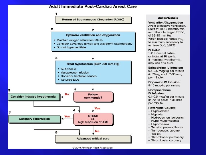
�Post–Cardiac Care Arrest
� There is increasing recognition that systematic post–cardiac arrest care after return of spontaneous circulation (ROSC) can improve the likelihood of patient survival with good quality of life.
The initial objectives of post– cardiac arrest care to ● Optimize cardiopulmonary function and vital organ perfusion. ● After out-of-hospital cardiac arrest, transport patient to an appropriate hospital with a comprehensive post– cardiac arrest treatment system of care that includes acute coronary interventions, neurological care, goaldirected critical care, and hypothermia. ● Transport the in-hospital post– cardiac arrest patient to an appropriate critical-care unit capable of providing comprehensive post– cardiac arrest care. ● Try to identify and treat the precipitating causes of the arrest and prevent recurrent arrest.
� Subsequent objectives of post– cardiac arrest care to � ● Control body temperature to optimize survival and neurological recovery ● Identify and treat acute coronary syndromes (ACS) ● Optimize mechanical ventilation to minimize lung injury ● Reduce the risk of multiorgan injury and support organ function if required ● Objectively assess prognosis for recovery ● Assist survivors with rehabilitation services when required � � �
� � � Systems of Care for Improving Post– Cardiac Arrest Outcomes Post–cardiac arrest care is a critical component of advanced life support. Most deaths occur during the first 24 hours after cardiac arrest.
A comprehensive, structured, multidisciplinary system of care should be implemented in a consistent manner for the treatment of post– cardiac arrest patients (Class I, LOE B). Programs should include as part of structured interventions therapeutic hypothermia; optimization of hemodynamics and gas exchange; immediate coronary reperfusion when indicated for restoration of coronary blood flow with percutaneous coronary intervention (PCI); glycemic control; and neurological diagnosis, management, and prognostication.
Although 100% oxygen may have been used during initial resuscitation, providers should titrate inspired oxygen to the lowest level required to achieve an arterial oxygen saturation of 94%, so as to avoid potential oxygen toxicity. It is recognized that titration of inspired oxygen may not be possible immediately after out-of-hospital cardiac arrest until the patient is transported to the emergency department or, in the case of inhospital arrest, the intensive care unit (ICU). Hyperventilation or “overbagging” the patient is common after cardiac arrest and should be avoided because of potential dverse hemodynamic effects.
� Hyperventilation increases intrathoracic pressure and inversely lowers cardiac output. The decrease in Pa. CO 2 seen with hyperventilation can also potentially decrease cerebral blood flow directly. Ventilation may be started at 10 to 12 breaths per minute and titrated to achieve a PETCO 2 of 35 to 40 mm Hg or a. Pa. CO 2 of 40 to 45 mm Hg.
� The clinician should assess vital signs and monitor for recurrent cardiac arrhythmias. � Continuous electrocardiographic (ECG) monitoring should continue after ROSC, during transport, and throughout ICU care until stability has been achieved. Intravenous (IV) access should be obtained if not already established and the position and function of any intravenous catheter verified. IV lines should be promptly established to replace emergent intraosseous access achieved during resuscitation. If the patient is hypotensive (systolic blood pressure 90 mm Hg), fluid boluses can be considered.
� � Cold fluid may be used if therapeutic hypothermia is elected. Vasoactive drug infusions such as dopamine, norepinephrine, or epinephrine may be initiated if necessary and titrated to achieve a minimum systolic blood pressure of 90 mm Hg or a mean arterial pressure of 65 mm Hg.
� Brain injury and cardiovascular instability are the major determinants of survival after cardiac arrest.
� � � Central Nervous System Brain injury is a common cause of morbidity and mortality in post–cardiac arrest patients. Brain injury is the cause of death in 68% of patients after out-of-hospital cardiac arrest and in 23% after in-hospital cardiac arrest. The pathophysiology of post– cardiac arrest brain injury involves a complex cascade of molecular events that are triggered by ischemia and reperfusion and then executed over hours to days after ROSC.
� � � Clinical manifestations of post–cardiac arrest brain injury include coma, seizures, myoclonus, various degrees of neurocognitive dysfunction (ranging from memory deficits to persistent vegetative state), and brain death.
� � Targeted Temperature Management Induced Hypothermia For protection of the brain and other organs, hypothermia is a helpful therapeutic approach in patients who remain comatose (usually defined as a lack of meaningful response to verbal commands) after ROSC. Questions remain about specific indications and populations, timing and duration of therapy, and methods for induction, maintenance, and subsequent reversal of hypothermia.
� Clinicians should continuously monitor the patient’s core temperature using an esophageal thermometer, bladder catheter in nonanuric patients, or pulmonary artery catheter if one is placed for other indications. Axillary and oral temperatures are inadequate for measurement of core temperature changes, especially during active manipulation of temperature for therapeutic hypothermia, and true tympanic temperature probes are rarely available and often unreliable.
� A number of potential complications are associated with cooling, including coagulopathy, arrhythmias, and hyperglycemia. The likelihood of pneumonia and sepsis may increase in patients treated with therapeutic hypothermia.
� In summary, we recommend that comatose (ie, lack of meaningful response to verbal commands) adult patients with ROSC after out -of-hospital VF cardiac arrest should be cooled to 32°C to 34°C (89. 6°F to 93. 2°F) for 12 to 24 hours (Class I, LOE B). Induced hypothermia also may be considered for comatose adult patients with ROSC after in-hospital cardiac arrest of any initial rhythm or after out-ofhospital cardiac arrest with an initial rhythm of pulseless electric activity or asystole (Class IIb, LOE B).
� � � Hyperthermia After resuscitation, temperature elevation above normal can impair brain recovery. The etiology of fever after cardiac arrest may be related to activation of inflammatory cytokines in a pattern similar to that observed in sepsis.
� � � Pulmonary System Pulmonary dysfunction after cardiac arrest is common. Etiologies: include hydrostatic pulmonary edema from left ventricular dysfunction; noncardiogenic edema from inflammatory, infective, or physical injuries; severe pulmonary atelectasis; atelectasis or aspiration occurring during cardiac arrest or resuscitation. Patients often develop regional mismatch of ventilation and perfusion, contributing to decreased arterial oxygen content.
� � Essential diagnostic tests in intubated patients include : a chest radiograph and arterial blood gas measurements. Other diagnostic tests may be added based on history, physical examination, and clinical circumstances. Evaluation of a chest radiograph should verify the correct position of the endotracheal tube and the distribution of pulmonary infiltrates or edema and identify complications from chest compressions (eg, rib fracture, pneumothorax, and pleural effusions) or pneumonia.
� � Sedation After Cardiac Arrest Patients with coma or respiratory dysfunction after ROSC are routinely intubated and maintained on mechanical ventilation for a period of time, which results in discomfort, pain, and anxiety. Intermittent or continuous sedation and/or analgesia can be used to achieve specific goals. Patients with post cardiac arrest cognitive dysfunction may display agitation or frank delirium with purposeless movement and are at risk of selfinjury
� � Opioids, anxiolytics, and sedative-hypnotic agents can be used in various combinations to improve patient-ventilator interaction and blunt the stress-related surge of endogenous catecholamines.
�Cardiovascular System
� � � Overall the most common cause of cardiac arrest is cardiovascular disease and coronary ischemia. Therefore, a 12 -lead ECG should be obtained as soon as possible to detect ST elevation or new or presumably new left bundle branch block. When there is high suspicion of acute myocardial infarction (AMI), local protocols for treatment of AMI and coronary reperfusion should be activated. Even in the absence of ST elevation, medical or interventional treatments may be considered for treatment of ACS and should not be deferred in the presence of coma or in conjunction with hypothermia. Concurrent PCI and hypothermia are safe, with good outcomes reported for some comatose patients who undergo PCI.
� � Because it is impossible to determine the final neurological status of comatose patients in the first hours after ROSC, aggressive treatment of ST-elevation myocardial infarction STEMI should begin as in non–cardiac arrest patients, regardless of coma or induced hypothermia.
� � Patients with cardiac arrest may receive antiarrhythmic drugs such as lidocaine or amiodarone during initial resuscitation. There is no evidence to support or refute continued or prophylactic administration of these medications.
�Vasoactive Drugs for Use in Post–Cardiac Arrest Patients
� Vasopressors � Vasoactive drugs may be administered after ROSC to support cardiac output, output especially blood flow to the heart and brain. � Drugs may be selected to improve heart rate (chronotropic effects), myocardial contractility (inotropic effects), or arterial pressure (vasoconstrictive effects), or to reduce afterload � (vasodilator effects). Unfortunately many adrenergic drugs are not selective and may increase or decrease heart rate and afterload, increase cardiac arrhythmias, and increase myocardial ischemia by creating a mismatch between myocardial oxygen demand delivery.
� � � � In general, adrenergic drugs should not be mixed with sodium bicarbonate or other alkaline solutions in the IV line because there is evidence that adrenergic agents are inactivated in alkaline solutions. Norepinephrine (levarterenol) and other catecholamines that activate – adrenergic receptors may produce t i s s u e n e c r o s if extravasation occurs. Therefore, administration through a central line is preferred whenever possible. If extravasation develops, infiltrate 5 to 10 mg of phentolamine diluted in 10 to 15 m. L of saline into the site of extravasation as soon as possible to prevent tissue death and sloughing.
� � � � � Common Vasoactive Drugs Epinephrine ● Useful for symptomatic bradycardia if atropine and transcutaneous pacing fail or if pacing is not available ● Used to treat severe hypotension (eg, systolic blood pressure< 70 mm Hg) ● Useful for anaphylaxis associated with hemodynamic instability or respiratory distress.
� � � Norepinephrine ● Used to treat severe hypotension (eg, systolic blood pressure< 70 mm Hg) and a low total peripheral resistance ● Relatively contraindicated in patients with hypovolemia. It may increase myocardial oxygen requirements, mandating cautious use in patients with ischemic heart disease ● Usually induces renal and mesenteric vasoconstriction; in sepsis, sepsis however, norepinephrine improves renal blood flow and urine output.
� � � Dopamine ● Used to treat hypotension, especially if it is associated with symptomatic bradycardia.
� � � Dobutamine ● The (+) isomer is a potent beta-adrenergic agonist, whereas the (–) isomer is a potent alpha-1 -agonist ● The vasodilating beta 2 -adrenergic effects of the (+) isomer counterbalance the vasoconstricting alpha-adrenergic effects, often leading to little change or a reduction in systemic vascular resistance.
� � � Use of Vasoactive Drugs After Cardiac Arrest Hemodynamic instability is common after cardiac arrest. Death due to multiorgan failure is associated with a persistently low cardiac index during the first 24 hours after resuscitation. Vasodilation may occur from loss of sympathetic tone and from metabolic acidosis.
� Fluid administration as well as vasoactive (eg, norepinephrine), inotropic (eg, dobutamine), agents should be titrated as needed to optimize blood pressure, cardiac output, and systemic perfusion (Class I , LOE B). Although human studies have not established ideal targets for blood pressure or blood oxygenation, a mean arterial pressure 65 mm Hg is generally considered reasonable goals.
� � Glucose Control The post– cardiac arrest patient is likely to develop metabolic abnormalities such as hyperglycemia that may be detrimental.
� In summary ; every organ system is at risk during this period, and patients are at risk of developing multiorgan dysfunction. The comprehensive treatment of diverse problems after cardiac arrest involves multidisciplinary aspects of critical care, cardiology and neurology For this reason, it is important to admit patients to appropriate critical-care units with a prospective plan of care to anticipate, monitor, and treat each of these diverse problems.