DEVELOPMENT OF INTERATRIAL AND INTERVENTRICULAR SEPTUM HIMANSHU GUPTA


 septum • Atrial septum •")


 RAP > LAP, oxygenated blood flows directly from right")


")


70 -80% � Muscular- 5 -20% Central- mid muscular Apical Marginal- along RV")


- Slides: 62
DEVELOPMENT OF INTERATRIAL AND INTERVENTRICULAR SEPTUM HIMANSHU GUPTA
Introduction to development of the heart � It develops early in the middle of 3 rd week , from aggregation of splanchnic mesodermal cells, in cardiogenic area, ventral to pericardial coelom, and dorsal to yolk sac. � They form 2 angioblastic cords that canalize to form 2 endocardial heart tubes.
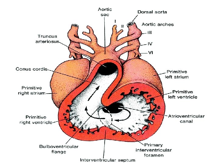
The Heart Tube � The two endocardial tubes fuse to form Single heart tube.
� The heart tube is differentiated into: 1 -truncus arteriosus. � 2 -bulbus cordis. � 3 -primitive ventricle. � 4 -primitive atrium. � 5 -sinus venosus. �
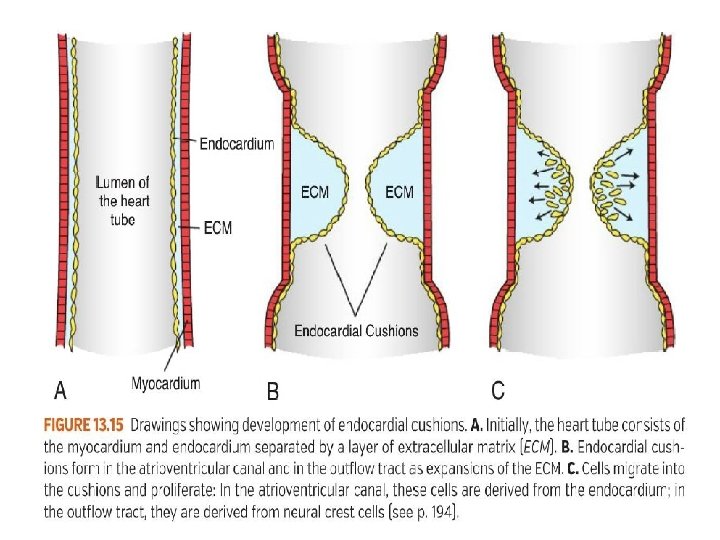
Folding of the heart tube � During the 4 th week, the folding of the heart tube takes place. � The formation of the AV canal and the endocardial cushions also take place around the same time.
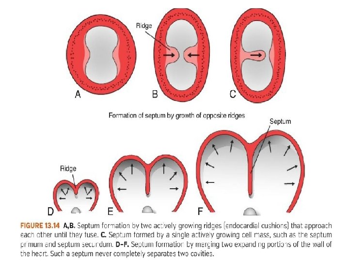
FORMATION OF CARDIAC SEPTA � The major septa of the heart are formed between the 27 th and 37 th days of development. � When the embryo grows in length from 5 mm to approx. 16 to 17 mm. � Septum formation can involves. Endocardial cushions formation Passive expansion of chambers
Formation of the Cardiac Septa • The Atrioventricular (AV) septum • Atrial septum • Interventricular septum • Aorticopulmonary septum
Molecular regulation of septal development � NKX 2. 5. -the master gene for heart development � BMPs 2 and 4 � WNT protein inhibitors-CRESCENT and CERBERUS � FGF 8 � Retinoic acid � TBX 5 - DNA- binding motif known as the T-box. Expressed later than NKX 2. 5, it plays an important role in septation.
Partitioning of the primitive Heart � Division of A-V canal , primitive atrium & primitive ventricle begins at the middle or end of 4 th week and completed by the end of 5 th week. � These processes occur concurrently. � At the end of 4 th week, 2 endocardial cushions on dorsal & ventral walls of atrioventricular canal , develop from mesenchymal cells of cardiac jelly.
ØDuring 5 th week, the AV- endocardial cushions meet and unite in the mid line to form a septum and divide the common A-V canal into right & left A-V canals. ØEndocardial cushions also form the AV- valves + membranous septa of interventricular septum. ØNote in D, coronal section , begining of development of interatrial & intervent. septa.
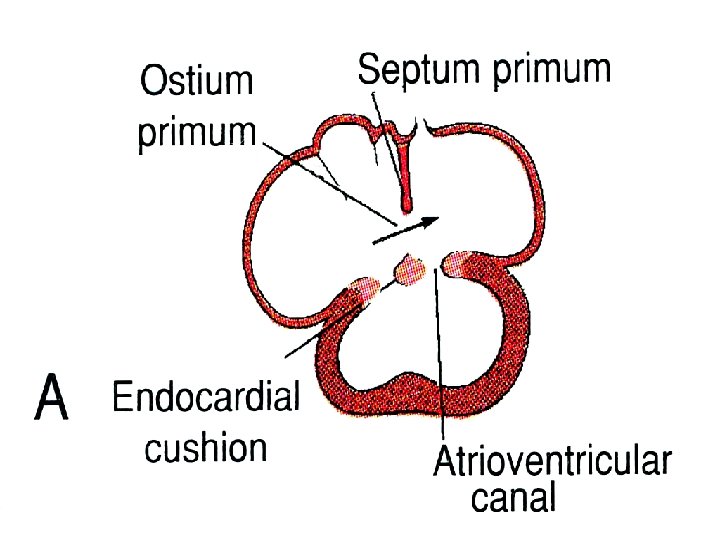
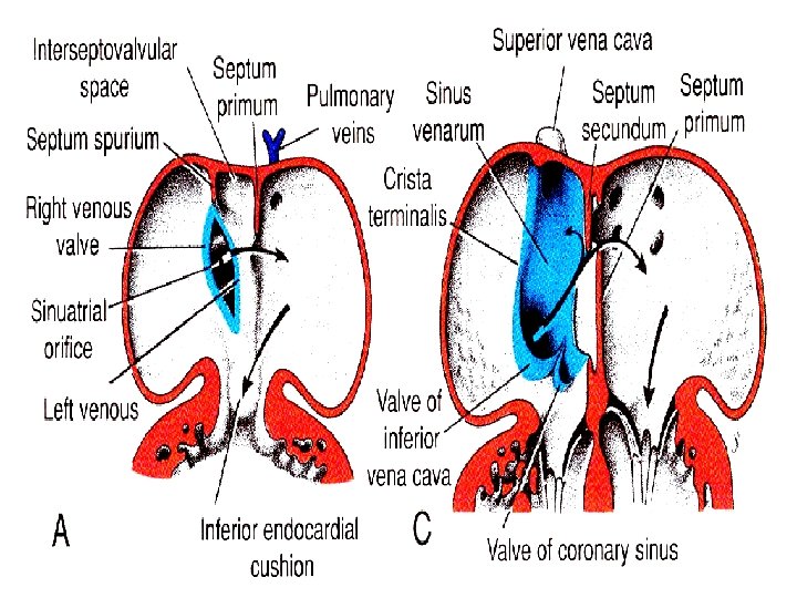
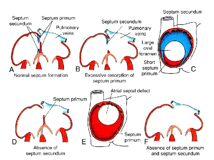
Formation of the Interatrial septum. � The septum is developed from three sources: � 1. Septum primum � 2. Septum intermedium � 3. Septum secundum
1 -Septum primum : • A thin crescent-shaped membrane grows from the roof of common atrium. • The lower margin of the septum is free and concave. • Anterior and posterior horns of the septum fuse respectively with the ventral and dorsal endocardial cushions of the primitive atrioventricular canal. 2 -Septum intermedium: • The ventral and dorsal cushions of the atrioventricular canal fuse to form a broad anterioposterior partition, which divide the canal into right and left atrioventricular orifices.
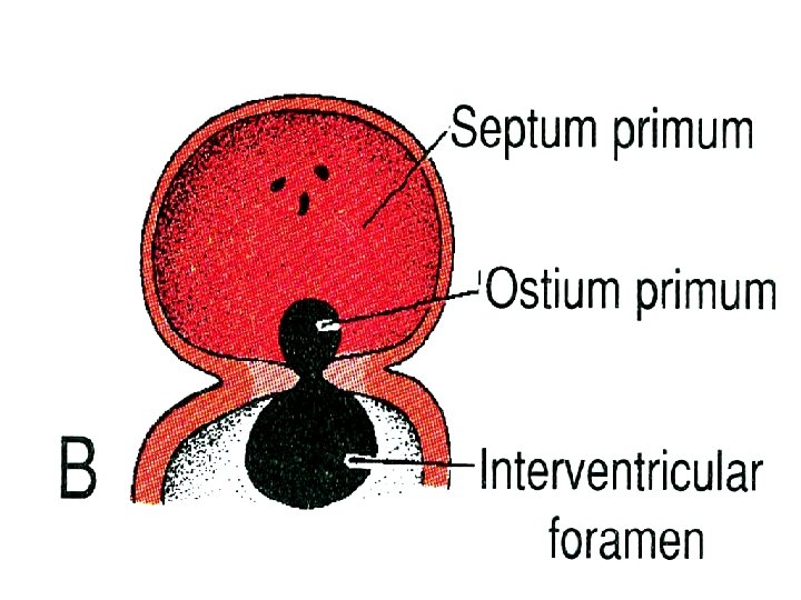
� A foramen known as ostium primum is formed between the upper surface of septum intermedium and lower border of septum primum. � Later, ostium primum is closed by the fusion of two septa. � Associated with the closure of ostium primum, the upper and dorsal part of septum disintegrates forming a foramen known as ostium secundum.
3 -Septum secundum • It arises on the right side of septum primum from the space of the right atrium which is interval between septum primum and septum spurium. • The septum secundum incorporates the whole of left venous valve and extends vertically downwards. • The lower margin grows sufficiently to overlap the upper margin of the septum primum. • The valvular opening formed between the lower margin of the septum secundum and upper margin of the septum primum is called foramen ovale.
ØIn the fetus (before birth) RAP > LAP, oxygenated blood flows directly from right atrium to left atrium through open foramen ovale. ØAfter birthØwhen pulmonary circulation begins, LAP rises and the upper edge of septum primum is pressed against the upper limb of septum secundum. ØThis closes the foramen ovale , forming a complete partition between the 2 atria. ØAn oval depression in the lower part of interatrial septum of right atrium…. The fossa ovalis is a remnant of the foramen ovale.
Features of the interatrial septum � On the right side: 1. Fossa ovalis: Oval depression in the lower part of the septum, and the floor is formed by the septum primum. 2. Limbus fossa ovalis: a sickle shaped fold that surrounds the upper, anterior and posterior margins of the fossa ovalis. It represents lower free margins of the septum secundum. 3. Foramina venarium minimarium: Venae cordis minimi open through these foramina. 4. Atrio-ventricular node: It is situated in the lower part of the septum above the opening of coronary sinus.
� On the left side: � 1. Presence of the semilunar fold with the concavity directed upwards; it is a remnant of the upper margin of the septum primum. � 2. Lunate impression above the fold is formed by septum secundum. � 3. Foramina venarium minimarium.
Development of IVS
Ventricular Septum Membranous Muscular Spiral (Aorticopulmonary)
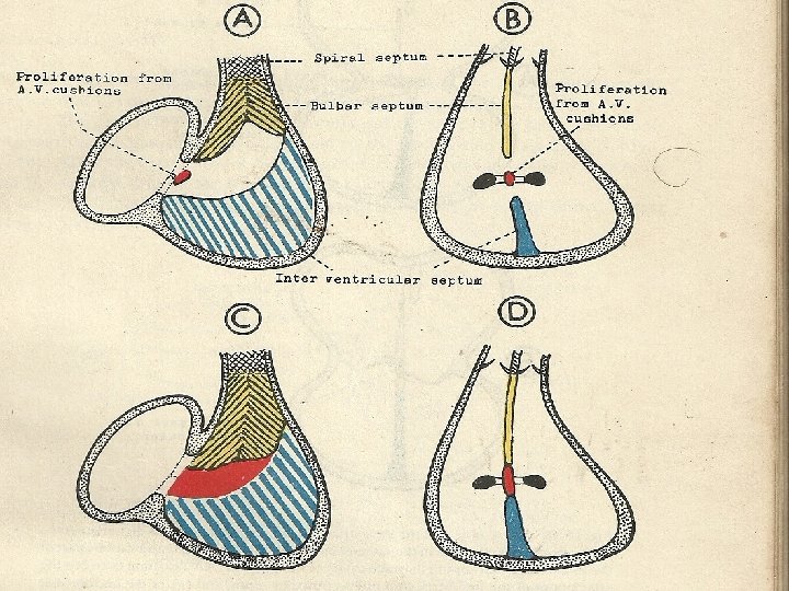
Development of muscular part of IVS: ØPrimordial muscular IVS arises in the floor of ventricle , as thick crescentic fold with concave free edge. ØThis septum subdivides the original ventricular cavity incompletely into right & left ventricles that communicate together through IV foramen. ØThis foramen closes by the end of 7 th week as the 2 bulbar ridges fuse with the endocadial cushion. ØA-sagittal section 5 th week. ØCoronal section. 6 th week.
Incorporation of the proximal part of bulbus cordis into the ventricles ØA sagittal s. at 5 th w. , showing the bulbus cordis in the primitive heart. ØB coronal s. at 6 th w. after incorporation of the proximal part of bulbus cordis into the ventricles to forms : ØIn right ventricle …Conus arteriosus (infundibulum), which gives origin of pulmonary trunk. ØIn left ventricle…. Aortic vestibule part of ventricular cavity just inferior to aortic valve.
Ø Closure of IV foramen & formation of membranous part of IV septum result from fusion of the following : Ø 1 -right bulbar ridge. Ø 2 -left bulbuar rige. Ø 3 -fused endocardial cushions.
ØB, coronal s. at 6 th w. after incorporation of the proximal part of bulbus cordis into the ventricles. ØC, 5 th w. , showing the bulbar ridges & fused endocardial cushions. ØD, 6 th w. , proliferation of endocardial cushions to diminish I V foramen. ØE, 7 th w. , fusion of bulbar ridges + extensions of endocardial cushions upward with aortico-pulmonary septum and down with muscular I V septum to close I V foramen , so memb. IV septum is formed
ATRIAL SEPTAL DEFECTS
Introduction � ASD detected in 1 child per 1500 live births, and accounts for 5 -10% of congenital heart defects. � Make up 30 -40% of all congenital heart disease detected in adults (second only to bicuspid aortic valve). � ASDs occur in women 2 -3 times as often as men.
Introduction � ASDs can occur in different anatomic portions of the atrial septum. � can be isolated or occur with other congenital cardiac anomalies. � Functional consequences of ASDs are related to the anatomic location of the defect, its size, and the presence or absence of other cardiac anomalies.
Classification � Primum ASD � Secundum ASD � Sinus venosus defects � Coronary sinus defects � Patent foramen ovale
Primum ASD � Make upto 15% of all ASDs. � Occur if the septum primum does not fuse with the endocardial cushions, leaving a large defect at the base of the interatrial septum. � Usually not isolated – primum ASDs are typically associated with anomalies of the AV valves (such as cleft mitral valve) and defects of the ventricular septum (VSDs) or a common AV canal.
Secundum ASD Make up ~70% of all ASDs. � Occur twice as often in females. � Typically located within the area bordered by the limbus of the fossa ovalis. � Defects vary in size, from <3 mm to >20 mm. �
Secundum ASD May be associated with other ASDs. � Multiple defects can be seen if the floor of the fossa ovalis (AKA valve of the foramen ovale) is fenestrated. � Ten to twenty percent have a functional mitral valve prolapse � May be related to changing LV geometry associated with RV volume overload �
Sinus venosus ASD Make up ~10% of ASDs. � Characterized by malposition of the insertion of the SVC or IVC straddling the atrial septum. � Often associated with anomalous pulmonary venous return – the RUL/RML pulmonary veins may connect with the junction of the SVC and RA in the setting of a superior sinus venosus ASD. �
Coronary Sinus Septal Defects � Less than 1% of ASDs � Defects in the inferior/anterior atrial septum region that includes the coronary sinus orifice. � Defect of at least a portion of the common wall separating the coronary sinus and the left atrium – AKA “unroofed coronary sinus” � Can be associated with a persistent left SVC draining into the coronary sinus.
Patent Foramen Ovale � Not truly an “ASD” because no septal tissue is missing. � Oxygenated blood from the IVC crosses the foramen ovale in utero. � At birth, the flap normally closes due to �Reduced right heart pressure and PVR �Elevated LA pressure. � Flap fusion is complete by age two in 7075% of children; the remainder have a PFO.
Other congenital anomalies � Probe patency of foramen ovale: It takes place when the foramen is closed functionally, but remains patent anatomically. � These subjects are considered as normal. � Biventricular mono-atrial heart: This is due to complete failure of the septation of primitive atrium. � Pre-natal closure of foramen ovale: This is a rare anomaly.
� Cor Triatum Dexter : � Complete persistence of the right venous valve produces a septum in the right atrium � Separates the intercaval part of the right atrium from the atrial body. The remaining opening may be quite small and restrictive � Persistent Left Superior Vena Cava : Persistence of the left common cardinal vein and left sinus horn results in a left superior vena cava draining into the coronary sinus
Cor Triatum Sinister � Incorporation of the common pulmonary vein into the left atrium does not take place � common pulmonary venous ostium remains narrow � results in a septum- that divides the left atrium into two components: One receives the pulmonary veins, and the other has access to MV and the left atrial appendage
VENTRICULAR SEPTAL DEFECT
� VSD is a defect in the ventricular septum Most common congenital cardiac anomalies. � 3 -3. 8 per 1000 live births � 30 -60% of all newborns with a CHD � � The membranous portion-most commonly affected in adults and older children � Prospective studies give a prevalence of 2 -5 per 100 births of trabecular VSDs that closes shortly after birth in 80 -90% of the cases
Classification Perimembranous(membranous/infracristal)70 -80% � Muscular- 5 -20% Central- mid muscular Apical Marginal- along RV septal junction Swiss cheese septum – multiple defects � Inlet/ AV canal type-5 -8% � Supracrital/ subaortic- 5 -7% �
Location of VSDs outlet Muscular perimembranous Inlet Swiss cheese
Types of VSD
Small VSD in infancy � <1/3 rd size of aortic root � shunt limited by size of the defect � Shunt entirely during ventricular systole � L R shunt <50% LV output � Pulmonary: systemic flow ratio < 2: 1
Medium sized VSD � VSD size about half – equal to the size of the aortic orifice � When PA & RVSP are > 50% of systemic arterial pressure � mod-large L R shunt develops
Large VSD � Size equal to the aortic root � Equalization of pressures in RV& LV � Increased LA pressure opening of foramen ovale
Hemodynamic classification Restrictive- resistance that limits the shunt at the site of vsd LVSP > RVSP pulm /aortic systolic pressure ratio < 0. 3 Qp / Qs < 1. 4/1 Moderately restrictive - RVSP high, but less than LVSP - Qp/Qs 1. 4 -2. 2 Non restrictive -Shunt not limited at the site of defect RVSP , LVSP, PA , Aortic systolic pressures equal Qp/Qs >2. 2 Flow determined by PVR
Natural history � Spontaneous closure : 75 -85 % all VSDs � : 35% perimemb ( 1 st 6/12) More frequent in small defects Decrease in size with age Inlet & outlet defects do not become smaller /close spont Large & nonrestrictive defects : 10 - 15% � Endocarditis – risk of endocarditis 4 -10% for the first 30 � � � years of life � High velocity turbulent jet into RV
CHF � Large VSDs � Mod sized VSDs survive into adulthood � Increased rt sided flow pulmonary vascular disease Eisenmenger’s physiology if left untreated
Mechanisms of closure � Growth & hypertrophy of septum around the defect � By development of subacute bacterial endocarditis � Adherence of septal tissue to the margins (Negative pressure effect exerted by a high velocity stream flowing through the defect ) � Ventricular septal aneurysm � prolapse of aortic cusp � intrusion of a sinus of valsalva aneurysm
VSD with AR � � � � Peri membranous VSD with AR - 5 -8% Subarterial VSDs – 30% Sagging or herniation of RCC or RCC+ NCC May cause RVOT obstruction Due to morphological abnormality of valve LV volume – regurgitant volume & shunt volume VSD murmur dates from infancy AR murmur appears (5 -9 yrs)
LV RA shunt � Gerbode defect � Shunt begins in utero � Usually restrictive � Rightward thoracic position of murmur � X ray – RA enlargement disproportionate to the size of pulmonary trunk
THANK YOU