Anomalies of the interatrial Septation There are many

 • At early stages, the")

 • Ventricular Septation: • After the 4 th week, the")

 • Septation of the truncus arteriosus and conus cordis: •")
- Slides: 10
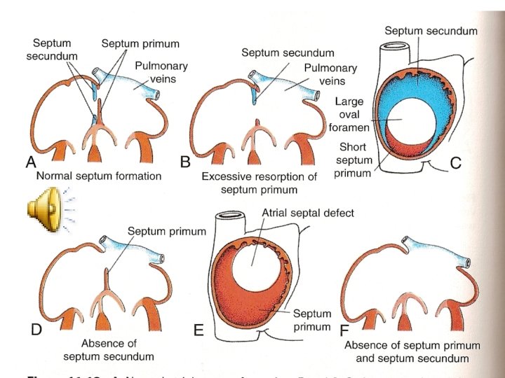
• Anomalies of the interatrial Septation: • There are many types of defected atrial Septation with Rt to Lt shunt of blood: • 1. ostium secondum defect: either by a large ostium secondum formation, or small septum secondum. • 2. ostium primum defect: by abnormal endocardial cushions closing the ostium primum. • 3. complete absence of the septum: common atrium is formed and is called triloculare biventriculare. • 4. premature closure of the foramen ovale (before birth): leading to increased pressure and hypertrophy of the Rt atrium and ventricle, with atrophy of the left atrium and ventricle. It causes death shortly after birth.
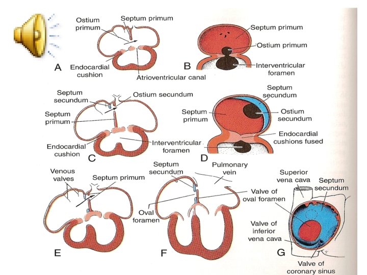
• Septation of the atrioventricular canal: (Easy Go) • At early stages, the canal connects the common atrium and the Lt ventricle primordium only. • Later on, the canal is enlarged on the Rt side to be connected with the Rt ventricle. After the 4 th week, the four atrioventricular endocardial cushions develop at the canal as anterior, posterior, and 2 lateral cushions. These cushions fuse with each other to from the Rt and Lt atrioventricular orifices. The mesenchymal proliferation developing around these orifices are hollowed by the blood flow forming the cusps of the mitral and tricuspid valves.
• Anomalies of atrioventricular Septation • 1. persistent atrioventricular canal: by failure of fusion of the endocardial cushions that is associated with defected atrial and ventricular Septation. • 2. ostium primum defect. • 3. tricuspid Artesia: associated with opened ovale foramen and defected ventricular septum, with hypertrophy of the Lt ventricle and atrophy of the Rt ventricle.
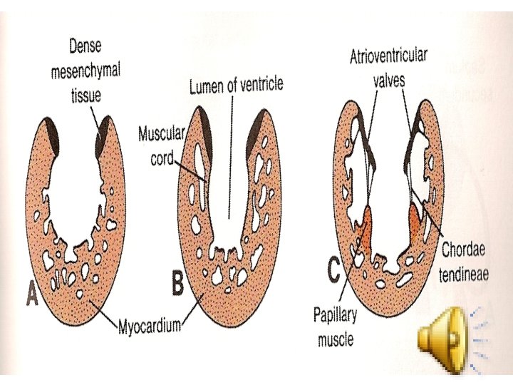
• (Easy Go) • Ventricular Septation: • After the 4 th week, the lateral walls of both ventricles grow on the outside and absorbed from the inside forming the trabeculae carnae and papillary muscles. The medial walls of both of the ventricles fail to grow and thus become opposed and fused with each other forming the muscular interventricular septum. The opening between the upper free end of this septum and the endocardial cushions is closed by the membranous interventricular septum that is derived from the inferior cushion. In addition, the conus and truncus swellings will contribute to the interventricular septum.
• Anomalies of the ventricular Septation: • 1. defected muscular and membranous interventricular Septation. • 2. persistent atrioventricular canal. • 3. tricuspid atresia. • 4. tetralogy of fallote. • 5. transposition of the great vessels.
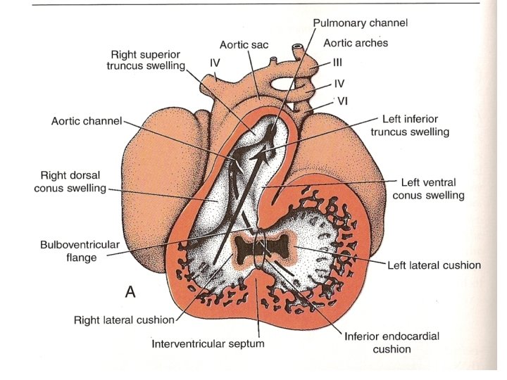
• (Easy Go) • Septation of the truncus arteriosus and conus cordis: • During the 5 th week, two swellings develop in the truncus arteriosus as a Rt superior and Lt inferior swellings. Similar swellings appear in the conus cordis as Rt dorsal and Lt ventral swellings. These four swellings grow toward each other in a spiral way to separate the aorta from the pulmonary artery by a spiral septum that is connected with the septum derived from the conus swellings separating the out flow (smooth part) of the Lt and Rt ventricles. Tubercles of the semilunar valves develop inside the truncal swellings forming the semilunar valves.