DEVELOPMENT OF THE HEART VESSELS CARDIOVASCULAR SYSTEM M
 M. MANSYUR ROMI Department Anatomy, Embryology")


 extra-embryonic blood vessels ---at the middle of 3 rd week, wall of the")

 intra-embryonic blood vessels ---by the 18 -20 th days, endothelial tube network appears")


")

 Aggregate & arrange side by side Two")
 Mesenchyme around tubes thickens")


 and ventral (superior) walls of the")


 septum grows as a ridge")


- Slides: 52
DEVELOPMENT OF THE HEART & VESSELS (CARDIOVASCULAR SYSTEM) M. MANSYUR ROMI Department Anatomy, Embryology and Anthropology Faculty of Medicine Universitas Gadjah Mada
OUTLINE 1. 2. 3. 4. 5. Introduction Early Development of the Cardiovascular/Circulatory System Development of the Primitive Heart Partitioning/Septation of the Heart Development of the Major Arteries & Veins
1. INTRODUCTION n n n The development of the cardiovascular system is an early embryological event. From fertilization, it takes eight weeks for the human heart to develop into its definitive fetal structure. During this period the system develops so it can 1) supply nutrients and oxygen to the fetus, and 2) immediately function after birth.
2. EARLY DEVELOPMENT OF THE CARDIOVASCULAR SYSTEM n n n Blood Islands Heart tube Primitive Cardiovascular Circuits
1) extra-embryonic blood vessels ---at the middle of 3 rd week, wall of the yolk sac, body stalk and chorion mesenchyma proliferate and form isolated cell clusters: angiobalstic blood islands; central located cells are detached and develop into primitive blood cells (hemopoetic/blood stem cells) peripheral cells become flattened and differentiate into endothelial cells to from endothelial tube,
---endothelial tube approach and fuse with each other to form an endothelial tube network ---endothelial tube network appears in chorionic membrane and body stalk, and connect to vitelline circulation
2) intra-embryonic blood vessels ---by the 18 -20 th days, endothelial tube network appears in intraembryonic mesenchyma to form intraembryonic endothelial tube network ---by the end of 3 rd week, intraembryonic and extraembryonic endothelial tube networks connect to each other to form a diffuse endothelial tube network ---endothelial tube networks fuse or disappear to form primitive cardiovascular system
Heart tube n blood islands from the splanchnic mesoderm appear and form a plexus of vessels lying deep to the horseshoe-shaped prospective pericardial cavity. These small vessels develop into paired endocardial heart tubes. The splanchnic mesoderm proliferates and develops into the myocardial mantle which gives rise to the myocardium. The epicardium develops from cells that migrate over the myocardial mantle from areas adjacent to the developing heart.
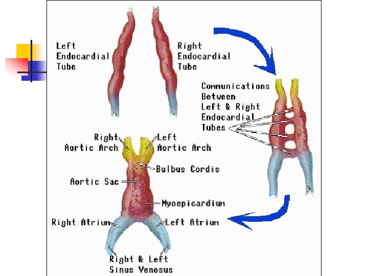
Primitive cardiovascular system: /heart tube: paired by the 4 th week which fuse /arteries: -dorsal aorta: -vitelline artery -umbilical artery -aortic arch: 6 pairs /veins: -anterior cardinal vein -posterior cardinal vein -common cardinal vein -vitelline vein -umbilical vein
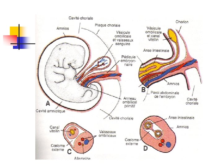
Primitive circuits n n One intraembryonic circuit Two extraembryonic circuits: Vitelline (omphalomesenteric, yolk sac) Umbilical (allantoic, placental)
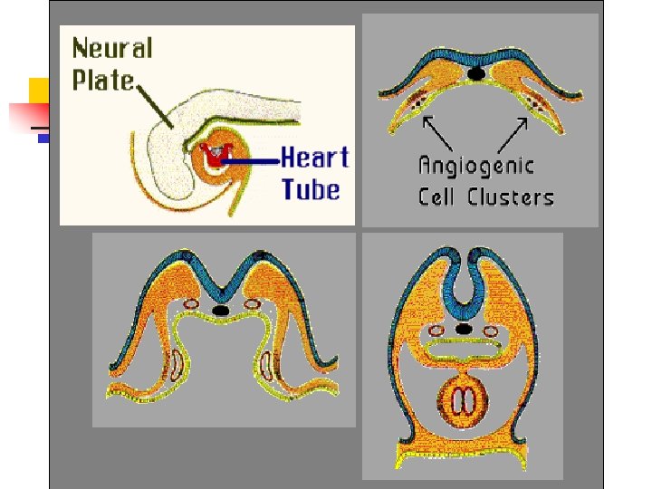
3. DEVELOPMENT OF PRIMITIVE HEART n n The earliest major organ to function is formed from at least 3 sources : n Angioblastic mesenchyme n Midline splanchnopleuric coelomic epithelium n Neural crest cells First indicated in embryos at 18 or 19 days Contraction of the heart begin by day 22
Splanchnic mesenchymal cell (in the cardiogenic area) Aggregate & arrange side by side Two longitudinal cellular strands (CARDIOGENIC CORDS) canalized Two thin-walled endothelial tubes (ENDOCARDIAL HEART TUBE) Gradually approach & fuse A SINGLE TUBE
endocardial heart tube inner Internal endothelial lining of heart (ENDOCARDIUM) Mesenchyme around tubes thickens MYOEPICARDIAL MANTLE Muscular wall (MYOCARDIUM) Visceral pericardium (EPICARDIUM)
HEART DEVELOPMENT n Clusters of angiogenic cells form a "horseshoe- shaped" cluster anterior and lateral to the brain plate.
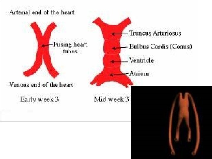
The cardiac tube folds under the gut tube…… THE EARLY DEVELOPMENT OF THE HEART - 1 B A DORSAL AORTA GUT TUBE ENDODERM CARDIAC TUBE PERICARDIAL CAVITY CARDIAC PRIMORDIUM (SPLANCHNIC MESODERM) The cardiac primordia are established in the early gastrula as regions of splanchnic mesoderm ahead of the embryo itself. As a result of the head fold, this region ends up beneath the pharynx. VITELLINE VEINS The heart is a U-shaped tube at this stage and the forming blood vessels are initially unconnected
HEART DEVELOPMENT n n Following cardiac mesoderm involution during gastrulation, the two endocardial tubes fuse along the embryonic midline. The single tubular heart develops many constrictions outlining future structure
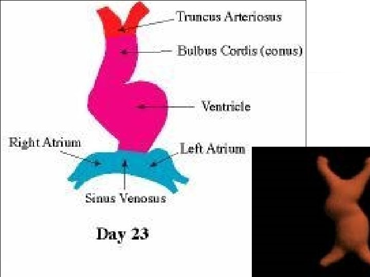
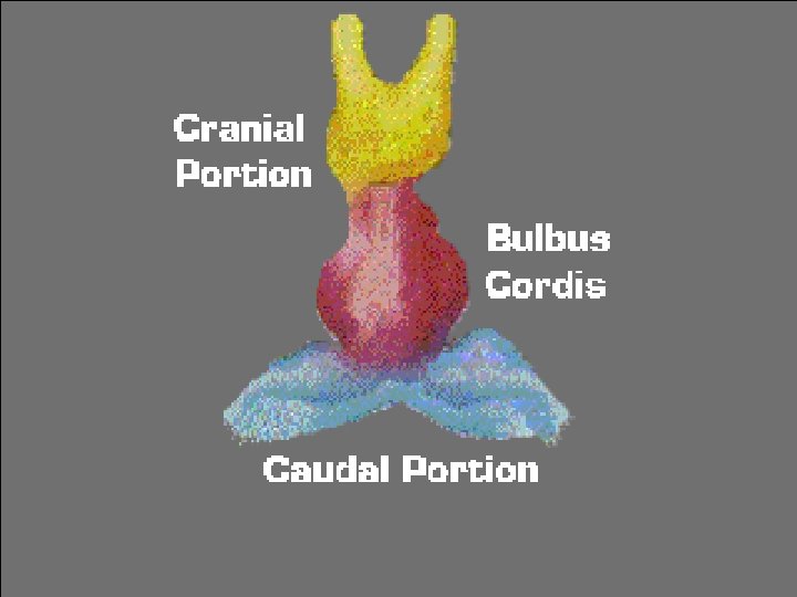
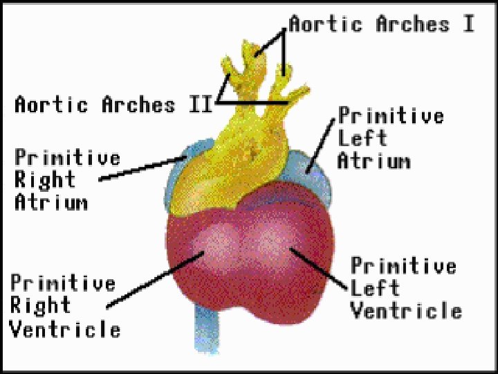
HEART DEVELOPMENT Prior to looping the heart tubes forms constrictions defining separate components of the heart (cranial to caudal): n Bulbus Cordis n Ventricle n Atrium n The primitive atrium is still paired and connects caudally to the paired sinus venosus. n The heart tube will bend ventrally, caudally and slightly to the right. n
HEART DEVELOPMENT n n At this stage the paired sinus venosus extend laterally and give rise to the sinus horns. As the cardiac looping is developing the paired atria form a common chamber and move into the pericardial sac.
Sinus venosus n n a large venous sinus Receives : n n n The umbilical vein from the chorion (primitive placenta) Vitelline vein from yolk sac Common cardinal vein from embryo
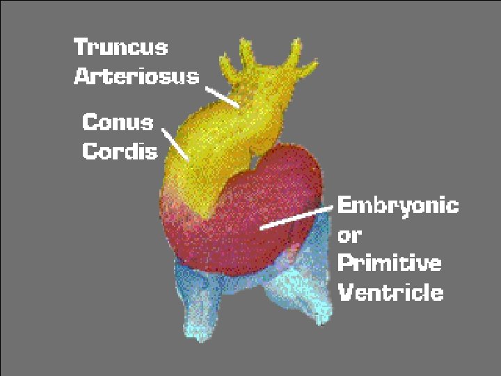
HEART DEVELOPMENT n The arterial and venous end of the heart tube are fixed by : n n n The branchial arches The septum transversum Bulbus cordis & ventricel grow faster than other region, the heart bends upon itself, n n forming a U-shaped bulboventricular loop Later an S-shaped heart
4. PARTITIONING n n During the second month, the heart begins to septate into two atria, two ventricles, the ascending aorta and the pulmonary trunk. Partitioning of the atrioventricular canal , the atrium and the ventricle Ø Ø begins around the middle of the fourth week essentially complete by the end of the fifth week
n Endocardial cushions develop in the dorsal (inferior) and ventral (superior) walls of the heart. These grow toward each other as the cardiac jelly mesenchyme proliferates deep to the endocardium. These cushions fuse and divide the common AV canal into the left and right AV canals.
Atrial Partitioning SAO = Sinoatrial oriface SS = Septum spurium S 1 = Septum primum Perf = Perforations O 1 = Ostium primum EC = Endocardial cushions
n At the same time there is a developing septum from the dorsocranial atrial wall that grows toward the cushions. This is the septum primum, and the intervening space is called the foramen primum. As the septum reaches the endocardial cushions closing foramen primum, a second opening, foramen secundum appears in septum primum.
n As foramen secundum enlarges, a second septum, septum secundum forms to the right of septum primum. Septum secundum forms an incomplete partition (lying to the right of foramen secundum) which leaves an opening, the foramen ovale. The remaining portions of septum primum become the valve of foramen ovale.
Atrial Partitioning O 1 = Ostium primum S 1 = Septum primum FO = Foramen ovale S 2 = Septum secundum
Ventricular septation n The muscular interventricular (I. V. ) septum grows as a ridge of tissue from the caudal heart wall toward the fused endocardial cushions. The remaining opening is the interventricular foramen. The IV foramen is closed by the conal ridges, outgrowth of the inferior endocardial cushion, the right tubercle, and connective tissue from the muscular interventricular septum.
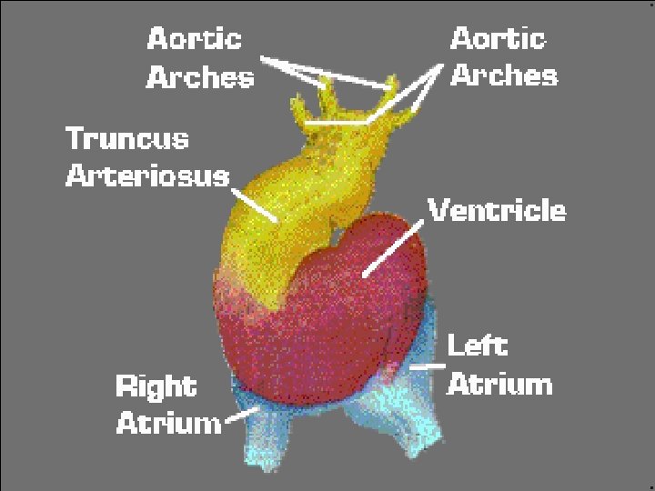
5. Development of the Major Arteries & Veins n n n The six pairs of aortic arches, develop in a cephalocaudal direction and interconnect the ventral aortic roots and the dorsal aorta. Of the six pairs of aortic arches, most of the first, second and fifth arches disappear. The veins develop from the three major vascular circuits.
The Circulatory System n n Functioning circulatory by the end of the first month of life. is vital for sustaining life because it nourishes tissues with oxygen-rich blood and removes waste products from the body.
Second month n n n Throughout the second month of gestation the two atria are formed first, while there still remains just one large ventricle. At this point, the atria are only partially separate, joined by a hole called the foramen ovale. The ventricles are slightly slower to separate, and their separation marks the end of the development of the fetal heart.
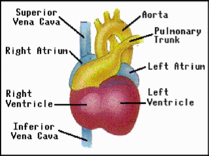
the end of the second month n n the fetal heart has n four chambers n four valves begun to beat, about 148 beats per minute the fetal lungs have formed but are not yet functioning Instead, the fetus receives oxygen-rich blood from its mother through a vein in the umbilical cord attached to the placenta
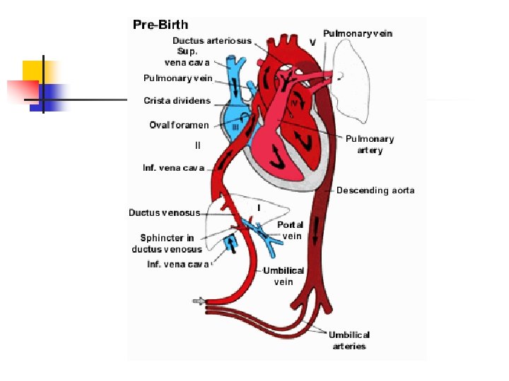
This oxygen-rich maternal blood enters the right atrium through the inferior vena cava, where it mixes with oxygen-poor fetal blood returning from the upper body through the superior vena cava. n Some of the blood then travels to the left atrium through the foramen ovale — a normal prenatal hole in the wall between the upper chambers of the heart (atria). n The blood in the right atrium is thus a mixture of oxygen-rich and oxygen-poor blood. n The blood travels from the upper to the lower chambers of the heart (the ventricles). n n
Before birth n n n The left ventricle pumps the blood out through the aorta to nourish the upper body. The right ventricle pumps the blood through the pulmonary artery (which will one day carry blood to the lungs), through a prenatal passageway called the ductus arteriosus and out through the aorta to nourish the lower body. The ductus arteriosus is responsible for bypassing the fetal lungs, which are not yet functioning.
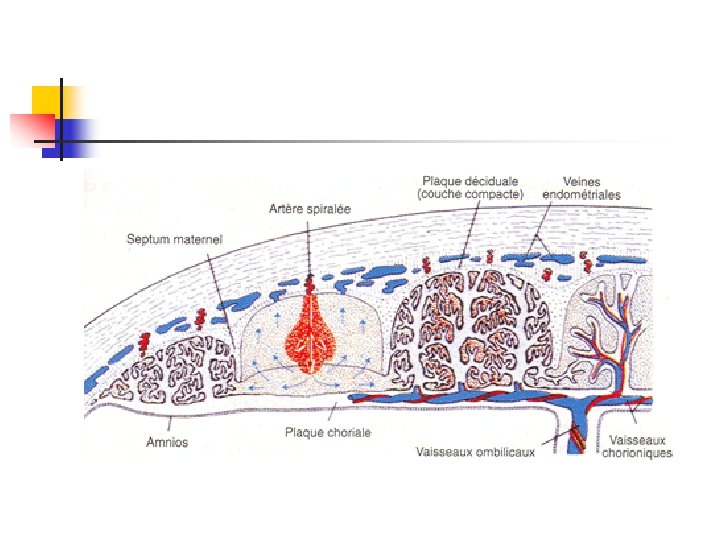
Before birth n n Waste products from the developing fetus are carried in the blood back to the placenta via two arteries in the umbilical cord. These waste products diffuse into the mother’s blood and are carried away and excreted by the maternal circulation.
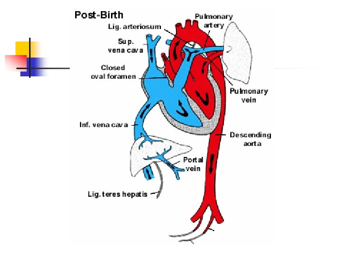
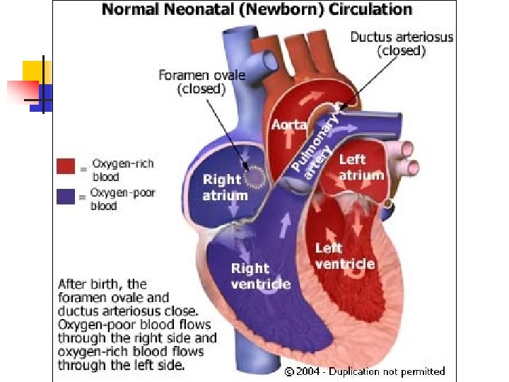
After Birth n n n During and immediately after birth, changes take place in the fetal heart that prepare it for independent life. 1 st: the foramen ovale closes, which prevents blood from freely mixing between the left and right atria. 2 nd: after birth, the baby draws its first breath, and the lungs go to work for the first time.
After Birth n n In healthy newborns, the lungs expand with air, and oxygen sensors in the ductus arteriosus muscles detect the rising levels of oxygen. This causes the ductus arteriosus muscles to contract, squeezing the ductus arteriosus (connects the pulmonary artery to the aorta) closed.