The Cardiovascular System Heart and vessels brings oxygen

The Cardiovascular System Heart and vessels, brings oxygen, nutrients, removes waste

The Heart n Hollow n Cone shaped n Muscular n In thoracic cavity (specifically in mediastinum) n Over diaphragm n Average adult heart: 14 cm. long and 9 cm wide

Coverings of the Heart n Pericardium encloses heart n Fibrous pericardium is the outer fibrous bag within which the heart lies n Epicardium is inner layer n Parietal pericardium at the base of the heart n Pericardial cavity is between the pericardium layers

Wall of the Heart n Epicardium: Outer layer: protects heart by reducing friction: Serous membrane n Myocardium: middle layer, muscular layer which forces blood out of heart chambers, richly supplied with blood vessels n Endocardium: consists of epithelial and connective tissue, some blood vessels and some specialized cardiac muscle, like Pekinje fibers, continuous with inner blood vessel lining.
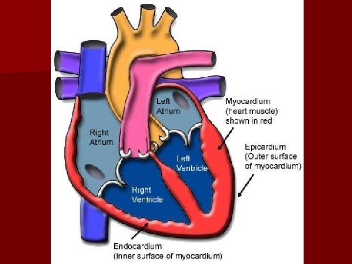

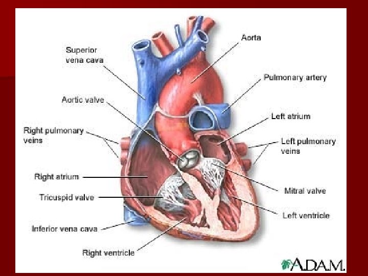
Heart Chambers n Atria: Thinner, upper chambers, receive blood from outside the heart. Auricles extend outward from the Atria. n Ventricles: Thicker (especially the left), lower chambers, receives blood from atria n Septum: separates left and right sides of heart.

Valves n Atriventricular ventricles. valves: between atria and – Tricuspid valve: on the right side – Bicuspid (mitral) Valve: On left side n The cusps of the valves are attached to strong fibrous strings called chordae tendineae n The chordae tendineae are attached to the papillary muscles: small mounds of cardiac muscle tissue


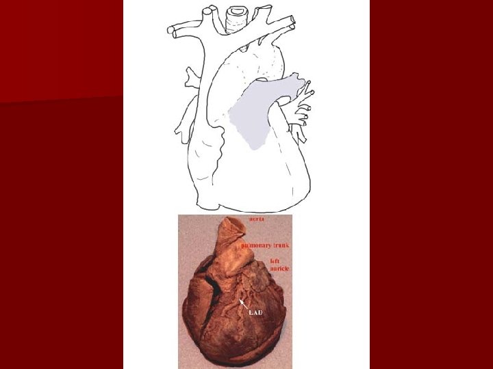
Pulmonary Trunk n On the right ventricle n Divides into left and right pulmonary arteries n At the base there is a pulmonary valve with 3 cusps

Left Side Blood flow n The left atrium gets blood from lungs via 4 pulmonary veins. n Blood flows through the bicuspid valve into the left ventricle n Blood flows through the aortic valve into the aorta
- Slides: 12