Cardiovascular system Heart Vascular system blood vessels Heart
")
Cardiovascular system • Heart • Vascular system (blood vessels)

Heart • Anatomy Consist of 2 separate sides, each composed of 2 chambers separated by septum, that has valve allowing the passage of blood from the atria to the ventricle in one direction. The left side contains oxygenated blood coming from the lungs, while the right side contains deoxygenated blood coming from the whole body.
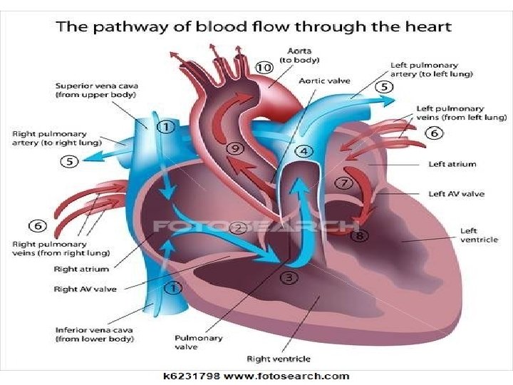

Vascular system The oxygenated blood pumped from the heart into the aorta which transfer the cardiac output to the vital organs in the body except the lungs. The two coronary arteries arise from the aorta supply the myocardium. The deoxygenated blood collected from the body collected in right atrium then pushed to the lungs through the pulmonary artery.
,")
Diseases affecting the coronary arteries cause ischemic heart disease (myocardial infarction or angina pectoris), while disease affecting the peripheral circulatory system may cause hypertension , deep venous thrombosis(DVT).

Signs and symptoms Symptoms = chest pain, palpitation, dyspnea. Signs = cyanosis , sweating , clubbing, edema.

Diagnostic tests 1. 2. 3. 4. 5. 6. 7. Cardiac enzymes. ECG. Holter monitoring. Echocardiography. Stress tests (ECG, Echo). C-T angiography. Cardiac catheterization.
, Aspartate aminotransferase (AST), Lactate dehydrogenase (LDH). Cardiac")
Cardiac enzymes troponin , Creatinin phosphokinase (CPK), Aspartate aminotransferase (AST), Lactate dehydrogenase (LDH). Cardiac enzymes increase in myocardial infarction.

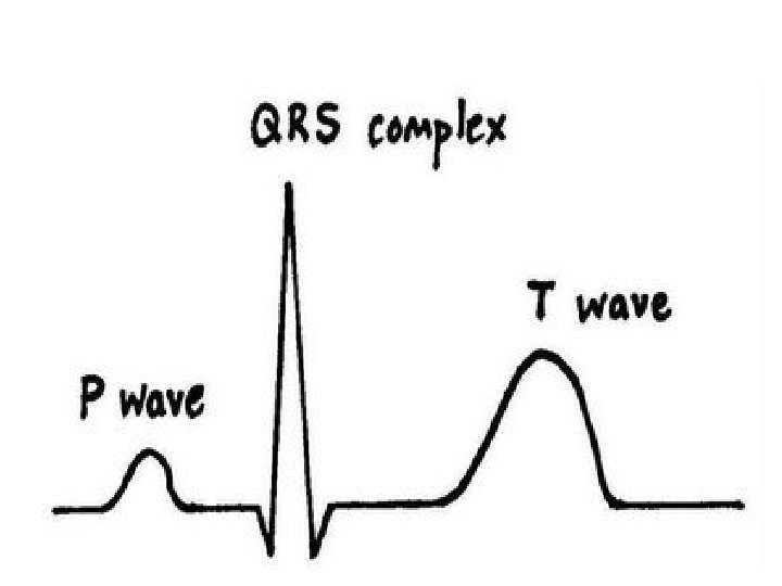
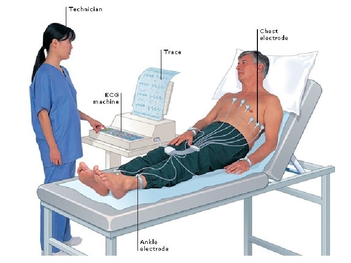
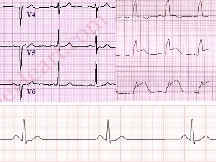
ECG ECG= is a graphic plotting of the electrical cardiac activity.

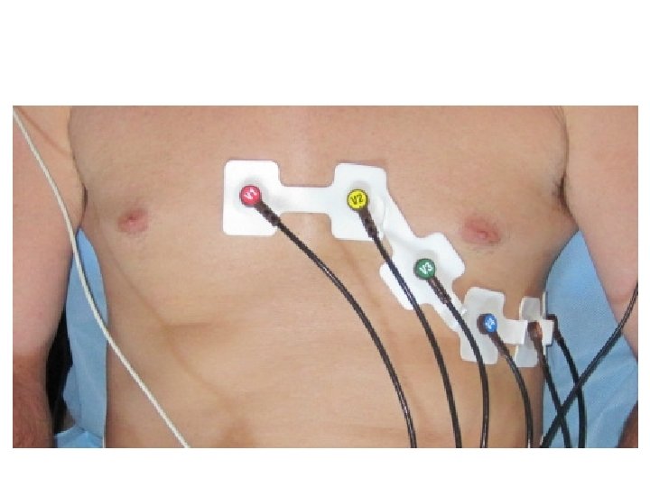
How to perform ECG The usual 12 leads ECG can be done by using 10 electrodes, 4 for the limbs , and 6 for the chest. Limb electrodes = I , III, a. VR , a. VL , a. VF. Chest electrodes = V 1 , V 2 , V 3 , V 4 , V 5 , V 6

Benefits Diagnosis of some cardiac problems especially ischemic heart disease and arrhythmia.

Holter monitor Similar to ordinary ECG , but is used to monitor cardiac activity for long time (1 -2 days). The instrument carried by the patient and usually taken home. It is useful for monitoring of infrequent events of cardiac arrhythmias (paroxysmal arrhythmia).

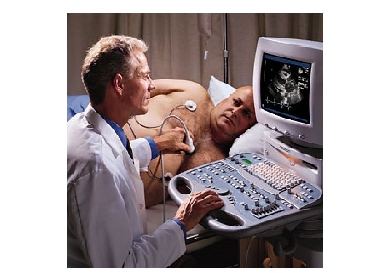
Echocardiography Ultra sound based test used to monitor cardiac function in real time state. It is a bed site examination. Can detect structural heart defects, abnormality in function, and the blood flow in and around the heart.

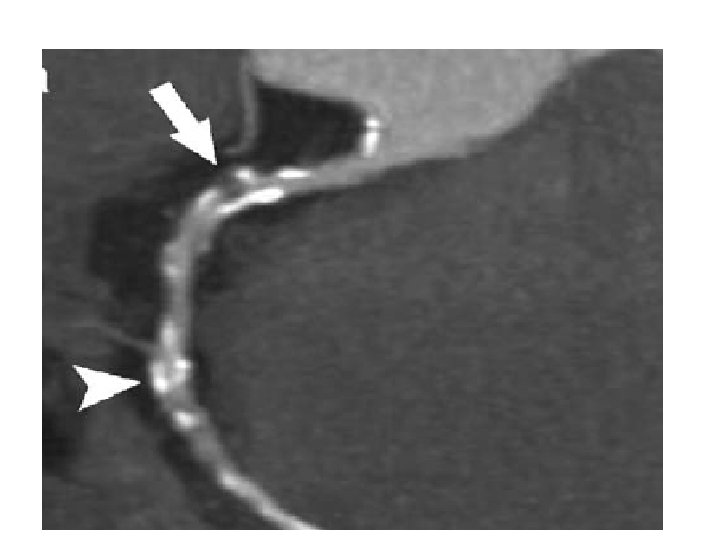
CT – angiography CT scan of the heart to detect plaque or calcium deposition in the coronary arteries, which indicates ischemia.

Cardiac catheterization Involve the insertion of plastic catheter within the femoral artery, and progressing the catheter to reach the aorta, the by selecting the coronary artery, dye is injected inside to make the artery visualized on a fluoroscopic screen. Normal flow of the dye indicates normal coronaries, while narrowing or obstruction of the flow indicating ischemic coronary arteries.
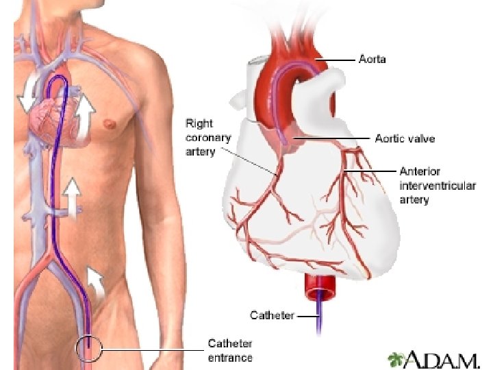
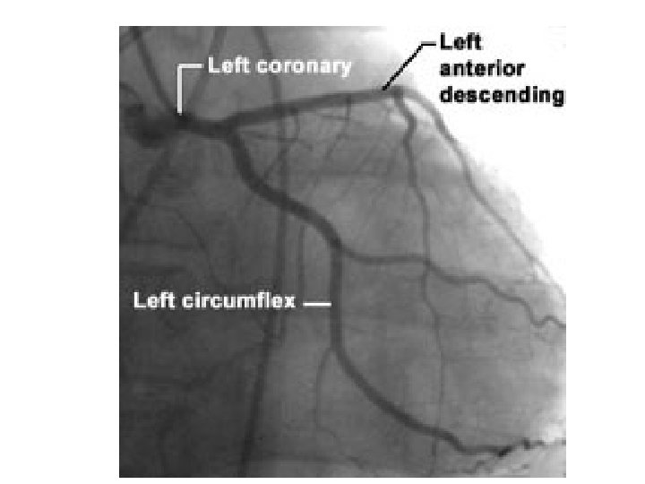
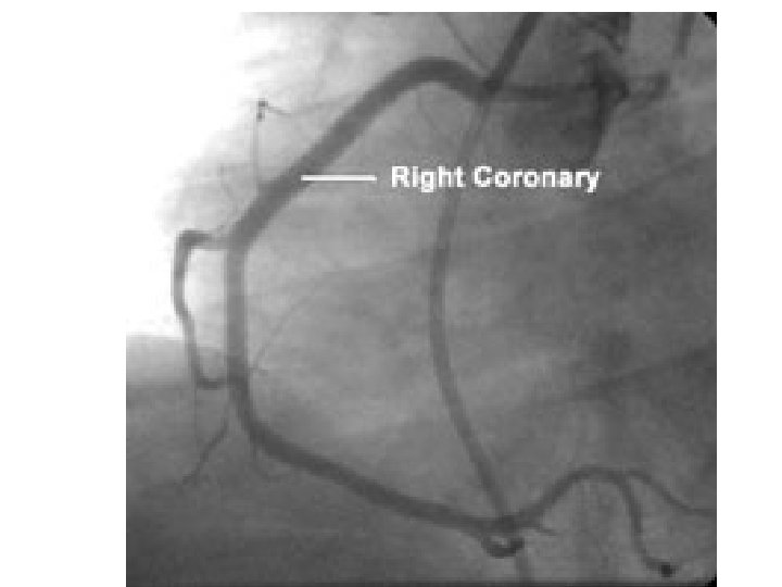

Procedure After good skin sterilization insert a plastic catheter percutaneously inside the femoral artery at the groin, then progress the catheter upward under fluoroscopic monitoring until reaching the aortic arch, from that position localizing the coronary arteries should be done, followed by selective insertion of the catheter inside either the left or right coronary artery to inject a dye making the coronary artery more clear to fluoroscopic vision.

Coronary cath. Should be done under anticoagulant cover, and removal of the femoral sheath should only be done after ensuring that the anticoagulant effect is ended to avoid excessive bleeding from the site of the catheter insertion. Legs should be kept straight until the catheter is removed. After removal apply local pressure on the site of puncture for appropriate time.

Patient should be kept flat for several hours. Patient can be discharge home in the next day. Post discharge follow up include monitoring the puncture site for bleeding , infection, or swelling (hematoma collection), and also changing dressing if required.

Clinical application 1. Diagnostic catheterization - only detect coronary patency. 2. Therapeutic catheterization – involve dilatation of the stenosed coronary artery or insertion of a coronary stent.
- Slides: 28