
Testregek elklnlse A bursa omentalis sebszetnek anatmiai alapjai
Testüregek elkülönülése A bursa omentalis sebészetének anatómiai alapjai Nemeskéri Ágnes 2018 Semmelweis Egyetem Anatómiai, Szövet- és Fejlődéstani Intézet nemeskeri. agnes@med. semmelweis-univ. hu
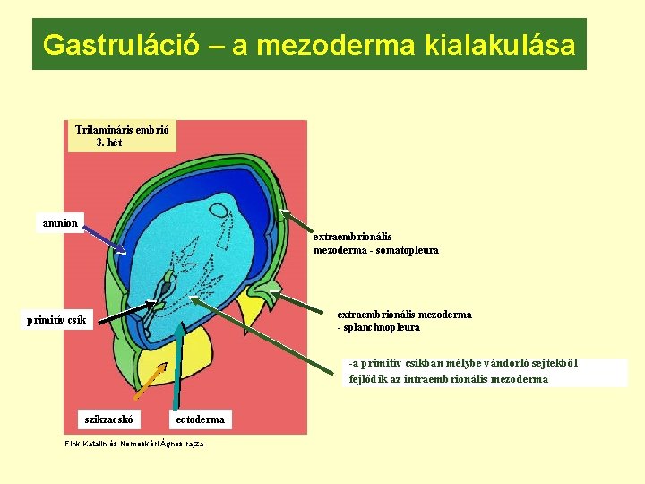
Gastruláció – a mezoderma kialakulása Trilamináris embrió 3. hét amnion extraembrionális mezoderma - somatopleura extraembrionális mezoderma - splanchnopleura primitív csík -a primitív csíkban mélybe vándorló sejtekből fejlődik az intraembrionális mezoderma szikzacskó ectoderma Fink Katalin és Nemeskéri Ágnes rajza
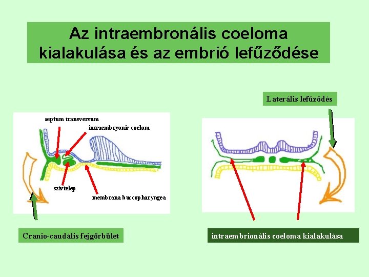
Az intraembronális coeloma kialakulása és az embrió lefűződése Laterális lefűződés septum transversum intraembryonic coelom szívtelep membrana buccopharyngea Cranio-caudális fejgörbület intraembrionális coeloma kialakulása
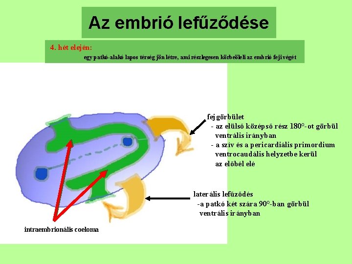
Az embrió lefűződése 4. hét elején: egy patkó-alakó lapos térség jön létre, ami részlegesen körbeöleli az embrió feji végét fejgörbület - az elülső középső rész 180°-ot görbül ventrális irányban - a szív és a pericardiális primordium ventrocaudális helyzetbe kerül az előbél elé laterális lefűződés -a patkó két szára 90°-ban görbül ventrális irányban intraembrionális coeloma
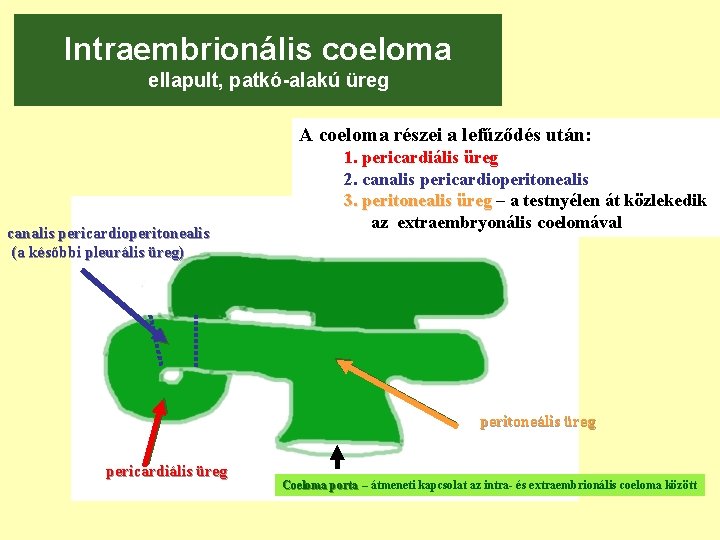
Intraembrionális coeloma ellapult, patkó-alakú üreg A coeloma részei a lefűződés után: canalis pericardioperitonealis (a későbbi pleurális üreg) 1. pericardiális üreg 2. canalis pericardioperitonealis 3. peritonealis üreg – a testnyélen át közlekedik az extraembryonális coelomával peritoneális üreg pericardiális üreg Coeloma porta – átmeneti kapcsolat az intra- és extraembrionális coeloma között
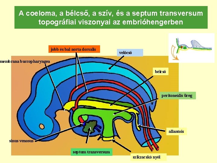
A coeloma, a bélcső, a szív, és a septum transversum topográfiai viszonyai az embrióhengerben jobb és bal aorta dorsalis membrana buccopharyngea * velőcső bélcső peritoneális üreg allantois sinus venosus sinus ven. septum transversum szikzacskó nyél
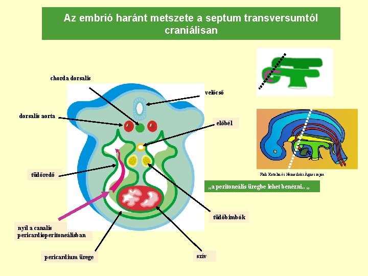
Az embrió haránt metszete a septum transversumtól craniálisan chorda dorsalis velőcső dorsalis aorta előbél tüdőredő Fink Katalin és Nemeskéri Ágnes rajza „a peritoneális üregbe lehet benézni. . „ tüdőbimbók nyíl a canalis pericardioperitoneálisban pericardium ürege szív
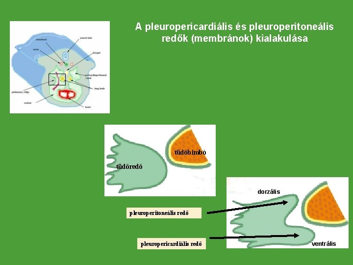
A pleuropericardiális és pleuroperitoneális redők (membránok) kialakulása tüdőbimbó tüdőredő dorzális pleuroperitoneális redő pleuropericardiális redő ventrális
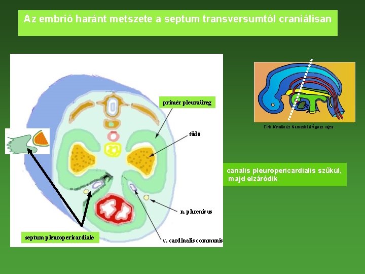
Az embrió haránt metszete a septum transversumtól craniálisan primér pleuraüreg Fink Katalin és Nemeskéri Ágnes rajza tüdő canalis pleuropericardialis szűkül, majd elzáródik n. phrenicus septum pleuropericardiale v. cardinalis communis
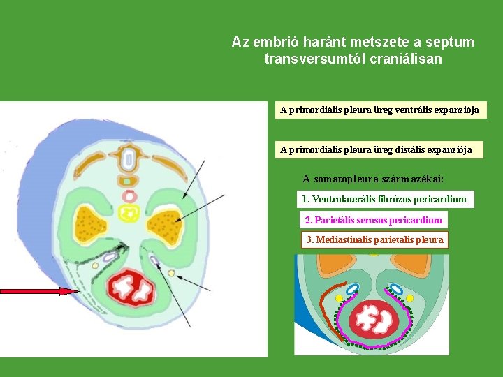
Az embrió haránt metszete a septum transversumtól craniálisan A primordiális pleura üreg ventrális expanziója A primordiális pleura üreg distális expanziója A somatopleura származékai: 1. Ventrolaterális fibrózus pericardium 2. Parietális serosus pericardium 3. Mediastinális parietális pleura
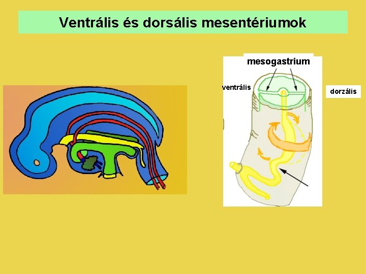
Ventrális és dorsális mesentériumok mesogastrium ventrális dorzális
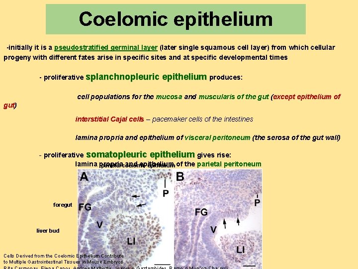
Coelomic epithelium -initially it is a pseudostratified germinal layer (later single squamous cell layer) from which cellular progeny with different fates arise in specific sites and at specific developmental times - proliferative splanchnopleuric epithelium produces: cell populations for the mucosa and muscularis of the gut (except epithelium of gut) interstitial Cajal cells – pacemaker cells of the intestines lamina propria and epithelium of visceral peritoneum (the serosa of the gut wall) - proliferative somatopleuric epithelium gives rise: lamina propria and epithelium parietal coelomic epithelium of the parietal peritoneum foregut liver bud Cells Derived from the Coelomic Epithelium Contribute to Multiple Gastrointestinal Tissues in Mouse Embryos
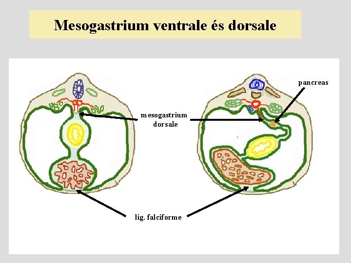
Mesogastrium ventrale és dorsale pancreas mesogastrium dorsale lig. falciforme
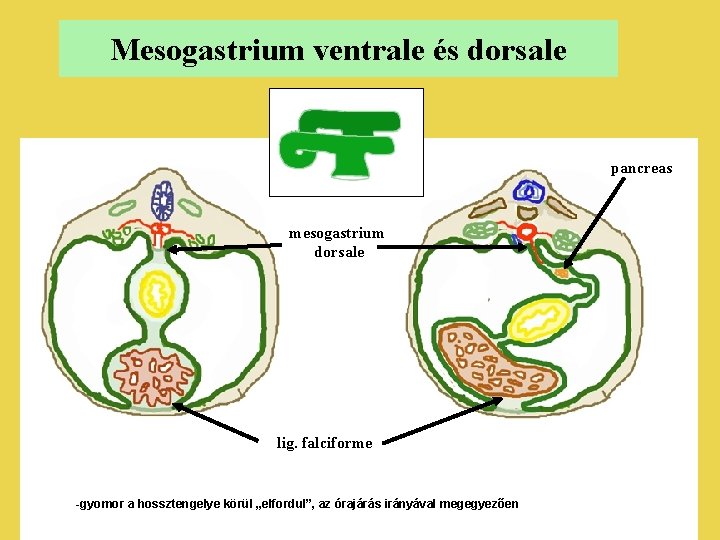
Mesogastrium ventrale és dorsale pancreas mesogastrium dorsale lig. falciforme -gyomor a hossztengelye körül „elfordul”, az órajárás irányával megegyezően
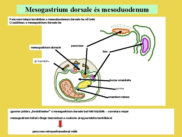
Mesogastrium dorsale és mesoduodenum Pancreas telepe kezdetben a mesoduodenum dorsale-ba nő bele Craniálisan a mesogastrium dorsale-ba mesogastrium dorsale pancreas lien gl. supraren. bursa omentalis gyomor omentum minus -gyomor jobbra „fordulásakor” a mesogastrium dorsale bal felé húzódik – curvatura major -mesogastrium hátsó rétege összeolvad a coeloma üreg parietalis boritékával pancreas retroperitonealissá válik
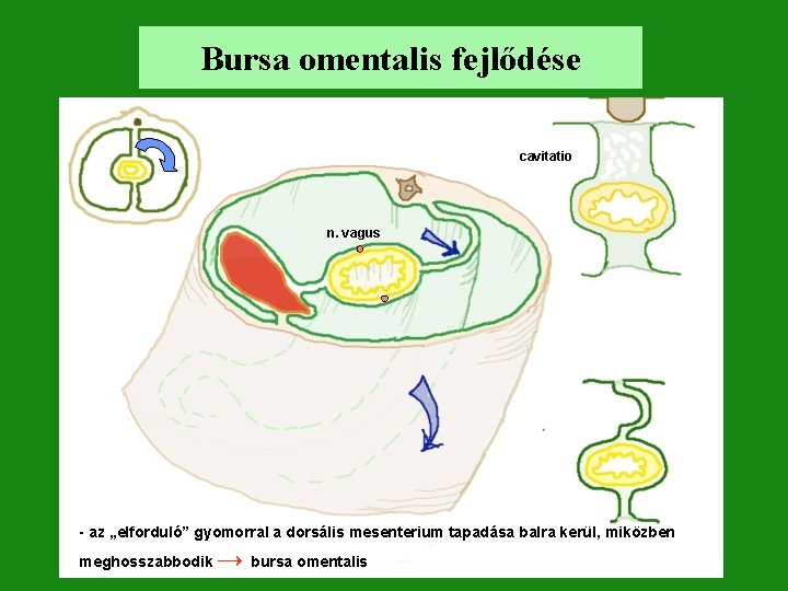
Bursa omentalis fejlődése cavitatio n. vagus - az „elforduló” gyomorral a dorsális mesenterium tapadása balra kerül, miközben meghosszabbodik → bursa omentalis
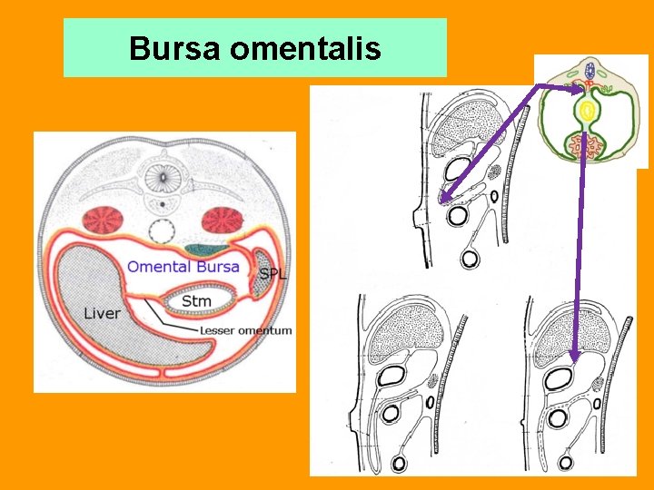
Bursa omentalis
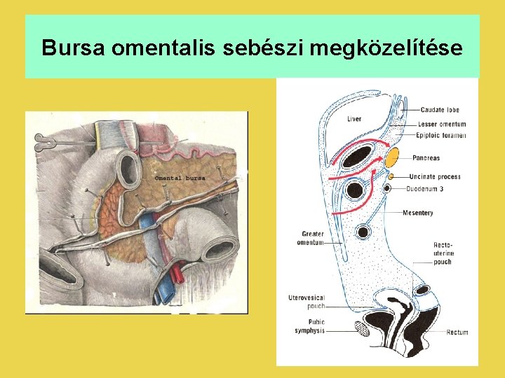
Bursa omentalis sebészi megközelítése
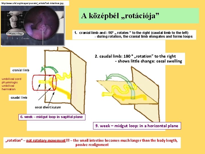
http: //www. ehd. org/images/prenatal_article/7 wk-intestines. jpg A középbél „rotációja” 1. cranial limb and : 90º „ rotates ” to the right (caudal limb to the left) - during rotation, the cranial limb elongates and forms loops 2. caudal limb: 180 º „rotation” to the right - shows little change: cecal swelling cranial limb umbilical cord physiologic umbilical herniation caudal limb cecal diverticulum 6. week – midgut loop in sagittal plane 9. week – midgut loop: in a horizontal plane „rotation” – not rotatory movement!!! – the small intestine becomes much longer than the body length, passive realignment
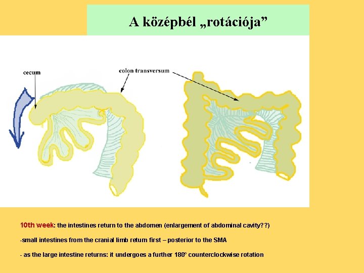
A középbél „rotációja” 10 th week: the intestines return to the abdomen (enlargement of abdominal cavity? ? ) -small intestines from the cranial limb return first – posterior to the SMA - as the large intestine returns: it undergoes a further 180° counterclockwise rotation
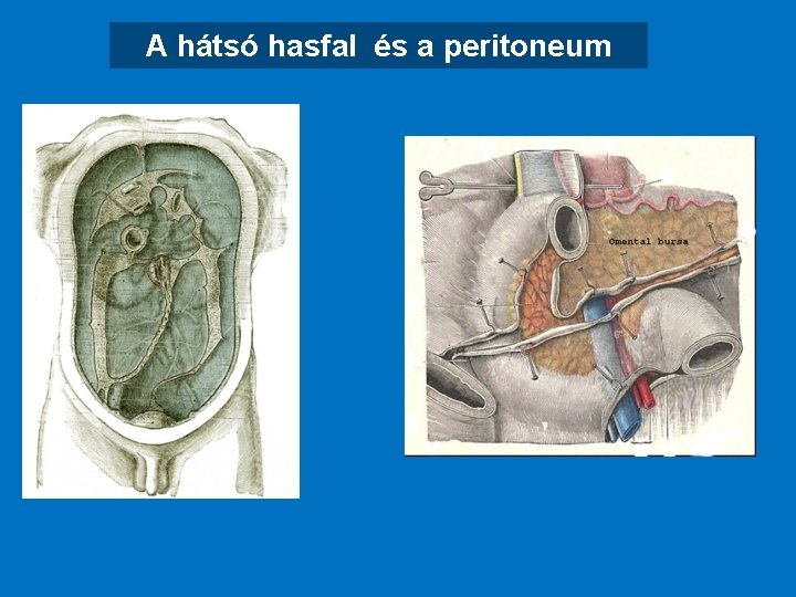
A hátsó hasfal és a peritoneum
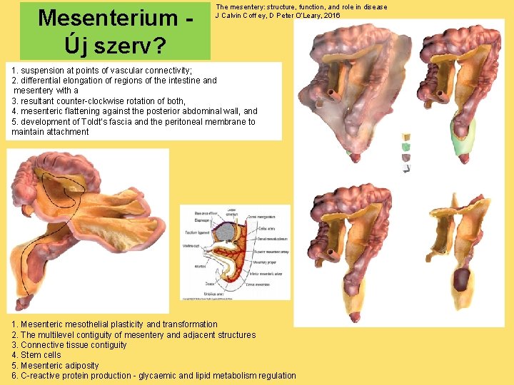
Mesenterium Új szerv? The mesentery: structure, function, and role in disease J Calvin Coff ey, D Peter O’Leary, 2016 1. suspension at points of vascular connectivity; 2. differential elongation of regions of the intestine and mesentery with a 3. resultant counter-clockwise rotation of both, 4. mesenteric flattening against the posterior abdominal wall, and 5. development of Toldt’s fascia and the peritoneal membrane to maintain attachment 1. Mesenteric mesothelial plasticity and transformation 2. The multilevel contiguity of mesentery and adjacent structures 3. Connective tissue contiguity 4. Stem cells 5. Mesenteric adiposity 6. C-reactive protein production - glycaemic and lipid metabolism regulation
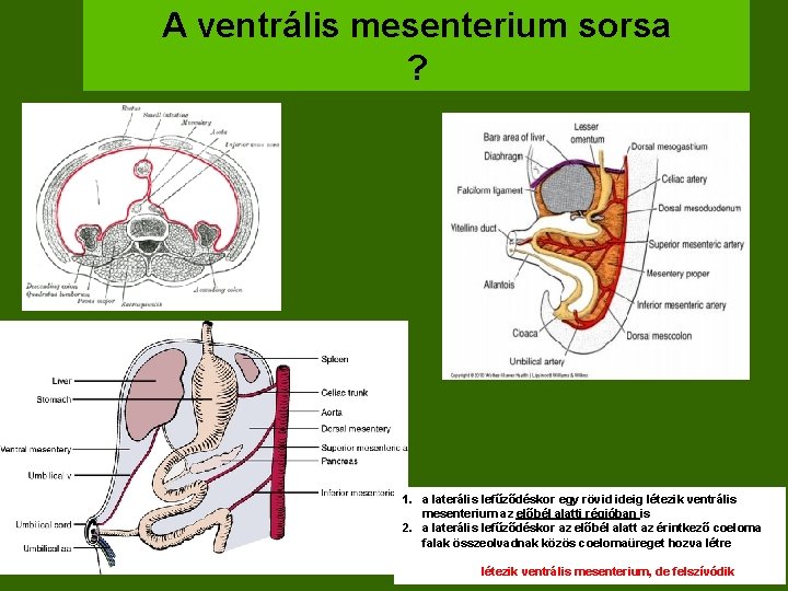
A ventrális mesenterium sorsa ? 1. a laterális lefűződéskor egy rövid ideig létezik ventrális mesenterium az előbél alatti régióban is 2. a laterális lefűződéskor az előbél alatt az érintkező coeloma falak összeolvadnak közös coelomaüreget hozva létre létezik ventrális mesenterium, de felszívódik
Létezik ventrális mesenterium az előbél szintje alatt? ? ? Surg Laparosc Endosc Percutan Tech. 2010 Feb; 20(1): e 34 -7. doi: 10. 1097/SLE. 0 b 013 e 3181 cdb 89 a. Laparoscopy in children with acute intestinal obstruction by aberrant congenital bands. Chang YT 1, Chen BH, Shih HH, Hsin YM, Chiou CS. Aberrant congenital bands are a rare cause of acute intestinal obstruction and usually present a diagnostic challenge. In 2008, the authors encountered 2 children with acute terminal ileal herniation. 1. In the first case, it was caused by a mesodiverticular band, and numerous freely hanging membranes attached to the antimesenteric side of the small intestine. 2. An anomalous band from the distal ileum to the cecum was the leading cause in the second case. The vascularity of both herniated intestines was not compromised. Instead of cohesive adhesions, both of the causes related to a single vascular band. The case with a mesodiverticular band filmy membranes is the first case report with incomplete regression of both the vitelline circulation and the ventral mesentery.
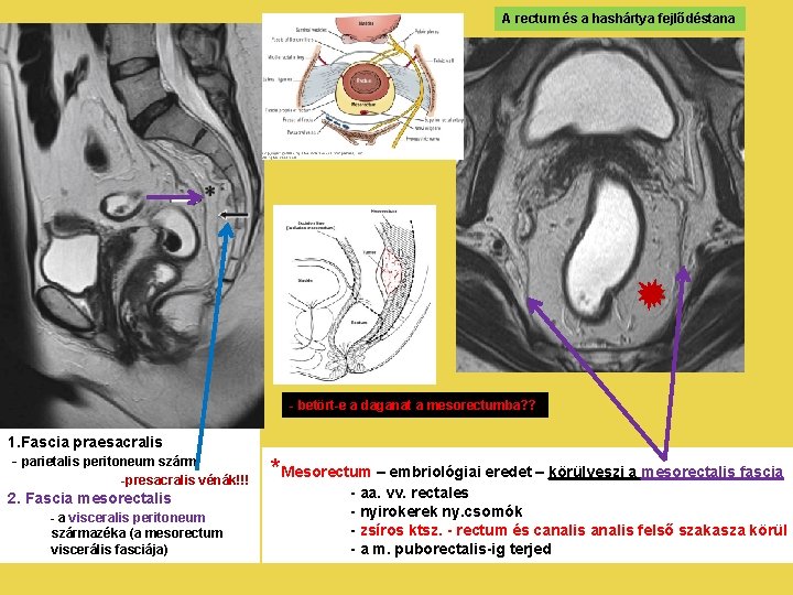
A rectum és a hashártya fejlődéstana - betört-e a daganat a mesorectumba? ? 1. Fascia praesacralis - parietalis peritoneum szárm. -presacralis vénák!!! 2. Fascia mesorectalis - a visceralis peritoneum származéka (a mesorectum viscerális fasciája) *Mesorectum – embriológiai eredet – körülveszi a mesorectalis fascia - aa. vv. rectales - nyirokerek ny. csomók - zsíros ktsz. - rectum és canalis felső szakasza körül - a m. puborectalis-ig terjed
A peritoneum fejlődési rendellenességei Omentum majus agenesia - complex peritoneális anomália - szívfejlődési rendellenességekkel együtt - a gyomor és a colon transversum között egy nagy széles ligamentum helyettesíti a lig. gastrocolicumot - majd folytatódik felfelé mint mesocolon transversum - az omentum majus teljesen hiányzik - ezt a rendellenességet mint a bursa omentalis fejlődésének hiányát lehet értelmezni (agenesia bursae omentalis) - a primitív mesocolon és a bursa omentalis falának inkomplett fúziója - a primitive mesocolon excessiv növekedése
Omentectomy - partially or completely removed - performed on patients suffering from certain types of cancer - in extremely obese patients - Women comprise a large of number of omentectomy patients - cancer of ovary - cancer cells from the ovaries can spread to the surrounding omentum, the fatty tissue removed - In men, intestinal cancer is the main reason for needing an omentectomy - length and size of the small and large intestines can affect a large area of the omentum, significantly increasing the risk of the cancer spreading
Felhasznált irodalom UNSW Embryology http: //php. med. unsw. edu. au/embryology/index. php? title=File: Bailey 301 -303. jpg http: //php. med. unsw. edu. au/embryology/index. php? title=File: Gray 1040. jpg
- Slides: 28