Content Consultants George A Cioffi MD Chief of





- Slides: 115
Content Consultants George A. Cioffi, MD Chief of Ophthalmology Legacy Health System Director, Glaucoma Services Director, Discoveries in Sight Devers Eye Institute Portland, Oregon Dr. Cioffi is now chairman of the Department of Ophthalmology at Columbia University College of Physicians and Surgeons and Ophthalmologist-in. Chief at New York-Presbyterian Hospital/Columbia University Medical Center. Anne L. Coleman, MD, Ph. D Professor of Ophthalmology Jules Stein Eye Institute David Geffen School of Medicine Los Angeles, California Ronald L. Gross, MD Professor of Ophthalmology and Clifton R. Mc. Michael Chair in Ophthalmology Cullen Eye Institute Baylor College of Medicine Houston, Texas Dr. Gross is now chairman of the Department of Ophthalmology at West Virginia University and director of the West Virginia University Eye Institute. Jeffrey M. Liebmann, MD Clinical Professor of Ophthalmology Director, Glaucoma Services Manhattan Eye, Ear & Throat Hospital New York University Medical Center New York, New York Robert N. Weinreb, MD Chairman of the Department of Ophthalmology Director, Shiley Eye Center Director, Hamilton Glaucoma Center University of California San Diego La Jolla, CA, USA 2
Reframing Disease Severity in Glaucoma • Incidence of open-angle glaucoma • Consequences of poorly managed glaucoma • Paradigm shift away from IOP for glaucoma diagnosis and evaluation • Evaluation of optic nerve (ON)/retinal nerve fiber layer (RNFL) • Controlling IOP in glaucoma treatment • Diagnosis & management of glaucoma in clinical practice IOP=intraocular pressure; ON=optic nerve; RNFL=retinal nerve fiber layer 3
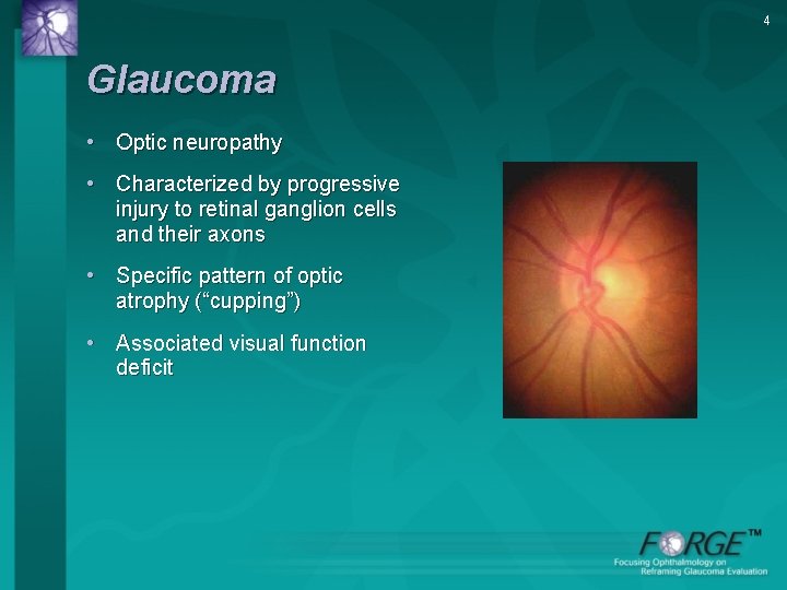
4 Glaucoma • Optic neuropathy • Characterized by progressive injury to retinal ganglion cells and their axons • Specific pattern of optic atrophy (“cupping”) • Associated visual function deficit
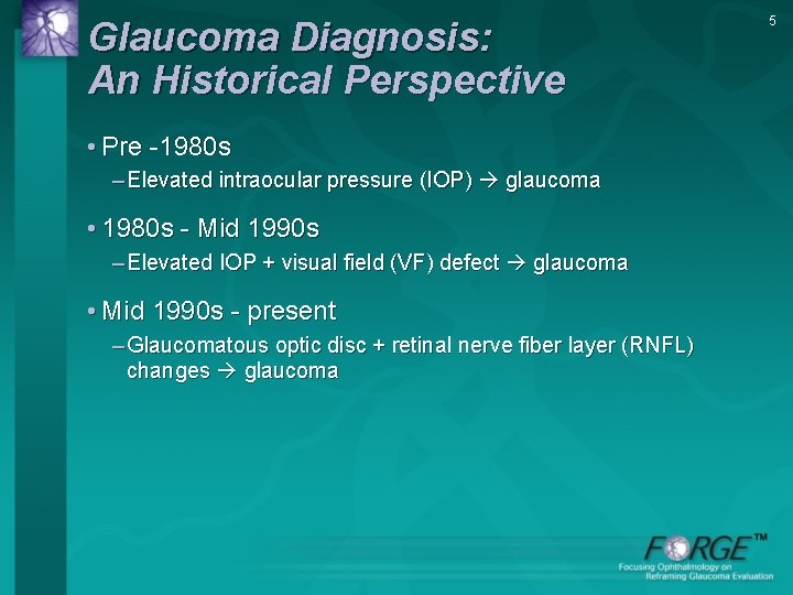
Glaucoma Diagnosis: An Historical Perspective • Pre -1980 s – Elevated intraocular pressure (IOP) glaucoma • 1980 s - Mid 1990 s – Elevated IOP + visual field (VF) defect glaucoma • Mid 1990 s - present – Glaucomatous optic disc + retinal nerve fiber layer (RNFL) changes glaucoma 5
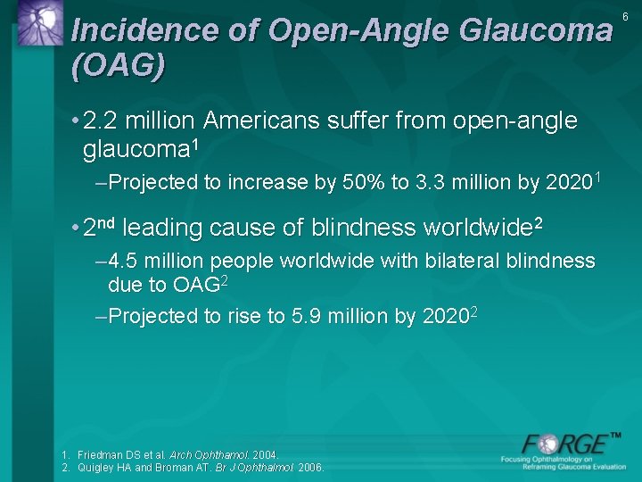
Incidence of Open-Angle Glaucoma (OAG) • 2. 2 million Americans suffer from open-angle glaucoma 1 –Projected to increase by 50% to 3. 3 million by 20201 • 2 nd leading cause of blindness worldwide 2 – 4. 5 million people worldwide with bilateral blindness due to OAG 2 –Projected to rise to 5. 9 million by 20202 1. Friedman DS et al. Arch Ophthamol. 2004. 2. Quigley HA and Broman AT. Br J Ophthalmol. 2006. 6
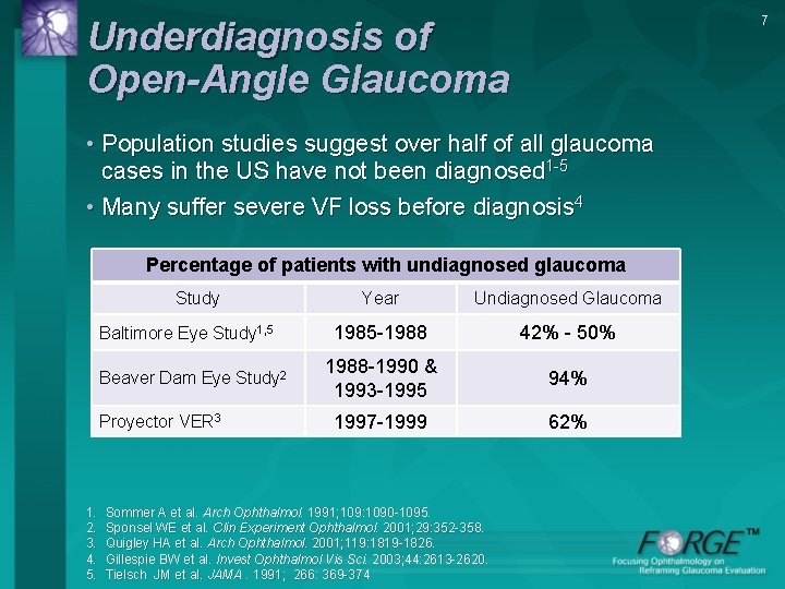
7 Underdiagnosis of Open-Angle Glaucoma • Population studies suggest over half of all glaucoma cases in the US have not been diagnosed 1 -5 • Many suffer severe VF loss before diagnosis 4 Percentage of patients with undiagnosed glaucoma Study Baltimore Eye Study 1, 5 Beaver Dam Eye Study 2 Proyector VER 3 1. 2. 3. 4. 5. Year Undiagnosed Glaucoma 1985 -1988 42% - 50% 1988 -1990 & 1993 -1995 94% 1997 -1999 62% Sommer A et al. Arch Ophthalmol. 1991; 109: 1090 -1095. Sponsel WE et al. Clin Experiment Ophthalmol. 2001; 29: 352 -358. Quigley HA et al. Arch Ophthalmol. 2001; 119: 1819 -1826. Gillespie BW et al. Invest Ophthalmol Vis Sci. 2003; 44: 2613 -2620. Tielsch JM et al. JAMA. 1991; 266: 369 -374
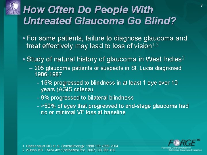
How Often Do People With Untreated Glaucoma Go Blind? • For some patients, failure to diagnose glaucoma and treat effectively may lead to loss of vision 1, 2 • Study of natural history of glaucoma in West Indies 2 – 205 glaucoma patients or suspects in St. Lucia diagnosed 1986 -1987 - 16% progressed to blindness in at least 1 eye over 10 years (AGIS criteria) - 9% progressed to bilateral blindness - >50% of eyes that progressed to end-stage glaucoma had no or minimal VF loss at baseline 1. Hattenhauer MG et al. Ophthalmology. 1998; 105: 2099 -2104. 2. Wilson MR. Trans Am Ophthalmol Soc. 2002; 100: 365 -410. 8
Lack of Effective Treatment Can Result in Blindness • In a 1996 retrospective study of 295 patients with newly diagnosed open-angle glaucoma between 1965 -1980 in Olmsted County, MN – Probability of blindness after 20 years - 27% in one eye; 9% in both eyes – Of 114 patients initially treated for OHT - Probability of blindness after 20 years · 14% in one eye and 4% in both eyes OHT=ocular hypertension. Hattenhauer MG et al. Ophthalmology. 1998; 105: 2099 -104. 9
Risk Factors for Development of Glaucoma • Ocular Factors 1, 2 – IOP – CCT – ON structure (C/D ratio) – Disc hemorrhage – Other ocular disorders • Non-ocular Factors – Age – Race – Family history/genetic predisposition – Vascular disease (HTN, vasospasm) CCT=central corneal thickness; HTN=hypertension; ON=optic nerve. 1. Wilson MR. In: Clinical Guide to Glaucoma Management. Butterworth Heinemann: 2004. 2. Medeiros FA et al. Am J Ophthalmol. 2003; 136: 805 -813. 10
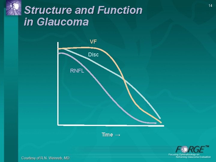
Structural Damage Precedes Functional Change • NFL injury can be observed up to 6 years before VF defects 1 – Mean number of axons 2 in normal ON ~800, 000– 1, 200, 000 – 25 -40% of ON fibers can be lost from an eye that retains a normal visual field 2, 3 1. Sommer A et al. Arch Ophthalmol. 1991; 109: 77 -83. 2. Quigley HA et al. Arch Ophthalmol. 1982; 100: 135 -46. 3. Kerrigan-Baumrind LA et al. Invest Ophthalmol Vis Sci. 2000; 41: 741 -748. 11
Structural Damage Precedes Functional Change (contd. ) • VF loss by SAP does NOT mean early disease – By the time VF loss is detected by SAP, substantial structural damage may exist 1, 2 – Functional loss may be detected earlier using selective tests (eg, FDT, SWAP)2 FDT=frequency doubing technology; SAP=standard automated perimetry; SWAP=short wavelength automated perimetry. 1. Sommer A et al. Arch Ophthalmol. 1991; 109: 77 -83. 2. Bowd C et al. Invest Ophthalmol Vis Sci. 2001; 42: 1993 -2003. 12
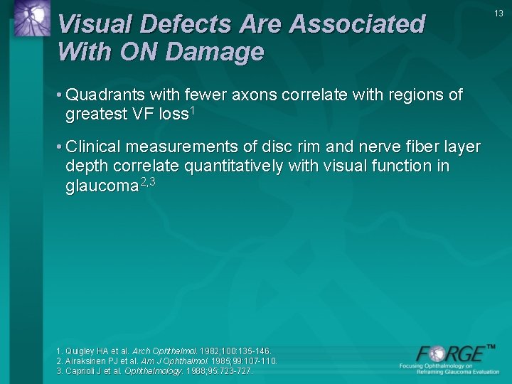
Visual Defects Are Associated With ON Damage • Quadrants with fewer axons correlate with regions of greatest VF loss 1 • Clinical measurements of disc rim and nerve fiber layer depth correlate quantitatively with visual function in glaucoma 2, 3 1. Quigley HA et al. Arch Ophthalmol. 1982; 100: 135 -146. 2. Airaksinen PJ et al. Am J Ophthalmol. 1985; 99: 107 -110. 3. Caprioli J et al. Ophthalmology. 1988; 95: 723 -727. 13
Structure and Function in Glaucoma VF Disc RNFL Time → 14
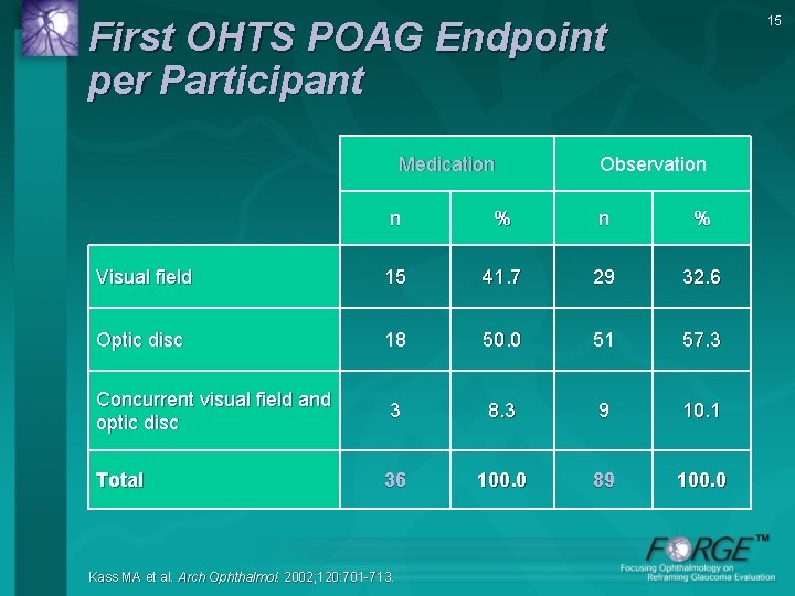
15 First OHTS POAG Endpoint per Participant Medication Observation n % Visual field 15 41. 7 29 32. 6 Optic disc 18 50. 0 51 57. 3 Concurrent visual field and optic disc 3 8. 3 9 10. 1 Total 36 100. 0 89 100. 0 Kass MA et al. Arch Ophthalmol. 2002; 120: 701 -713.
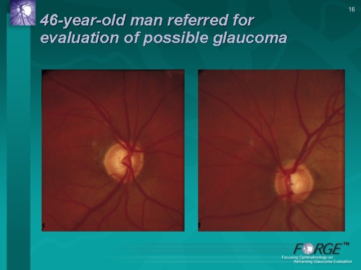
46 -year-old man referred for evaluation of possible glaucoma 16
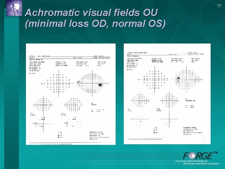
Achromatic visual fields OU (minimal loss OD, normal OS) 17
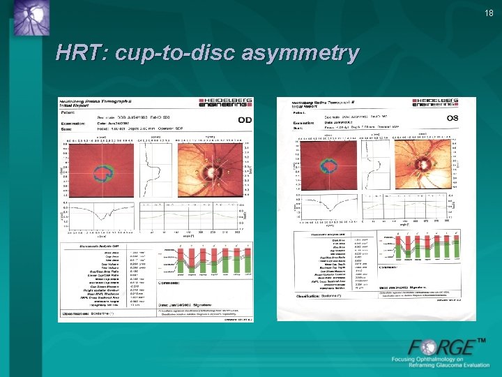
18 HRT: cup-to-disc asymmetry
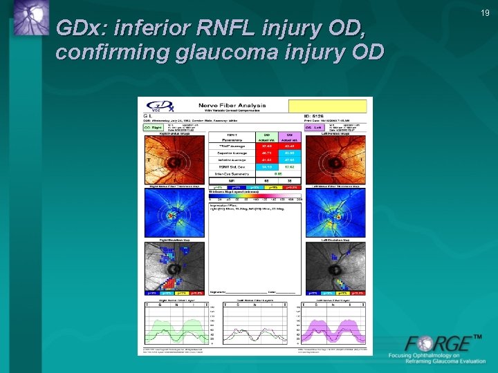
GDx: inferior RNFL injury OD, confirming glaucoma injury OD 19
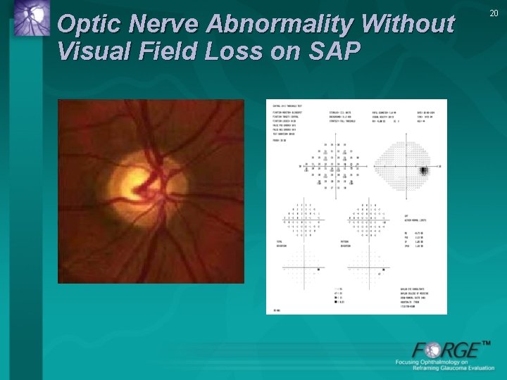
Optic Nerve Abnormality Without Visual Field Loss on SAP 20
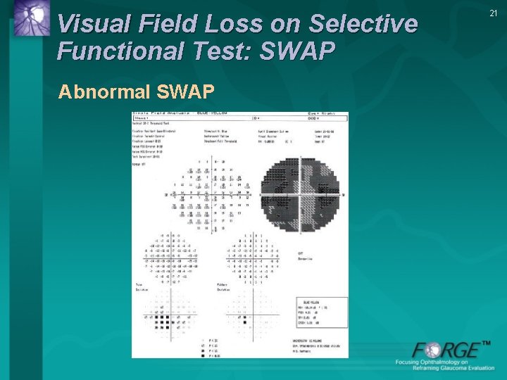
Visual Field Loss on Selective Functional Test: SWAP Abnormal SWAP 21
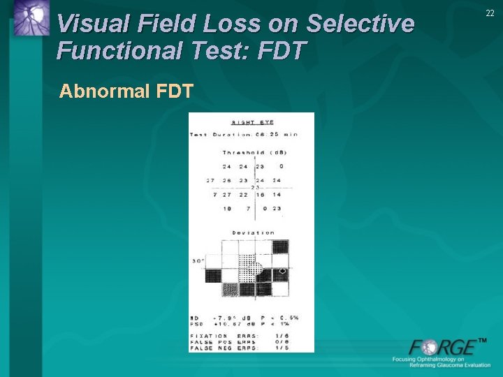
Visual Field Loss on Selective Functional Test: FDT Abnormal FDT 22
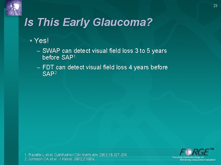
23 Is This Early Glaucoma? • Yes! – SWAP can detect visual field loss 3 to 5 years before SAP 1 – FDT can detect visual field loss 4 years before SAP 2 1. Racette L et al. Ophthalmol Clin North Am. 2003; 16: 227 -236. 2. Johnson CA et al. J Vision. 2002; 2: 100 a.
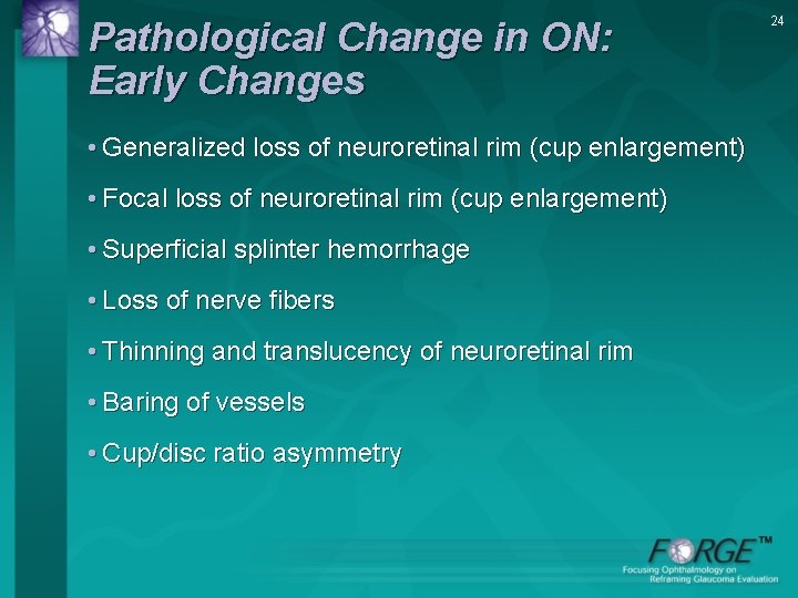
Pathological Change in ON: Early Changes • Generalized loss of neuroretinal rim (cup enlargement) • Focal loss of neuroretinal rim (cup enlargement) • Superficial splinter hemorrhage • Loss of nerve fibers • Thinning and translucency of neuroretinal rim • Baring of vessels • Cup/disc ratio asymmetry 24
Reframing Glaucoma Disease Severity • Redefine early disease – Include eyes with structural damage ± early functional loss – “Pre-achromatic glaucoma” - Glaucomatous optic neuropathy (GON) without SAP field loss 1 • Moderate/severe disease – Reproducible defects in white-on-white perimetry – Enlargement of blind spot – Nasal scotomas (nasal step) – Arcuate defects – Further constriction of visual field and threat to fixation 1. Medeiros FA et al. Am J Ophthalmol. 2003; 136: 805 -813. 25
Periodic ON Exam Advocated in Glaucoma Management Guidelines • Evidence-based guidelines for glaucoma agree 1 -3 -3 – Identification and follow-up of glaucoma requires careful evaluation of ON and RNFL as well as VF – Optic disc photographs or detailed drawings to be obtained at initial visit and at regular intervals during follow-up 1. EGS. Terminology and Guidelines for Glaucoma. 2008. 2. Damji KF, Behki R, Wang L. Can J Ophthalmol. 2003; 38: 189 -197. 3. AAO Preferred Practice Pattern® Guidelines. 2010. 26
27 Optic Disc Evaluation • AAO Preferred Practice Pattern – Detailed drawing – Disc photography • Best technique to detect early changes – Examination of ONH by high magnification, high intensity light stereo view
Lack of Compliance With AAO Guidelines • Chart review studies suggest lack of compliance with ON examination guidelines – 193 POAG patients followed for ≥ 2 years in 8 private practices in LA in 1996 - Almost all patients had photograph or drawing of optic disc at initial examination - At final follow-up visit · 33. 2% had not had ON drawing or photograph within 2 years · Another 37. 8% had not had ON drawing or photograph since initial examination Hertzog et al. Ophthalmology. 1996; 103: 1009 -1013. 28
Lack of Compliance With AAO Guidelines (contd. ) • More recent chart review study in 2003 – 395 POAG patients in 6 managed care plans - Only 53% of patients had optic nerve photograph or drawing at initial evaluation Fremont AM et al. Arch Ophthalmol. 2003; 121: 777 -783. 29
Is the Issue Examination or Documentation of the Nerve? • Are there obstacles to ON examination in clinical practice? 30
“Costs” of Examining the Optic Nerve • Dilation of pupils – Patient and technician time – Patient movement • Increased examination time – 3 to 5 minutes • Increased documentation • Patient discomfort • Others 31
“Benefits” of Examining the Optic Nerve • At baseline examination – Diagnosis - Detect disc hemorrhages - Detect nerve fiber layer defect - Diagnose early glaucomatous ON damage • At follow-up examinations – Diagnosis or Progression - Detect changes in ON appearance - Detect disc hemorrhages - Identify patients at risk of disease progression 32
Benefits of ON Examination Outweigh Costs • Compliance with AAO Preferred Practice Pattern guidelines will – Improve diagnosis of glaucoma – Detect more cases, miss fewer cases – Help more patients with glaucoma – Reduce disease progression 33
Integrating ON Examination Into Clinical Practice • Evaluate and document ON • Use ON evaluation to assess disease severity • Set and achieve lower targets based on risks and appearance of ON • Reevaluate structure & function critically as patient is followed • Reset and achieve lower IOP targets 34
35 Optic Disc/RNFL Examination “The 5 Rs”
36 This section was developed by Robert N. Weinreb, MD Felipe Medeiros, MD Hamilton Glaucoma Center University of California, San Diego Remo Susanna Jr. , MD University of San Paulo, Brazil
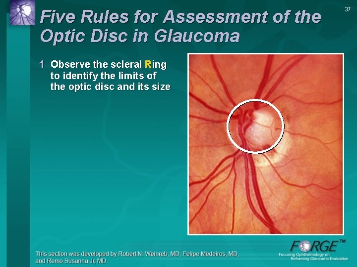
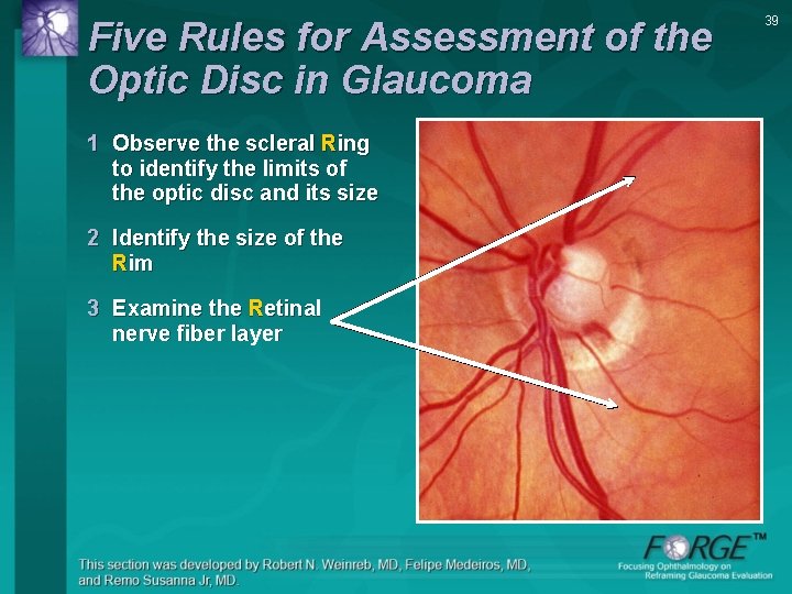
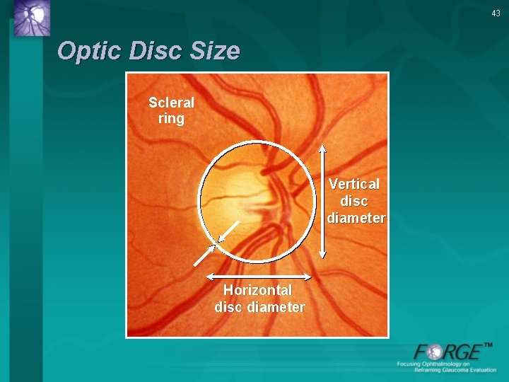
Five Rules for Assessment of the Optic Disc in Glaucoma 1 Observe the scleral Ring to identify the limits of the optic disc and its size 37
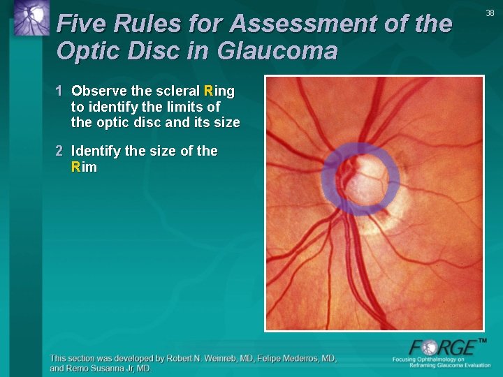
Five Rules for Assessment of the Optic Disc in Glaucoma 1 Observe the scleral Ring to identify the limits of the optic disc and its size 2 Identify the size of the Rim 38
Five Rules for Assessment of the Optic Disc in Glaucoma 1 Observe the scleral Ring to identify the limits of the optic disc and its size 2 Identify the size of the Rim 3 Examine the Retinal nerve fiber layer 39
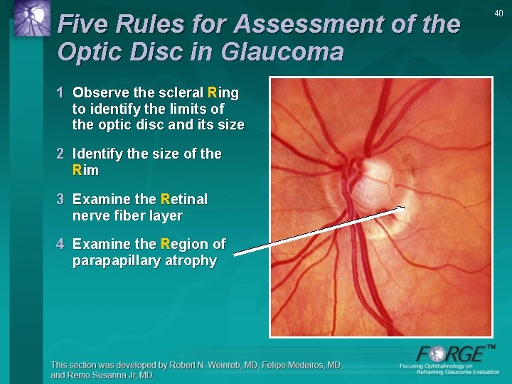
Five Rules for Assessment of the Optic Disc in Glaucoma 1 Observe the scleral Ring to identify the limits of the optic disc and its size 2 Identify the size of the Rim 3 Examine the Retinal nerve fiber layer 4 Examine the Region of parapapillary atrophy 40
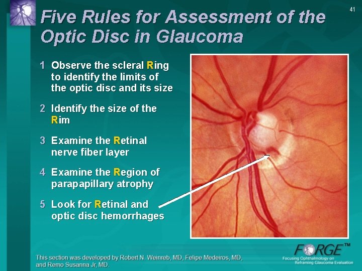
Five Rules for Assessment of the Optic Disc in Glaucoma 1 Observe the scleral Ring to identify the limits of the optic disc and its size 2 Identify the size of the Rim 3 Examine the Retinal nerve fiber layer 4 Examine the Region of parapapillary atrophy 5 Look for Retinal and optic disc hemorrhages 41
42 Rule #1 Observe the scleral Ring to identify the limits and the size of the optic disc
43 Optic Disc Size Scleral ring Vertical disc diameter Horizontal disc diameter
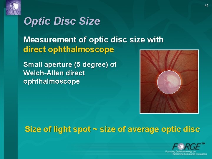
44 Optic Disc Size Measurement of optic disc size with direct ophthalmoscope Small aperture (5 degree) of Welch-Allen direct ophthalmoscope Size of light spot ~ size of average optic disc
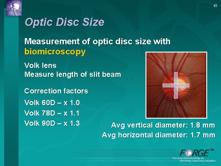
45 Optic Disc Size Measurement of optic disc size with biomicroscopy Volk lens Measure length of slit beam Correction factors Volk 60 D – x 1. 0 Volk 78 D – x 1. 1 Volk 90 D – x 1. 3 Avg vertical diameter: 1. 8 mm Avg horizontal diameter: 1. 7 mm
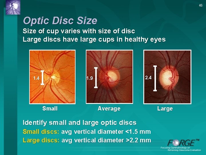
46 Optic Disc Size of cup varies with size of disc Large discs have large cups in healthy eyes 2. 4 1. 9 1. 4 Small Average Identify small and large optic discs Small discs: avg vertical diameter <1. 5 mm Large discs: avg vertical diameter >2. 2 mm Large
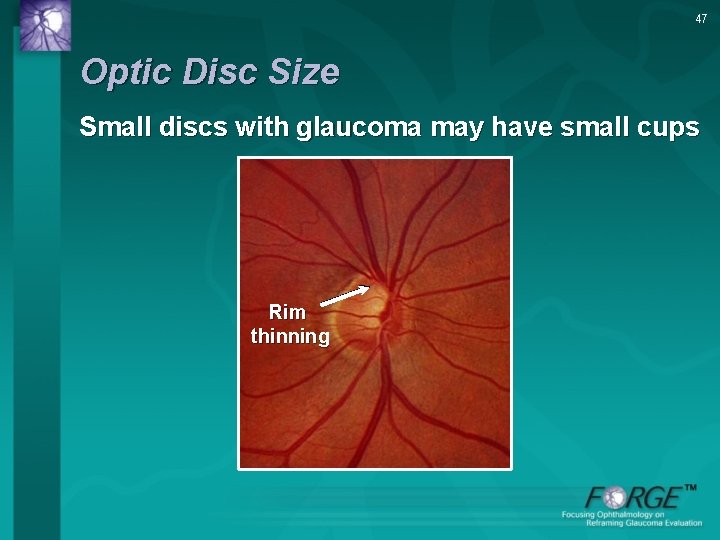
47 Optic Disc Size Small discs with glaucoma may have small cups Rim thinning
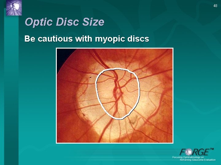
48 Optic Disc Size Be cautious with myopic discs
49 Rule #2 Identify the size of the neuroretinal Rim
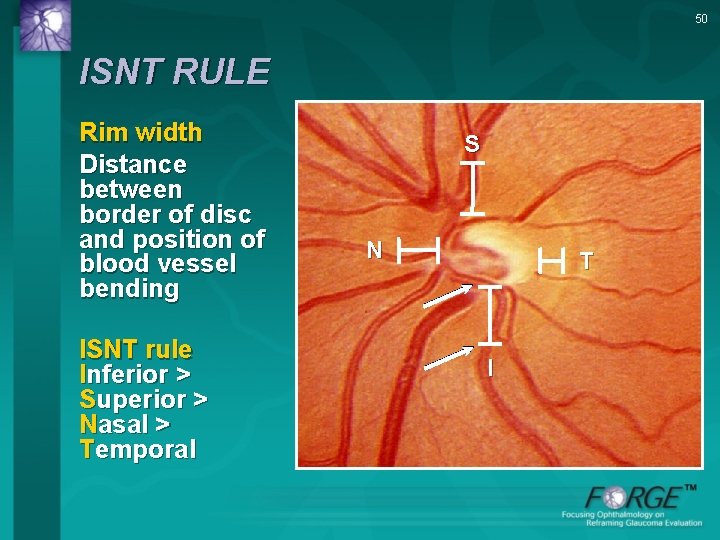
50 ISNT RULE Rim width Distance between border of disc and position of blood vessel bending ISNT rule Inferior > Superior > Nasal > Temporal S N T I
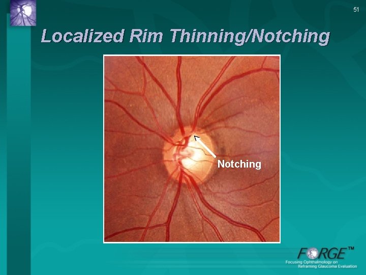
51 Localized Rim Thinning/Notching
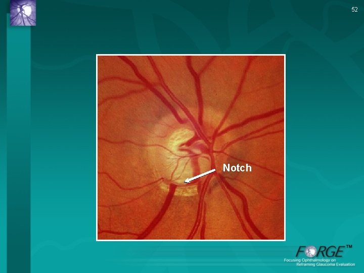
52 Notch
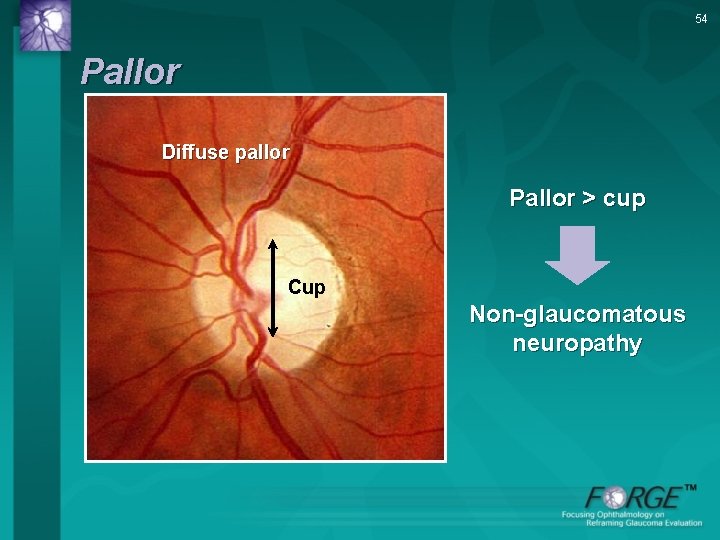
53 Observe the color of the rim to identify pallor A pale rim increases the likelihood for a non-glaucomatous optic neuropathy
54 Pallor Diffuse pallor Pallor > cup Cup Non-glaucomatous neuropathy
55 Rule # 3 Examine the Retinal nerve fiber layer (RNFL)
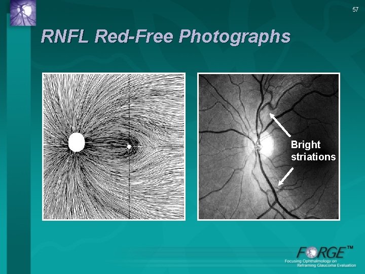
56 RNFL Examination • Best performed using red-free light (red-free photographs or green light) • Look at Striations Brightness Visibility of parapapillary retinal vessels • Look for diffuse and localized RNFL loss
57 RNFL Red-Free Photographs Bright striations
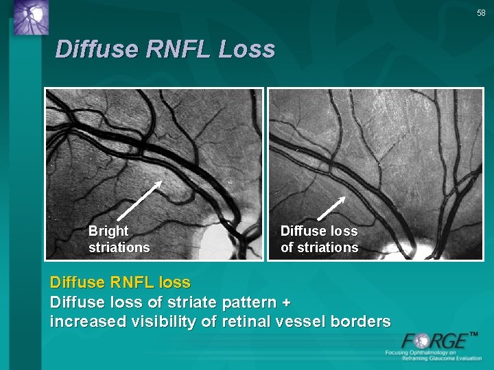
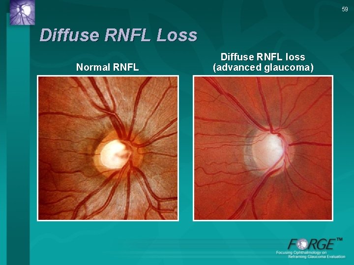
58 Diffuse RNFL Loss Bright striations Diffuse loss of striations Diffuse RNFL loss Diffuse loss of striate pattern + increased visibility of retinal vessel borders
59 Diffuse RNFL Loss Normal RNFL Diffuse RNFL loss (advanced glaucoma)
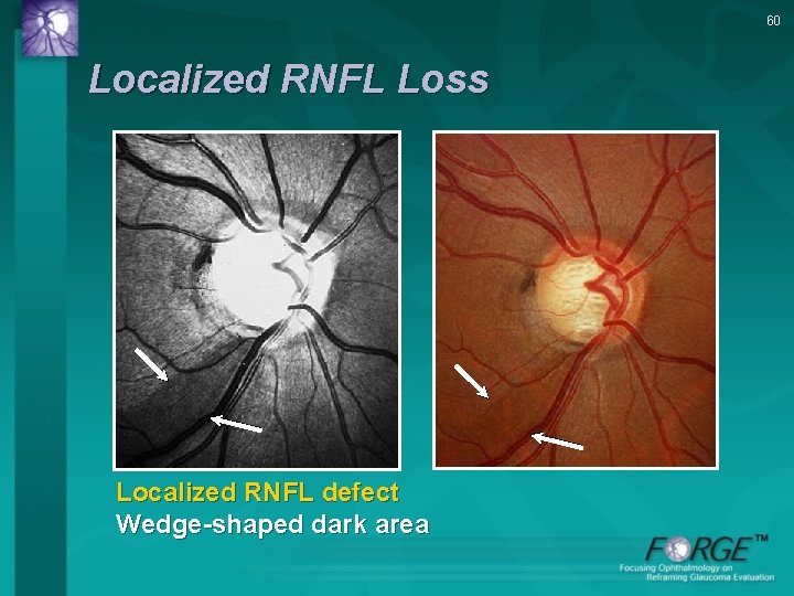
60 Localized RNFL Loss Localized RNFL defect Wedge-shaped dark area
61 Rule # 4 Examine the Region of parapapillary atrophy (PPA)
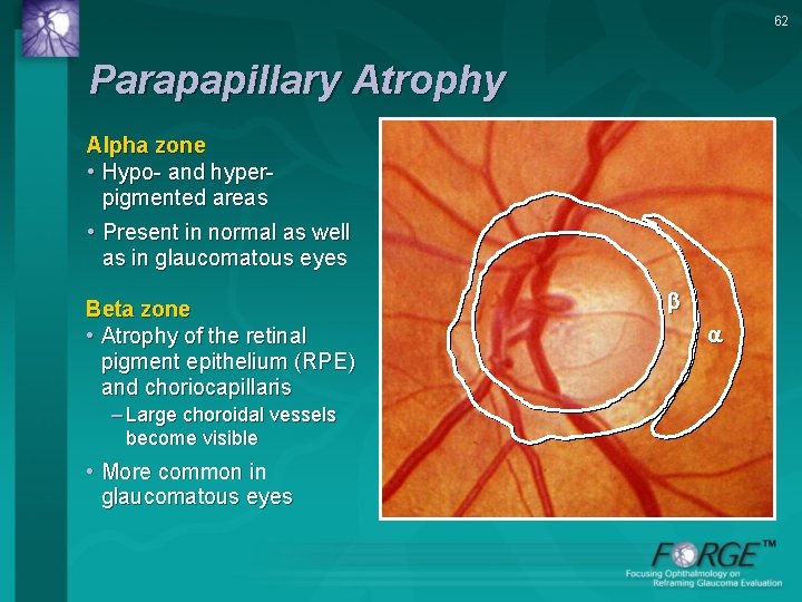
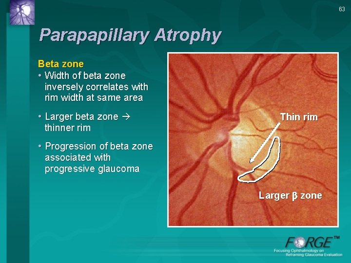
62 Parapapillary Atrophy Alpha zone • Hypo- and hyperpigmented areas • Present in normal as well as in glaucomatous eyes Beta zone • Atrophy of the retinal pigment epithelium (RPE) and choriocapillaris – Large choroidal vessels become visible • More common in glaucomatous eyes
63 Parapapillary Atrophy Beta zone • Width of beta zone inversely correlates with rim width at same area • Larger beta zone thinner rim Thin rim • Progression of beta zone associated with progressive glaucoma Larger zone
64 Rule # 5 Look for Retinal and optic disc hemorrhages
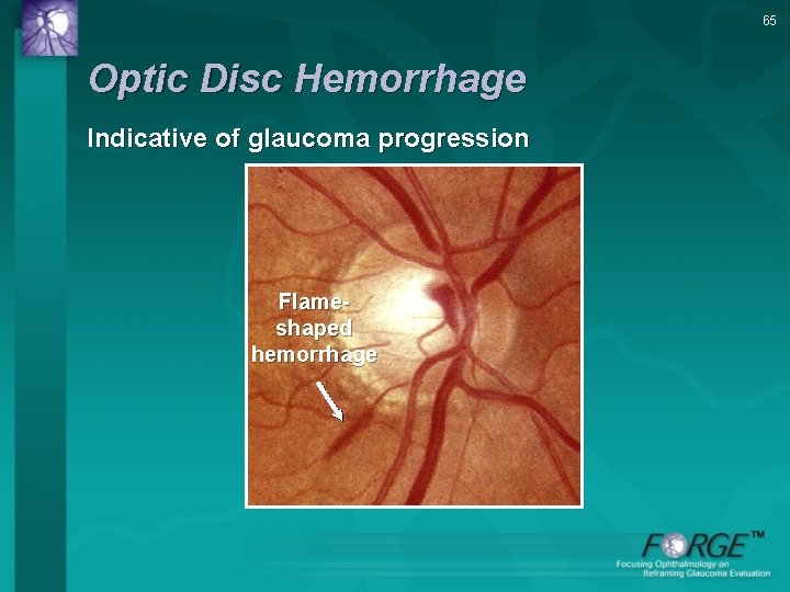
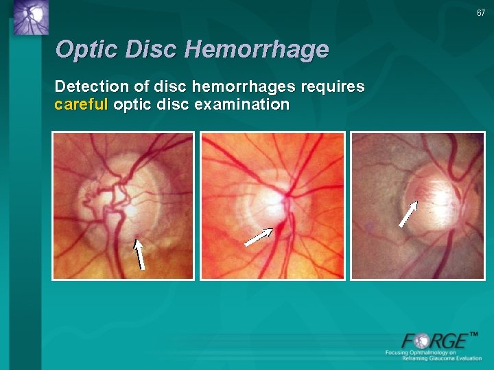
65 Optic Disc Hemorrhage Indicative of glaucoma progression Flameshaped hemorrhage
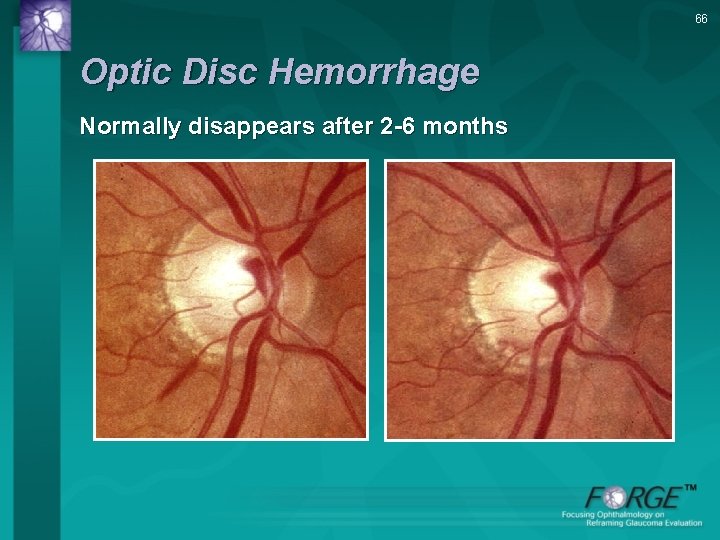
66 Optic Disc Hemorrhage Normally disappears after 2 -6 months
67 Optic Disc Hemorrhage Detection of disc hemorrhages requires careful optic disc examination
68 EXAMPLES
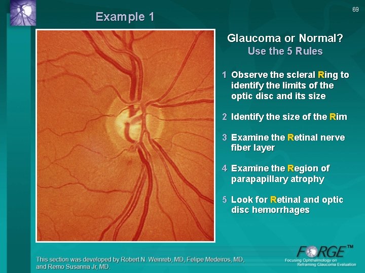
69 Example 1 Glaucoma or Normal? Use the 5 Rules 1 Observe the scleral Ring to identify the limits of the optic disc and its size 2 Identify the size of the Rim 3 Examine the Retinal nerve fiber layer 4 Examine the Region of parapapillary atrophy 5 Look for Retinal and optic disc hemorrhages
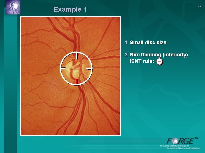
70 Example 1 1 Small disc size 2 Rim thinning (inferiorly) ISNT rule: –
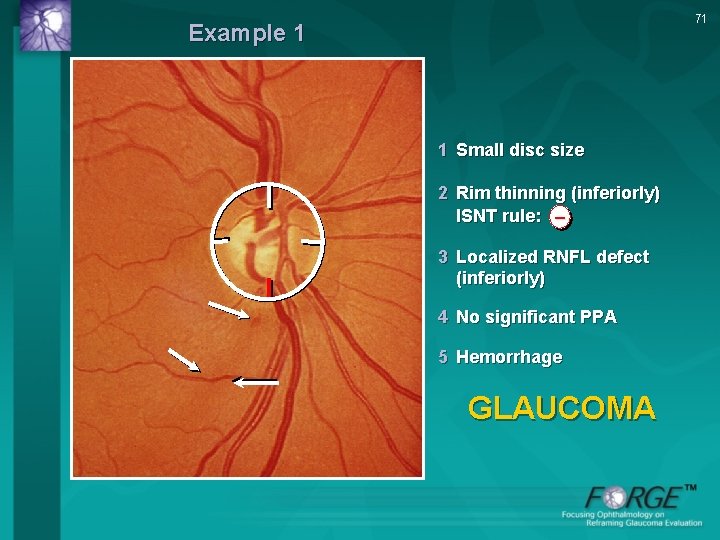
71 Example 1 1 Small disc size 2 Rim thinning (inferiorly) ISNT rule: − – 3 Localized RNFL defect (inferiorly) 4 No significant PPA 5 Hemorrhage GLAUCOMA
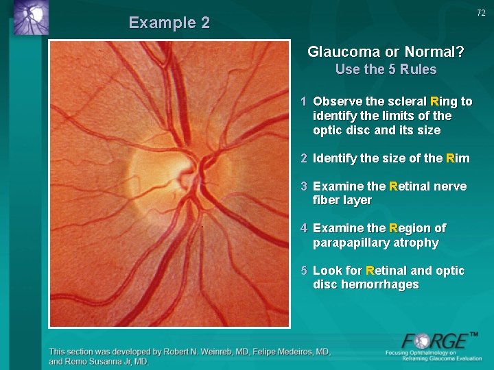
72 Example 2 Glaucoma or Normal? Use the 5 Rules 1 Observe the scleral Ring to identify the limits of the optic disc and its size 2 Identify the size of the Rim 3 Examine the Retinal nerve fiber layer 4 Examine the Region of parapapillary atrophy 5 Look for Retinal and optic disc hemorrhages
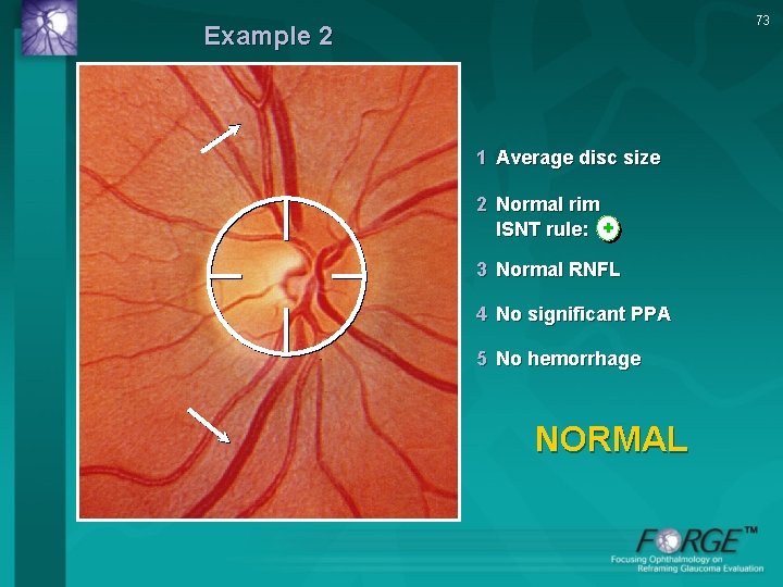
73 Example 2 1 Average disc size 2 Normal rim ISNT rule: + 3 Normal RNFL 4 No significant PPA 5 No hemorrhage NORMAL
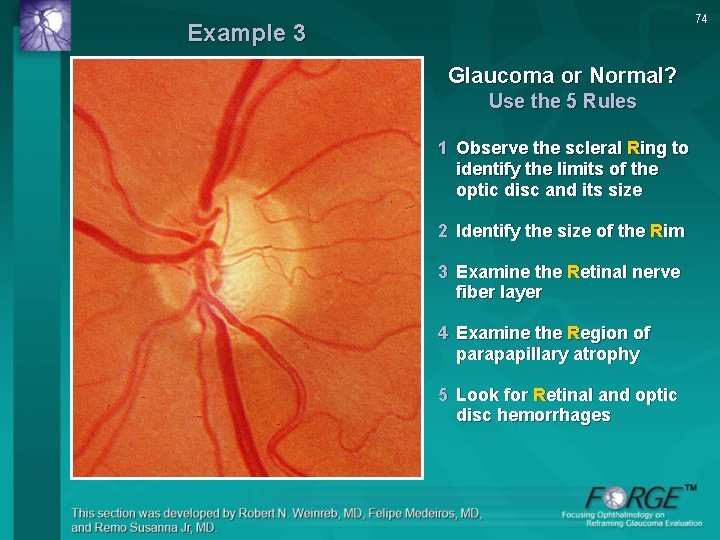
74 Example 3 Glaucoma or Normal? Use the 5 Rules 1 Observe the scleral Ring to identify the limits of the optic disc and its size 2 Identify the size of the Rim 3 Examine the Retinal nerve fiber layer 4 Examine the Region of parapapillary atrophy 5 Look for Retinal and optic disc hemorrhages
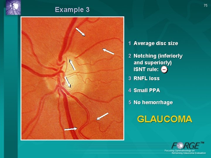
75 Example 3 1 Average disc size 2 Notching (inferiorly and superiorly) ISNT rule: – 3 RNFL loss 4 Small PPA 5 No hemorrhage GLAUCOMA
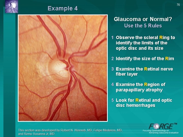
76 Example 4 Glaucoma or Normal? Use the 5 Rules 1 Observe the scleral Ring to identify the limits of the optic disc and its size 2 Identify the size of the Rim 3 Examine the Retinal nerve fiber layer 4 Examine the Region of parapapillary atrophy 5 Look for Retinal and optic disc hemorrhages
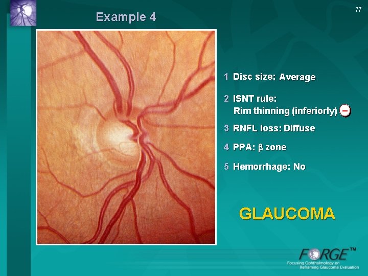
77 Example 4 1 Disc size: Average 2 ISNT rule: Rim thinning (inferiorly) – 3 RNFL loss: Diffuse 4 PPA: zone 5 Hemorrhage: No GLAUCOMA
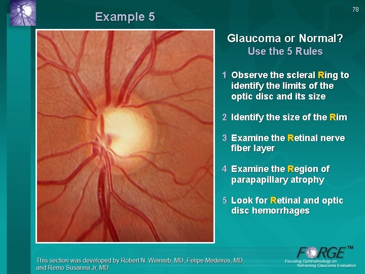
78 Example 5 Glaucoma or Normal? Use the 5 Rules 1 Observe the scleral Ring to identify the limits of the optic disc and its size 2 Identify the size of the Rim 3 Examine the Retinal nerve fiber layer 4 Examine the Region of parapapillary atrophy 5 Look for Retinal and optic disc hemorrhages
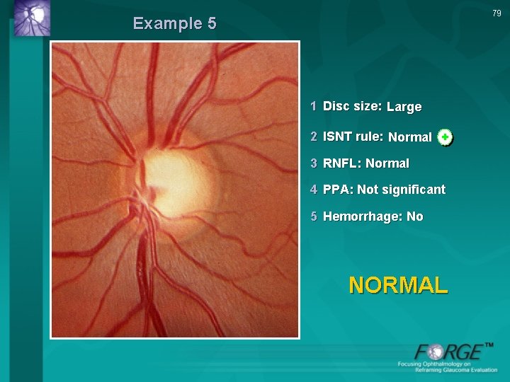
79 Example 5 1 Disc size: Large 2 ISNT rule: Normal + 3 RNFL: Normal 4 PPA: Not significant 5 Hemorrhage: No NORMAL
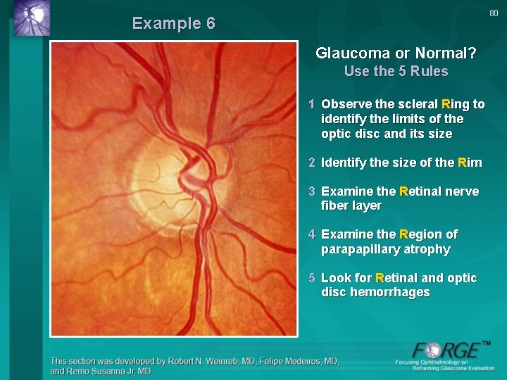
80 Example 6 Glaucoma or Normal? Use the 5 Rules 1 Observe the scleral Ring to identify the limits of the optic disc and its size 2 Identify the size of the Rim 3 Examine the Retinal nerve fiber layer 4 Examine the Region of parapapillary atrophy 5 Look for Retinal and optic disc hemorrhages
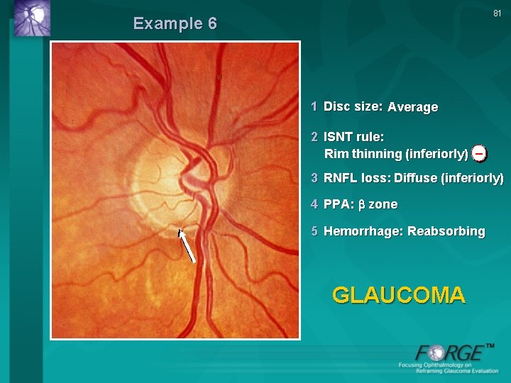
81 Example 6 1 Disc size: Average 2 ISNT rule: Rim thinning (inferiorly) – 3 RNFL loss: Diffuse (inferiorly) 4 PPA: zone 5 Hemorrhage: Reabsorbing GLAUCOMA
82 Clinical Practice Today: Diagnosis & Follow-Up of Glaucoma • Need to evaluate ON, VF, and IOP • Focus on ON damage in early disease – Disc or RNFL changes usually observed prior to functional damage – Examination of IOP alone, or IOP plus VF, not sufficient • VF testing – Reproducible VF defects in early disease can confirm glaucoma or progression in cases with an unremarkable or suspicious disc – Critical in more advanced disease
IOP Control in Glaucoma Management • Population studies indicate 1 -3 – Incidence, severity, & progression of glaucoma consistently correlate with elevated IOP • IOP reduction is currently the preferred treatment for decreasing risk of disease progression 4 • Accurate understanding of individual patient IOP affects treatment decisions and guides treatment plan 1. Quigley HA et al. Am J Ophthalmol. 1996; 122: 355 -363. 2. Leske MC et al. Arch Ophthalmol. 2001; 119: 89 -95. 3. Heijl A et al. Arch Ophthalmol. 2002; 120: 1268 -1279. 4. Lichter PR et al. Ophthalmology. 2001; 108: 1943 -1953. 83
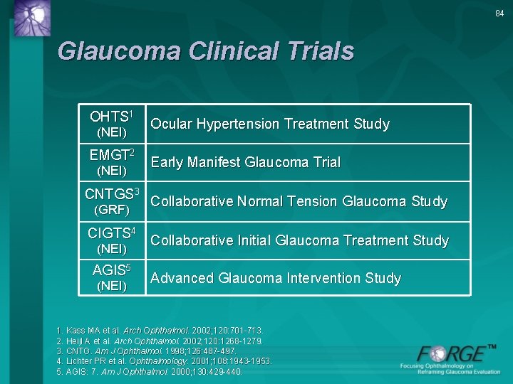
84 Glaucoma Clinical Trials OHTS 1 (NEI) EMGT 2 (NEI) Ocular Hypertension Treatment Study Early Manifest Glaucoma Trial CNTGS 3 Collaborative Normal Tension Glaucoma Study (GRF) CIGTS 4 Collaborative Initial Glaucoma Treatment Study (NEI) AGIS 5 (NEI) Advanced Glaucoma Intervention Study 1. Kass MA et al. Arch Ophthalmol. 2002; 120: 701 -713. 2. Heijl A et al. Arch Ophthalmol. 2002; 120: 1268 -1279. 3. CNTG. Am J Ophthalmol. 1998; 126: 487 -497. 4. Lichter PR et al. Ophthalmology. 2001; 108: 1943 -1953. 5. AGIS: 7. Am J Ophthalmol. 2000; 130: 429 -440.
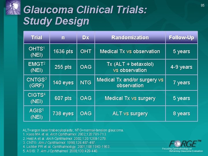
85 Glaucoma Clinical Trials: Study Design Trial n Dx Randomization Follow-Up OHTS 1 (NEI) 1636 pts OHT Medical Tx vs observation 5 years EMGT 2 (NEI) 255 pts OAG Tx (ALT + betaxolol) vs observation 4 -9 years CNTGS 3 (GRF) 140 eyes NTG Medical Tx and/or surgery vs observation 7 years CIGTS 4 (NEI) 607 pts OAG Medical Tx vs surgery 5 years AGIS 5 (NEI) 738 eyes OAG ALT vs surgery 8 years ALT=argon laser trabeculoplasty; NTG=normal-tension glaucoma. 1. Kass MA et al. Arch Ophthalmol. 2002; 120: 701 -713. 2. Heijl A et al. Arch Ophthalmol. 2002; 120: 1268 -1279. 3. CNTG. Am J Ophthalmol. 1998; 126: 487 -497. 4. Lichter PR et al. Ophthalmology. 2001; 108: 1943 -1953. 5. AGIS: 7. Am J Ophthalmol. 2000; 130: 429 -440.
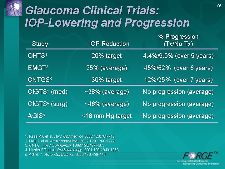
Glaucoma Clinical Trials: IOP-Lowering and Progression Study IOP Reduction % Progression (Tx/No Tx) OHTS 1 20% target 4. 4%/9. 5% (over 5 years) EMGT 2 25% (average) 45%/62% (over 6 years) 30% target 12%/35% (over 7 years) CIGTS 4 (med) ~38% (average) No progression (average) CIGTS 4 (surg) ~46% (average) No progression (average) <18 mm Hg target No progression (average) CNTGS 3 AGIS 5 1. Kass MA et al. Arch Ophthalmol. 2002; 120: 701 -713. 2. Heijl A et al. Arch Ophthalmol. 2002; 120: 1268 -1279. 3. CNTG. Am J Ophthalmol. 1998; 126: 487 -497. 4. Lichter PR et al. Ophthalmology. 2001; 108: 1943 -1953. 5. AGIS: 7. Am J Ophthalmol. 2000; 130: 429 -440. 86
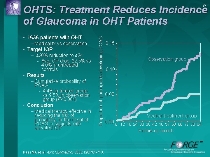
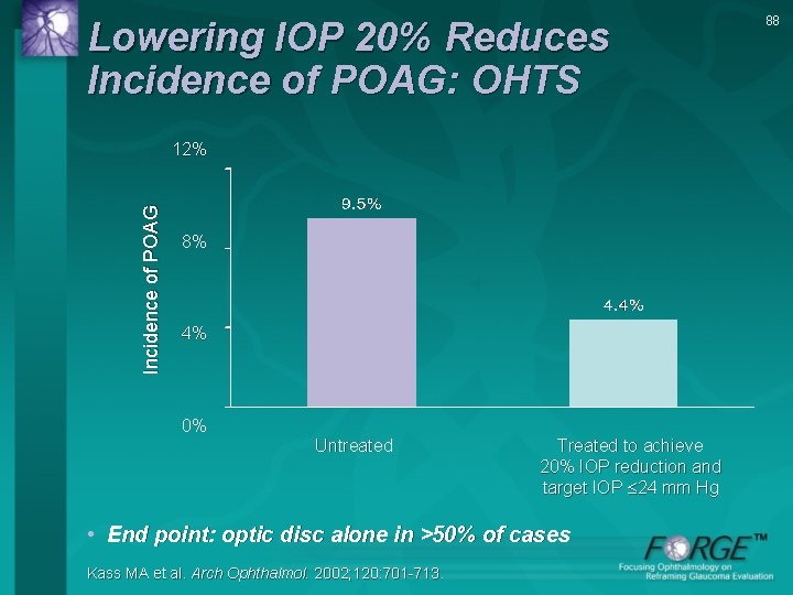
87 • 1636 patients with OHT – Medical tx vs observation • Target IOP – ≥ 20% reduction to ≤ 24 - Avg IOP drop: 22. 5% vs 4. 0% in untreated controls • Results – Cumulative probability of POAG - 4. 4% in treated group vs 9. 5% in observation group (P<0. 001) • Conclusion – Medical therapy effective in reducing the risk of probability for the onset of POAG in subjects with elevated IOP Proportion of participants developing POAG OHTS: Treatment Reduces Incidence of Glaucoma in OHT Patients Kass MA et al. Arch Ophthalmol. 2002; 120: 701 -713. 0. 15 Observation group 0. 10 0. 05 Medical treatment group 0. 00 6 12 18 24 30 36 42 48 54 60 66 72 78 84 Follow-up month
Lowering IOP 20% Reduces Incidence of POAG: OHTS Incidence of POAG 12% 8% 4% 0% Untreated Treated to achieve 20% IOP reduction and target IOP 24 mm Hg • End point: optic disc alone in >50% of cases Kass MA et al. Arch Ophthalmol. 2002; 120: 701 -713. 88
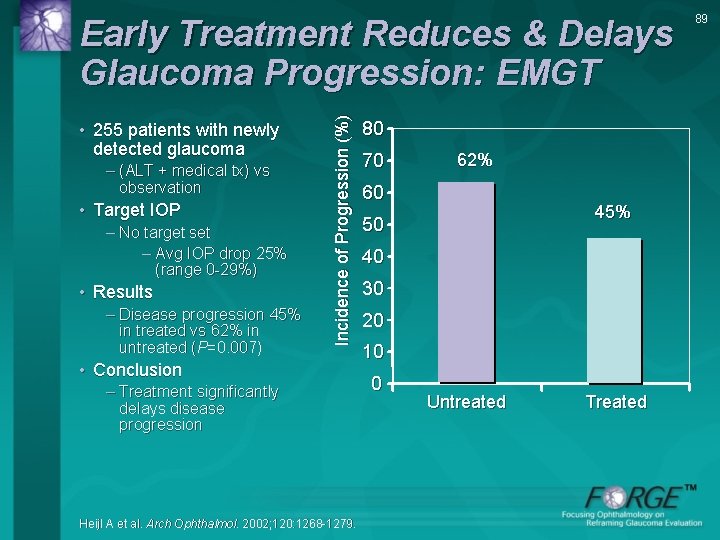
• 255 patients with newly detected glaucoma – (ALT + medical tx) vs observation • Target IOP – No target set – Avg IOP drop 25% (range 0 -29%) • Results – Disease progression 45% in treated vs 62% in untreated (P=0. 007) Incidence of Progression (%) Early Treatment Reduces & Delays Glaucoma Progression: EMGT • Conclusion – Treatment significantly delays disease progression Heijl A et al. Arch Ophthalmol. 2002; 120: 1268 -1279. 80 70 62% 60 45% 50 40 30 20 10 0 Untreated Treated 89
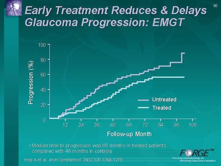
Early Treatment Reduces & Delays Glaucoma Progression: EMGT Progression (%) 100 80 60 40 Untreated Treated 20 0 12 24 36 48 60 72 84 Follow-up Month • Median time to progression was 66 months in treated patients compared with 48 months in controls Heijl A et al. Arch Ophthalmol. 2002; 120: 1268 -1279. 96 108 90
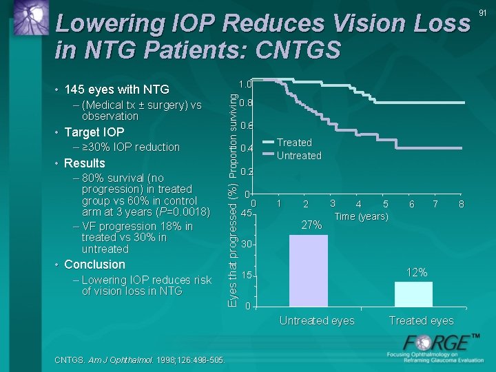
Lowering IOP Reduces Vision Loss in NTG Patients: CNTGS – (Medical tx ± surgery) vs observation • Target IOP – ≥ 30% IOP reduction • Results – 80% survival (no progression) in treated group vs 60% in control arm at 3 years (P=0. 0018) – VF progression 18% in treated vs 30% in untreated • Conclusion – Lowering IOP reduces risk of vision loss in NTG 1. 0 Eyes that progressed (%) Proportion surviving • 145 eyes with NTG 0. 8 0. 6 0. 4 Treated Untreated 0. 2 0 0 45 1 2 27% 3 4 5 Time (years) 7 30 12% 15 0 Untreated eyes CNTGS. Am J Ophthalmol. 1998; 126: 498 -505. 6 Treated eyes 8 91
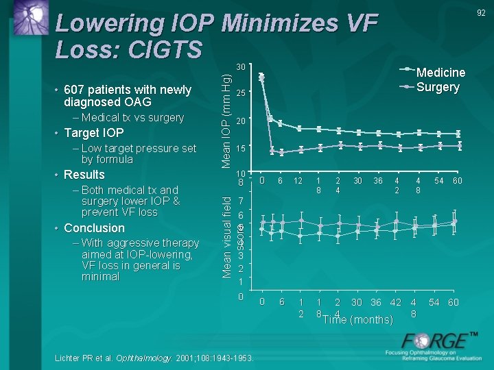
92 Lowering IOP Minimizes VF Loss: CIGTS – Medical tx vs surgery • Target IOP – Low target pressure set by formula • Results – Both medical tx and surgery lower IOP & prevent VF loss • Conclusion – With aggressive therapy aimed at IOP-lowering, VF loss in general is minimal Medicine Surgery 25 20 15 10 8 0 6 12 1 8 2 4 30 36 42 4 8 54 60 7 6 5 4 3 2 1 Mean visual field score • 607 patients with newly diagnosed OAG Mean IOP (mm Hg) 30 0 Lichter PR et al. Ophthalmology. 2001; 108: 1943 -1953. Time (months) 4 8 54 60
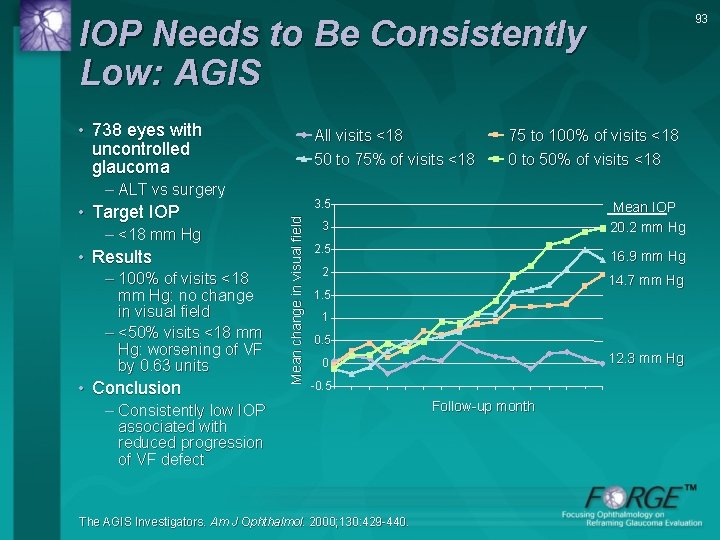
93 IOP Needs to Be Consistently Low: AGIS • 738 eyes with uncontrolled glaucoma All visits <18 50 to 75% of visits <18 – ALT vs surgery – <18 mm Hg • Results – 100% of visits <18 mm Hg: no change in visual field – <50% visits <18 mm Hg: worsening of VF by 0. 63 units • Conclusion 3. 5 Mean change in visual field • Target IOP 75 to 100% of visits <18 0 to 50% of visits <18 Mean IOP 20. 2 mm Hg 3 2. 5 16. 9 mm Hg 2 14. 7 mm Hg 1. 5 1 0. 5 12. 3 mm Hg 0 -0. 5 – Consistently low IOP associated with reduced progression of VF defect The AGIS Investigators. Am J Ophthalmol. 2000; 130: 429 -440. Follow-up month
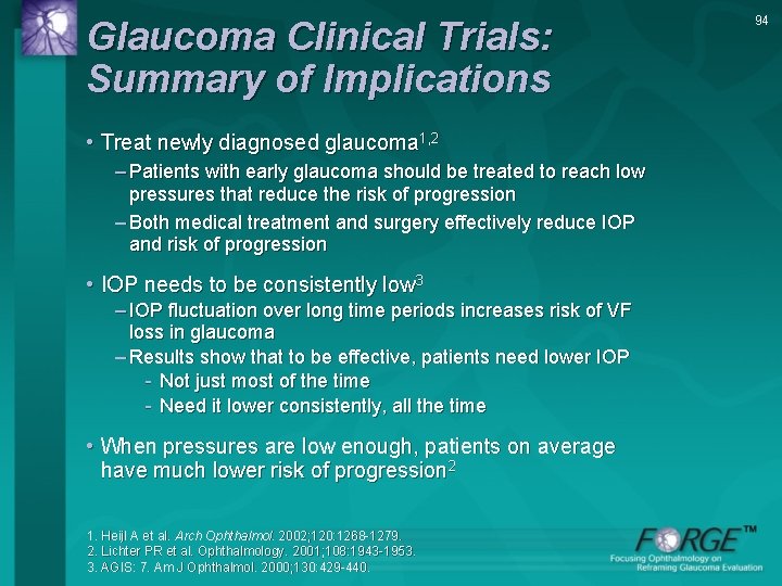
Glaucoma Clinical Trials: Summary of Implications • Treat newly diagnosed glaucoma 1, 2 – Patients with early glaucoma should be treated to reach low pressures that reduce the risk of progression – Both medical treatment and surgery effectively reduce IOP and risk of progression • IOP needs to be consistently low 3 – IOP fluctuation over long time periods increases risk of VF loss in glaucoma – Results show that to be effective, patients need lower IOP - Not just most of the time - Need it lower consistently, all the time • When pressures are low enough, patients on average have much lower risk of progression 2 1. Heijl A et al. Arch Ophthalmol. 2002; 120: 1268 -1279. 2. Lichter PR et al. Ophthalmology. 2001; 108: 1943 -1953. 3. AGIS: 7. Am J Ophthalmol. 2000; 130: 429 -440. 94
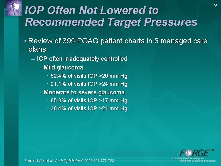
IOP Often Not Lowered to Recommended Target Pressures • Review of 395 POAG patient charts in 6 managed care plans – IOP often inadequately controlled - Mild glaucoma · 52. 4% of visits IOP >20 mm Hg · 21. 1% of visits IOP >24 mm Hg - Moderate to severe glaucoma · 65. 3% of visits IOP >17 mm Hg · 30. 4% of visits IOP >21 mm Hg Fremont AM et al. Arch Ophthalmol. 2003; 121: 777 -783. 95
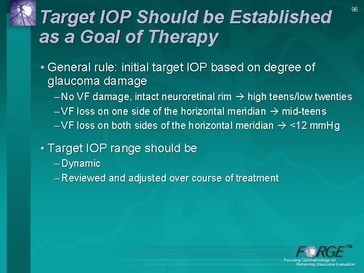
Target IOP Should be Established as a Goal of Therapy 96 • General rule: initial target IOP based on degree of glaucoma damage – No VF damage, intact neuroretinal rim high teens/low twenties – VF loss on one side of the horizontal meridian mid-teens – VF loss on both sides of the horizontal meridian <12 mm. Hg • Target IOP range should be – Dynamic – Reviewed and adjusted over course of treatment
Target IOP: How Low Is Low Enough? • Individualize • Factors – Damaging IOP – Severity of Damage – Other – Age – Family History 97
Target IOP: Recommendations • Larger IOP reductions associated with decreased risk of glaucoma & progression (OHTS, EMGT, CIGTS)1 -3 • For some patients, IOP should be reduced to levels lower than were considered acceptable in past • Guidelines for setting target pressures in glaucoma developed by the AAO, EGS, and others – Based on evidence from clinical trials and clinical experience 1. Kass MA et al. Arch Ophthalmol. 2002; 120: 701 -713. 2. Heijl A et al. Arch Ophthalmol. 2002; 120: 1268 -1279. 3. Lichter PR et al. Ophthalmology. 2001; 108: 1943 -1953. 98
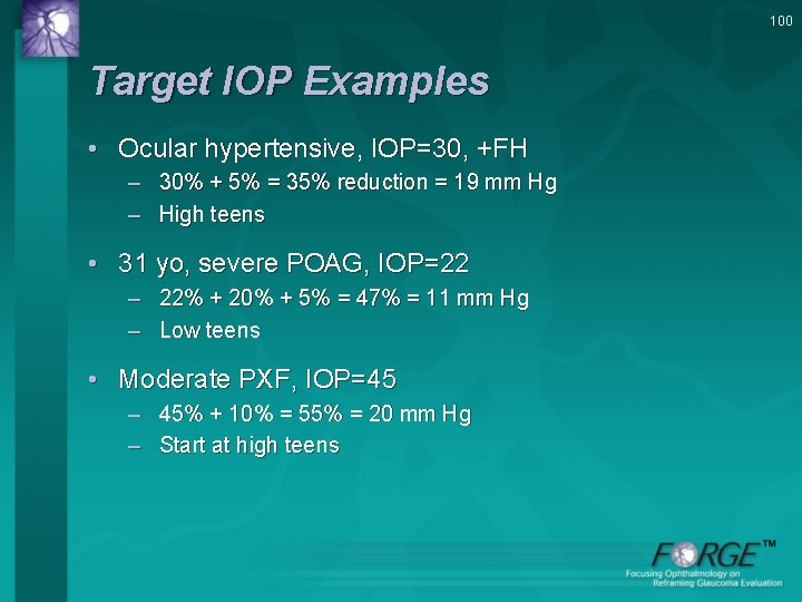
99 Target IOP in Clinical Practice • Set an individualized target IOP range for each glaucoma eye – Do not need to record target • Target often more aggressive than in past • Get IOP down to target • Keep IOP down to target • May need to adjust target over time
100 Target IOP Examples • Ocular hypertensive, IOP=30, +FH – 30% + 5% = 35% reduction = 19 mm Hg – High teens • 31 yo, severe POAG, IOP=22 – 22% + 20% + 5% = 47% = 11 mm Hg – Low teens • Moderate PXF, IOP=45 – 45% + 10% = 55% = 20 mm Hg – Start at high teens
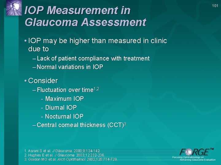
IOP Measurement in Glaucoma Assessment • IOP may be higher than measured in clinic due to – Lack of patient compliance with treatment – Normal variations in IOP • Consider – Fluctuation over time 1, 2 - Maximum IOP - Diurnal IOP - Nocturnal IOP – Central corneal thickness (CCT)3 1. Asrani S et al. J Glaucoma. 2000; 9: 134 -142. 2. Hughes E et al. J Glaucoma. 2003; 12: 232 -236. 3. Gordon MO et al. Arch Ophthalmol. 2002; 120: 714 -720. 101
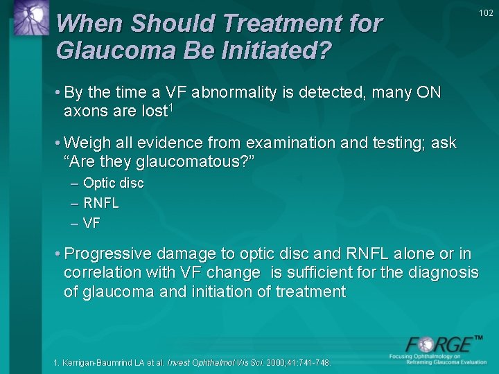
When Should Treatment for Glaucoma Be Initiated? 102 • By the time a VF abnormality is detected, many ON axons are lost 1 • Weigh all evidence from examination and testing; ask “Are they glaucomatous? ” – Optic disc – RNFL – VF • Progressive damage to optic disc and RNFL alone or in correlation with VF change is sufficient for the diagnosis of glaucoma and initiation of treatment 1. Kerrigan-Baumrind LA et al. Invest Ophthalmol Vis Sci. 2000; 41: 741 -748.
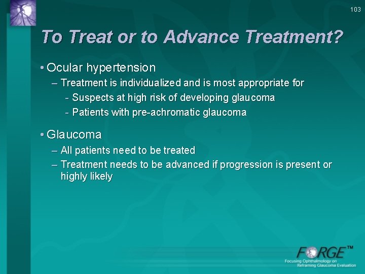
103 To Treat or to Advance Treatment? • Ocular hypertension – Treatment is individualized and is most appropriate for - Suspects at high risk of developing glaucoma - Patients with pre-achromatic glaucoma • Glaucoma – All patients need to be treated – Treatment needs to be advanced if progression is present or highly likely
104 Prevent Progression • Prescribe therapy most likely to achieve and maintain target IOP or lower • Monitor patients for progression with serial ON examination as well as VF testing • Patients at risk of disease progression – Need to get IOP even lower – Readjust for lower target IOP and treat more effectively AAO Preferred Practice Pattern® Guidelines. 2010.
105 Treatment Options to Lower IOP • Medications – Commonly used drug classes include - Prostaglandins/prostamides - Beta-blockers - Alpha-adrenergic agonists - Topical CAIs - Fixed combination product • Surgical Intervention – Laser trabeculoplasty (ALT - SLT) – Filtration surgery CAI = carbonic anhydrase inhibitor. AAO Preferred Practice Pattern® Guidelines. 2010.
106 Patient Education and Adherence • Patients need motivation to adhere to visionpreserving treatment; educate them about – Insidious nature of glaucoma – Need to lower IOP – Available treatment options - Should consider both efficacy & potential adverse effects of treatment
Reframing Disease Severity Means Reframing Disease Treatment • Glaucoma suspect – Treatment based on risk of developing glaucoma • Glaucoma – Abnormal ON, normal SAP, abnormal selective VF test (eg, SWAP, FDT) treat – Early VF defect moderate or even advanced glaucoma • Based on large population clinical trials – Treat aggressively with most effective therapy – Go lower - Reduce IOP and keep it low consistently 107
108 Conclusions • Evaluate and document ON to – Diagnose glaucoma – Determine disease severity – Evaluate progression • Set target pressure based on risk factors and optic nerve appearance – Aim for lower targets: every mm Hg counts • Prescribe therapy with best ability to achieve and maintain target IOP (or lower) • Re-evaluate structure/function critically as patient is followed • Readjust target IOP and treat more effectively when subtle progression is noted
109 Reference List The Advanced Glaucoma Intervention Study (AGIS). 7. The relationship between control of intraocular pressure and visual field deterioration. The AGIS Investigators. Am J Ophthalmol. 2000; 130: 429 -440. Airaksinen PJ, Drance SM, Douglas GR, Schulzer M. Neuroretinal rim areas and visual field indices in glaucoma. Am J Ophthalmol. 1985; 99: 107 -110. American Academy of Ophthalmology Glaucoma Panel. Preferred Practice Pattern® Guidelines. Primary Open-Angle Glaucoma. San Francisco, CA: American Academy of Ophthalmology; 2010. Asrani S, Zeimer R, Wilensky J, et al. Large diurnal fluctuations in intraocular pressure an independent risk factor in patients with glaucoma. J Glaucoma. 2000; 9: 134 -142. Bowd C, Zangwill LM, Berry CC, Blumenthal EZ, Vasile C, Sanchez-Galeana C, Bosworth CF, Sample PA, Weinreb RN. Detecting early glaucoma by assessment of retinal nerve fiber layer thickness and visual function. Invest Ophthalmol Vis Sci. 2001; 42: 1993 -2003. Caprioli J, Miller JM. Correlation of structure and function in glaucoma: quantitative measurements of disc and field. Ophthalmology. 1988; 95: 723 -727.
110 Reference List (continued) Collaborative Normal-Tension Glaucoma Study Group (CNTG). The effectiveness of intraocular pressure reduction in the treatment of normal-tension glaucoma. Am J Ophthalmol. 1998; 126: 498 -505. Collaborative Normal-Tension Glaucoma Study Group (CNTG). Comparison of glaucomatous progression between untreated patients with normal-tension glaucoma and patients with therapeutically reduced intraocular pressures. Am J Ophthalmol. 1998; 126: 487 -497. Damji KF, Behki R, Wang L, for the Target IOP Workshop participants. Canadian perspectives in glaucoma management: setting target intraocular pressure range. Can J Ophthalmol. 2003; 38: 189 -197. European Glaucoma Society. Terminology and guidelines for glaucoma. Savona, Italy. 2008. Fremont AM, Lee PP, Mangione CM, et al. Patterns of care for open-angle glaucoma in managed care. Arch Ophthalmol. 2003; 121: 777 -783.
111 Reference List (continued) Friedman DS, Wolfs RC, O'Colmain BJ, Klein BE, Taylor HR, West S, Leske MC, Mitchell P, Congdon N, Kempen J; Eye Diseases Prevalence Research Group. Prevalence of open-angle glaucoma among adults in the United States. Arch Ophthalmol. 2004(Apr); 122(4): 532 -8. Gillespie BW, Musch DC, Guire KE, Mills RP, Lichter PR, Janz NK, Wren PA; CIGTS (Collaborative Initial Glaucoma Treatment Study) Study Group. The collaborative initial glaucoma treatment study: baseline visual field and test-retest variability. Invest Ophthalmol Vis Sci. 2003; 44: 2613 -2620. Gordon MO, Beiser JA, Brandt JD, et al. The Ocular Hypertension Treatment Study: baseline factors that predict the onset of primary open-angle glaucoma. Arch Ophthalmol. 2002; 120: 714 -720. Hattenhauer MG, Johnson DH, Ing HH, Herman DC, Hodge DO, Yawn BP, Butterfield LC, Gray DT. The probability of blindness from open-angle glaucoma. Ophthalmology. 1998; 105: 2099 -2104. Heijl A, Leske MC, Bengtsson B, Hyman L, Bengtsson B, Hussein M; Early Manifest Glaucoma Trial Group. Reduction of intraocular pressure and glaucoma progression: results from the Early Manifest Glaucoma Trial. Arch Ophthalmol. 2002; 120: 12681279.
112 Reference List (continued) Hertzog LH, Albrecht KG, La Bree L, Lee PP. Glaucoma care and conformance with preferred practice patterns. Examination of the private, community-based ophthalmologist. Ophthalmology. 1996; 103: 1009 -1013. Hughes E, Spry P, Diamond J. 24 -hour monitoring of intraocular pressure in glaucoma management: a retrospective review. J Glaucoma. 2003; 12: 232 -236. Johnson CA, Takahashi G, Demirel S. The ability of frequency doubling technology (FDT) perimetry to predict the onset of glaucomatous visual field loss for standard automated perimetry (SAP) [Abstract]. Journal of Vision. 2002; 2(10): 100 a. Kass MA, Heuer DK, Higginbotham EJ, et al. The Ocular Hypertension Treatment Study. Arch Ophthalmol. 2002; 120: 701 -713. Kerrigan-Baumrind LA, Quigley HA, Pease ME, et al. Number of ganglion cells in glaucoma eyes compared with threshold visual field tests in the same persons. Invest Ophthalmol Vis Sci. 2000; 41: 741 -748. Leske MC, Connell AM, Wu SY, Nemesure B, Li X, Schachat A, Hennis A. Incidence of open-angle glaucoma: the Barbados Eye Studies. The Barbados Eye Studies Group. Arch Ophthalmol. 2001; 119: 89 -95.
113 Reference List (continued) Lichter PR, Musch DC, Gillespie BW, et al. Interim clinical outcomes in the Collaborative Initial Glaucoma Treatment Study comparing initial treatment randomized to medications or surgery. Ophthalmology. 2001; 108: 1943 -1953. Medeiros FA, Sample PA, Zangwill LM, Bowd C, Aihara M, Weinreb RN. Corneal thickness as a risk factor for visual field loss in patients with preperimetric glaucomatous optic neuropathy. Am J Ophthalmol. 2003; 136: 805 -813. Quigley HA, Addicks EM, Green WR, et al. Optic nerve damage in human glaucoma. III. Quantitative correlation of nerve fiber loss and visual field defect in glaucoma, ischemic neuropathy, papilledema, and toxic neuropathy. Arch Ophthalmol. 1982; 100: 135 -146. Quigley HA, Broman AT. The number of people with glaucoma worldwide in 2010 and 2020. Br J Ophthalmol. 2006(Mar); 90(3): 262 -7. Quigley HA, Tielsch JM, Katz J, Sommer A. Rate of progression in open-angle glaucoma estimated from cross-sectional prevalence of visual field damage. Am J Ophthalmol. 1996; 122: 355 -363. Quigley HA, West SK, Rodriguez J, Munoz B, Klein R, Snyder R. The prevalence of glaucoma in a population-based study of Hispanic subjects: Proyecto VER. Arch Ophthalmol. 2001; 119: 1819 -1826.
114 Reference List (continued) Racette L, Sample PA. Short-wavelength automated perimetry. Ophthalmol Clin North Am. 2003; 16: 227 -236. Sommer A, Katz J, Quigley HA, et al. Clinically detectable nerve fiber atrophy precedes the onset of glaucomatous field loss. Arch Ophthalmol. 1991; 109: 77 -83. Sommer A, Tielsch JM, Katz J, et al. Relationship between intraocular pressure and primary open-angle glaucoma among white and black Americans. Arch Ophthalmol. 1991; 109: 1090 -1095. Sponsel WE, Shoemaker J, Trigo Y, Mensah J, Rugwani R, Garrett B. Frequency of sustained glaucomatous-type visual field loss and associated optic nerve cupping in Beaver Dam, Wisconsin. Clin Experiment Ophthalmol. 2001(Dec); 29(6): 352 -8. Tielsch, JM, Sommer, A, et al. Racial Variations in the Prevalence of Primary Openangle Glaucoma. JAMA. 1991; 266: 369 -374. Wilson MR. Primary open-angle glaucoma: epidemiology and risk factors. In: Clinical Guide to Glaucoma Management. Higginbotham EJ, Lee DA, eds. Butterworth Heinemann: 2004. Wilson MR. Progression of visual field loss in untreated glaucoma patients and suspects in St Lucia, West Indies. Trans Am Ophthalmol Soc. 2002; 100: 365 -410.
115 Thank You © 2013 Allergan, Inc. , Irvine, CA 92612 APC 57 SC 13