ATHEROSCLEROSIS Atherosclerosis or hardening of the arteries is


- Slides: 30

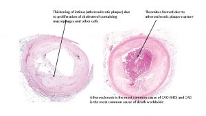
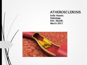
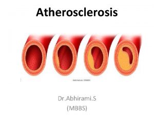
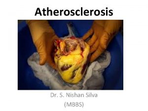
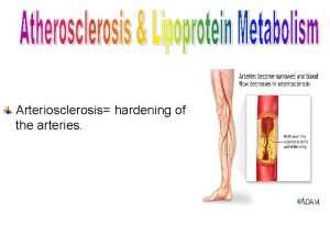
ATHEROSCLEROSIS Atherosclerosis or hardening of the arteries is a condition of the large and small arteries characterized by accumulation of fatty deposits, platelets, neutrophils, monocytes and microphages throughout tunica intima&tunica media. Arteries most often affected include the coronaries, the aorta &the cerebral arteries

ATHEROSCLEROSIS The development of atherosclerosis begins with dysfunction of the endothelial cells lining the lumen of the artery. This may occur following injury to the endothelial cells, or from other stimuli.

ATHEROSCLEROSIS Injury to the endothelial cells increases their permeability to various plasma components, including fatty acids and TGs, allowing these substances access to the inside of the artery.

ATHEROSCLEROSIS Oxidation of fatty acids produces oxygen free radicals that further damage the vessel. Injury to the endothelial cells also initiates inflammatory&immune reactions, including the attraction of WBC, especially neutrophils &monocytes, &platelets to the , area.

ATHEROSCLEROSIS The WBCs release potent proinflammatory cytokines that further aggravate the situation, drawing even more WCs & platelets that act as chemo attractants to activate the cycle of inflammation, clotting , and fibrosis stimulating clotting , activating T&B cells and releasing chemicals that act as chemo attractants to activate the cycle of inflammation , clotting and fibrosis.

ATHEROSCLEROSIS Once drawn to the area of injury, the WBCs are caught by activation of endothelial adhesion factors to make the endothelium especially sticky to WBCs. Once attached to the endothelial layer, the monocytes and neutrophils begin to emigrate between the endothelial cells, into the interstitial space.

ATHEROSCLEROSIS In the interstitium, the monocytes mature into macrophages and along with the neutrophils , continue to release cytokines, which further the inflammatory cycle. The proinflammatory cytokines also stimulate smooth muscle cell proliferation, causing smooth muscle cells to grow into the tunica intima.

ATHEROSCLEROSIS Additional plasma cholesterol and fats gain access to the tunica intima and media as permeability of the endothelial layer increases. With continued injury and inflammation, platelet aggregation increases and blood clot begins to form.

ATHEROSCLEROSIS Scar tissue replaces some of the vascular wall, changing the structure of the wall. The end results are cholesterol and fat buildup, scar tissue deposits, plateletderivated clots, and smooth muscle cell proliferation

ATHEROSCLEROSIS Even without direct injury to the endothelial cells, changes in the endothelial adhesion factors may occur, resulting in the accumulation of WBCs and release of inflammatory mediators and clot-forming substances. Why some individuals have especially active adhesion factors is unclear.

ATHEROSCLEROSIS It is likely that both genetic and environmental factors are involved. Regardless of the precipitating event, atherosclerosis leads to a decrease in the diameter of the artery and an increase in its stiffness. The atherosclerotic area of an artery is called a plaque.

CAUSES OF ATHEROSCLEROSIS High serum cholesterol The first hypothesis suggests that high serum cholesterol and high levels of circulating TGs can cause development of atherosclerosis. Fatty deposits, called atheromas, are found throughout and inside the tunica media in persons with atherosclerosis.

High serum cholesterol Cholesterol&TGs are carried in the blood encased in fat-carrying proteins called lipoproteins. High-density lipoprotein (HDL) carries fat away from cells to be degraded and is known to be protective against atherosclerosis.

High serum cholesterol Low-density lipoprotein (LDL) and very-low density lipoprotein (VLDL) carry fat to the cells of the body, including the endothelial cells of the arteries.

High serum cholesterol Stimulation of cholesterol efflux from macrophages in the atherosclerotic lesion, and the regulation of immune and inflammatory responses. In the arterial wall, oxidation of cholesterol&TGs leads to inflammation &production of free radicals known to damage endothelial cells.

High serum cholesterol The initial oxidation of LDL in the subendothelial layer of the arteries turns on various inflammatory reactions, which ultimately attract monocytes and neutrophils to the area. These WBCs become anchored to the endothelial layer by the adhesion molecules, and release additional inflammatory mediators that attract more WCs to the area and further stimulate LDL oxidation.

High serum cholesterol The monocytes move into the wall of the artery, where they mature into macrophages and internalize the LDL as fatty foam cells. Oxidized LDL is cytotoxic to vascular cells.

High serum cholesterol Patients with D. M often exhibit atherosclerosis caused by high cholesterol D. M is a major risk factor for atherosclerosis. Persons with diabetes have high plasma cholesterol and TGs. Poor circulation to most organs causes hypoxia and tissue injury, also stimulating inflammatory reactions that contribute to atherosclerosis.

High blood pressure Chronically hypertension produces shear forces that scrape away at the endothelial layer of the arteries and arterioles, intiating their injury. Shear forces especially occur in sites of arterial bifurcation or bending, a trait characteristic of the coronary arteries, aorta, and cerebral arteries.

High blood pressure With shearing of the endothelial layer, damage can occur repeatedly, leading to a cycle of inflammation, accumulation and adhesion of WBCs and platelets, and clot formation. Any thrombus that develops can be shared off the artery, leading to a thromboembolus downstream, or may grow large enough to obstruct blood flow.

Infection directly produces cell-damaging free radicals, it also initiates the cycle of inflammation, process associated with free radicals and adhesion factor activation. WBCs &platelets arrive in the area and cause clots and scarring. A specific organism that has been implicated in this infection is Chlamydia pneumonia common respiratory pathogen.

High blood iron levels Iron is rapidly oxidized and capable of producing artery damaging free radicals. This is suggested by some to explain the high incidence of coronary artery disease in men compared with premenopausal women, who typically have lower levels of iron.

High blood homocystein levels Homocysteine is an amino acid formed by the metabolism of methionine. Hyperhomocysteinemia is associated with endothelial dysfunction, specificically manifested by a decreased availability of endothelium-derived NO, a local VD. .

High blood homocystein levels Hyperhomocysteinemia also increases susceptibility to arterial thrombosis and accelerates the development of atherosclerosis in apolipoprotein E. Homocysteine may also increases oxidation of LDL. Nutritional deficient in folic acids and the B vitamins are associated with elevated homocysteine

Clinical manifestations -Intermittent claudication, an aching, cramping feeling in the lower extremities, occurs during of after exercise. -Cold sensitivity occurs with inadequate blood flow to the extremities. -Skin color changes occur as blood flow decreases to an area. With ischemia, the area becomes pale. This is followed by local auto regulatory responses, resulting in hyperemia to the area causing the skin to flush red.

Clinical manifestations -Reduce arterial pulses may be felt downstream from an atherosclerotic lesion. If blood is inadequate to support metabolic needs, cell necrosis and gangrene may develop.

Diagnostic tools -Elevated cholesterol and triglyceride levels may indicate a risk factor for atherosclerosis. Cholesterol levels higher than 180 mg100 of blood are considered elevated, and the individual is considered especially at risk of coronary artery disease. -Imaging of the arteries may allow visualization of atherosclerotic coronary or carotid artery CT, ultrasound, or MRI.

Complications -Hypertension (systolic &diastolic) -A thrombus (MI or stroke if the blood vessels of the brain are occluded) -Development of an aneurysm, weakening of the artery.

Treatment -Diet modification can lower LDL and improve HDL. High-fiber foods (fruits, vegetables, whole grains), fatty fish (omega 3 fatty acids) soy products (isoflavones), and garlic have been shown to lower LDL cholesterol. -Drug therapy is frequently used to lower total cholesterol and TG levels &improve HDL. Drugs known as statins.

Treatment -Aspirin or anti-clotting drugs reduce risk of thrombus formation. -Exercise may reduce LDL, increase HDL, and lower body weight. -Good control of plasma glucose level is essential in D. M. -Cessation of smoking(----damaging effects of smoke related compounds on the endothelial cell wall). -Antihypertensive medications. -NO or nitroglycerin.
 Atherosclerosis
 Arteries in human
 Atherosclerosis
 Atherosclerosis tunica intima
 Atherosclerosis plaque development
 Dna structure
 Atherosclerosis
 Superadded changes in coronary atherosclerosis
 Atherosclerosis
 Mild atherosclerosis
 Work conditioning definition
 Cisco router hardening
 Surface hardening process
 Extra rapid hardening cement setting time
 Hardening servidores windows
 Flame hardening mild steel
 Target hardening definition
 Swift hardening law
 Ansys bilinear isotropic hardening
 Milestone hardening guide
 Cisco device hardening
 Host hardening techniques
 Brake cam
 In cement hardening process instants are very important
 Wholesale hardening and tempering
 Waney edge defect
 Self-hardening sandbags
 Age hardening ppt
 Adenomalacia is the abnormal hardening of a gland.
 Firedroid
 Hardening openvpn
