
ISCHEMIC HEART DISEASE Akram Saleh MD FRCP Director

- Slides: 46
ISCHEMIC HEART DISEASE Akram Saleh MD, FRCP Director of cardiology unit Consultant Invasive Cardiologist 15 -Oct-2012
Ischemic Heart Disease (IHD) When to suspect patient with IHD Basic: coronary circulation Myocardial oxygen supply and demands Causes of IHD Management
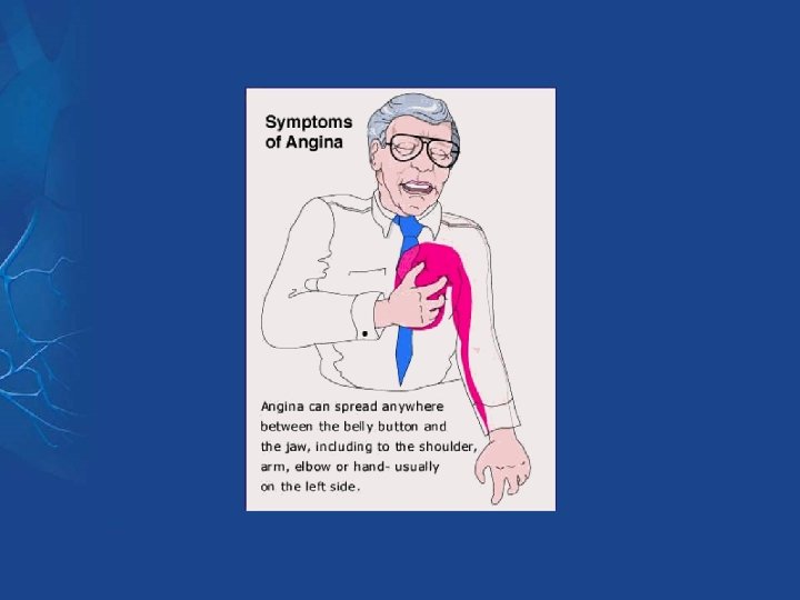
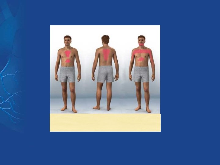
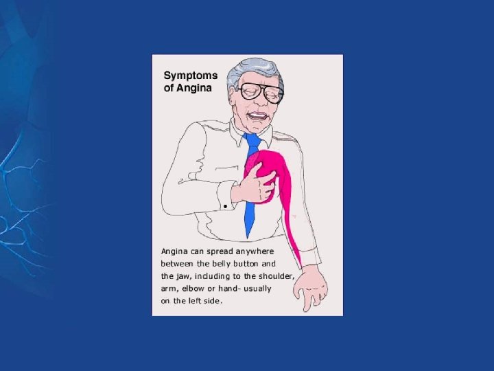
Case presentation A 65 Year old male, presented to outpatient clinic complaining of chest pain of 5 months duration. What are the possible anatomical causes of chest pain? The pain is retrosternal, compressive in nature, precipitated by wakening of 400 meter , relieved by rest, radiated to left shoulder, associated with sweating. Patient is diabetic And smoker On examination: Blood pressure: 160/100. pulse rate: 88 bpm Heart auscultation : normal WHAT IS THE PROBLEB? What is abnormal physical findings? What to do? Investigations
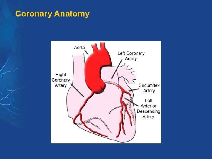
Coronary Anatomy
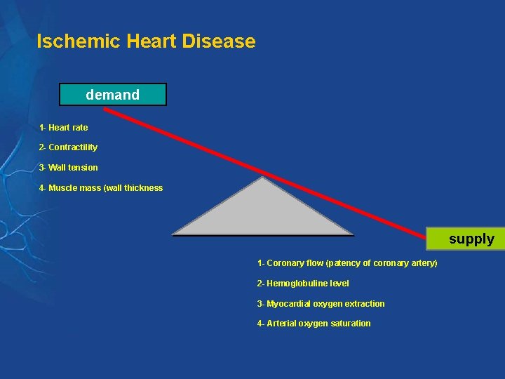
Ischemic Heart Disease demand 1 - Heart rate 2 - Contractility 3 - Wall tension 4 - Muscle mass (wall thickness supply 1 - Coronary flow (patency of coronary artery) 2 - Hemoglobuline level 3 - Myocardial oxygen extraction 4 - Arterial oxygen saturation
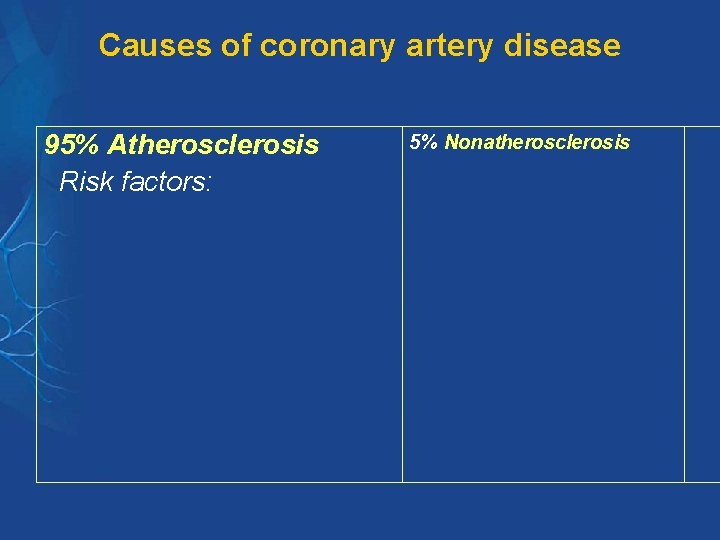
Causes of coronary artery disease 95% Atherosclerosis Risk factors: 5% Nonatherosclerosis
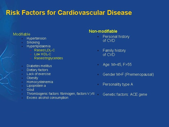
Risk Factors for Cardiovascular Disease Modifiable § § § Non-modifiable Hypertension Smoking Hyperlipidaemia - Raised LDL-C - Low HDL-C - Raised triglycerides § Personal history of CVD § Family history of CVD Diabetes mellitus Dietary factors Lack of exercise Obesity Homocysteinemia Lipoprotein a Gout Thrombogenic factors: fibrinogen, factors V, VII Excess alcohol consumption § Age: M>45, F>55 § Gender M>F (Premenopausal) § Personality type A § Genetic factors: ACE gene
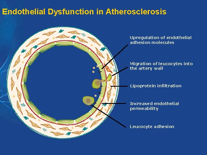
Endothelial Dysfunction in Atherosclerosis Upregulation of endothelial adhesion molecules Migration of leucocytes into the artery wall Lipoprotein infiltration Increased endothelial permeability Leucocyte adhesion
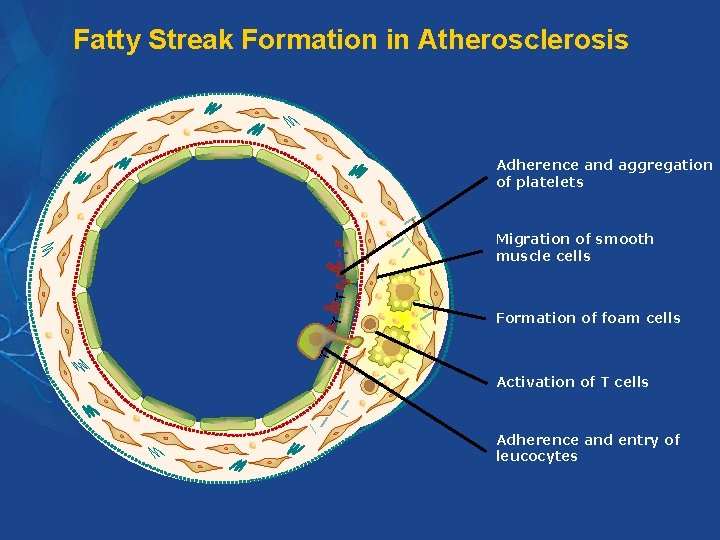
Fatty Streak Formation in Atherosclerosis Adherence and aggregation of platelets Migration of smooth muscle cells Formation of foam cells Activation of T cells Adherence and entry of leucocytes
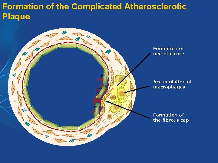
Formation of the Complicated Atherosclerotic Plaque Formation of necrotic core Accumulation of macrophages Formation of the fibrous cap
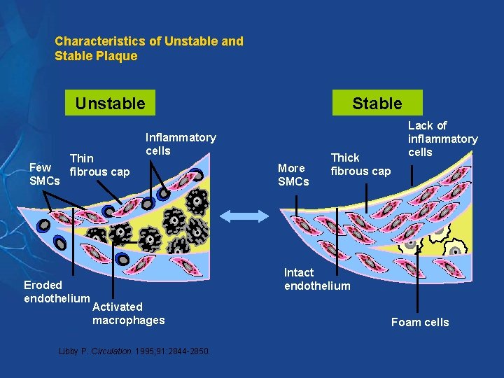
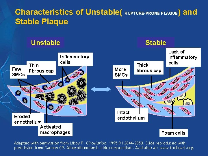
Characteristics of Unstable and Stable Plaque Unstable Few SMCs Thin fibrous cap Eroded endothelium Stable Inflammatory cells More SMCs Thick fibrous cap Lack of inflammatory cells Intact endothelium Activated macrophages Libby P. Circulation. 1995; 91: 2844 -2850. Foam cells
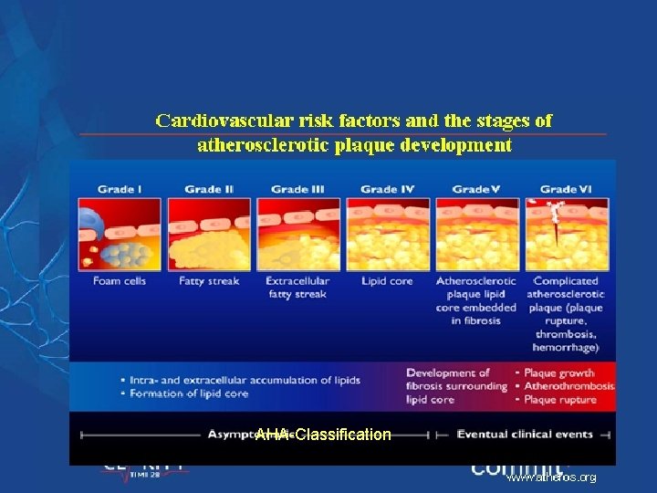
AHA-Classification
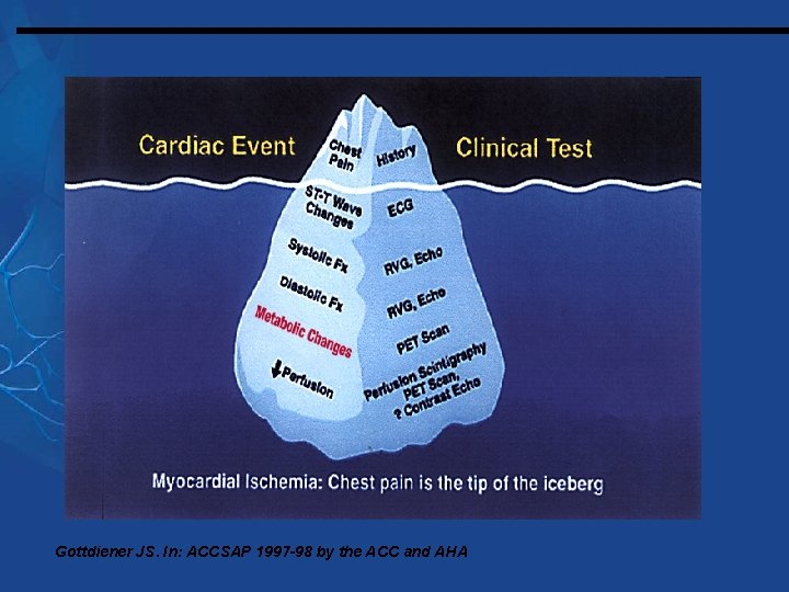
Gottdiener JS. In: ACCSAP 1997 -98 by the ACC and AHA
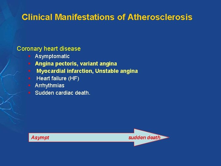
Clinical Manifestations of Atherosclerosis Coronary heart disease § § § Asymptomatic Angina pectoris, variant angina Myocardial infarction, Unstable angina Heart failure (HF) Arrhythmias Sudden cardiac death. Asympt sudden death
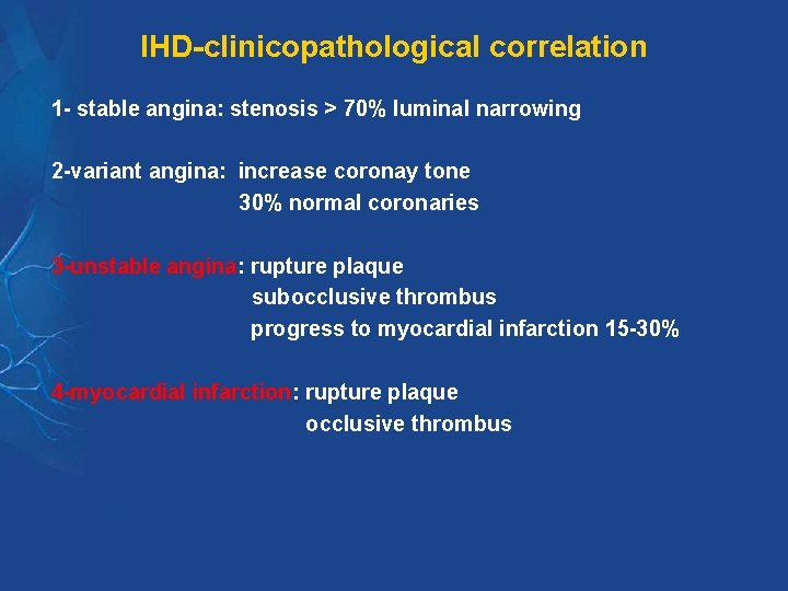
IHD-clinicopathological correlation 1 - stable angina: stenosis > 70% luminal narrowing 2 -variant angina: increase coronay tone 30% normal coronaries 3 -unstable angina: rupture plaque subocclusive thrombus progress to myocardial infarction 15 -30% 4 -myocardial infarction: rupture plaque occlusive thrombus
Angina Chest Pain: Clinical Diagnosis
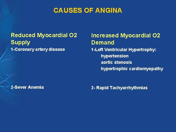
CAUSES OF ANGINA Reduced Myocardial O 2 Supply Increased Myocardial O 2 Demand 1 -Coronary artery disease 1 -Left Ventricular Hypertrophy: hypertension aortic stenosis hypertrophic cardiomyopathy 2 -Sever Anemia 2 - Rapid Tachyarrhythmias
Differential diagnosis of angina 1 - Neuromuscular disorder 2 - Respiratory disorders 3 -Upper GI disorder 4 - Psychological 5 - Syndrome X: Typical angina with normal coronary angio ? Increase tone or decrease coronary vasodilatation excellent prognosis antianginal therapy is rarely effective
Case presentation A 50 year old male presented to emergency room complaining of sudden sever chest pain of 1 hour duration. It is retrosternal, compressive, and radited to left shoulder and arm. Associated with sweating, nausea and vomiting On examination: patient is anxious, in pain, sweaty. BP: 100/60. PULSE: 120 BPM, RR: 26/min Chest: basal crepitations What is the most likely diagnosis pathophysiology
Characteristics of Unstable( RUPTURE-PRONE PLAQUE) and Stable Plaque Unstable Few SMCs Thin fibrous cap Stable Inflammatory cells Eroded endothelium Activated macrophages More SMCs Thick fibrous cap Lack of inflammatory cells Intact endothelium Foam cells Adapted with permission from Libby P. Circulation. 1995; 91: 2844 -2850. Slide reproduced with permission from Cannon CP. Atherothrombosis slide compendium. Available at: www. theheart. org.
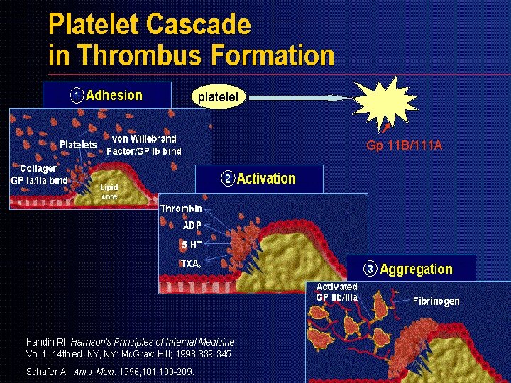
PATHOGENESIS OF ACS Plaque rupture-----Platelet adhesion---activation---aggregation THROMBOSIS 1 - Primary hemostasis: Initiated by platelets adhesion, activation, and aggregation---platelet plug 2 - Secondary hemostasis: activation of the coagulation system---fibrin clot. These two phases are dynamically interactive: Platelet can provide a surface for coagulation enzymes Thrombin is a potent platelet activator
platelet Gp 11 B/111 A
Thank you
Diagnosis of Myocardial Infarction 1 -History 2 -ECG (Electrocardiogram): STMI and NSTMI Hyperacute T wave ST-segment elevation Q- wave T- inversion ST-segment depresion normal ECG will not exclude MI 3 -Cardiac Marker: Troponin, CPK, myoglobulin, . . Troponin T, I: 4 -6 Hr last 10 -14 days CPK: 4 -6 Hr, peak 17 -24 hr, normal 72 hr MB(MM, BB) MB 2/MB 1 >1. 5
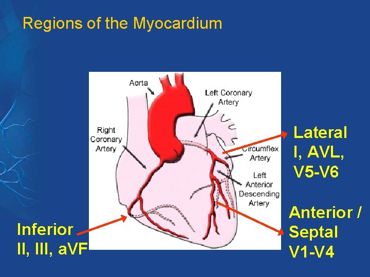
Regions of the Myocardium Lateral I, AVL, V 5 -V 6 Inferior II, III, a. VF Anterior / Septal V 1 -V 4
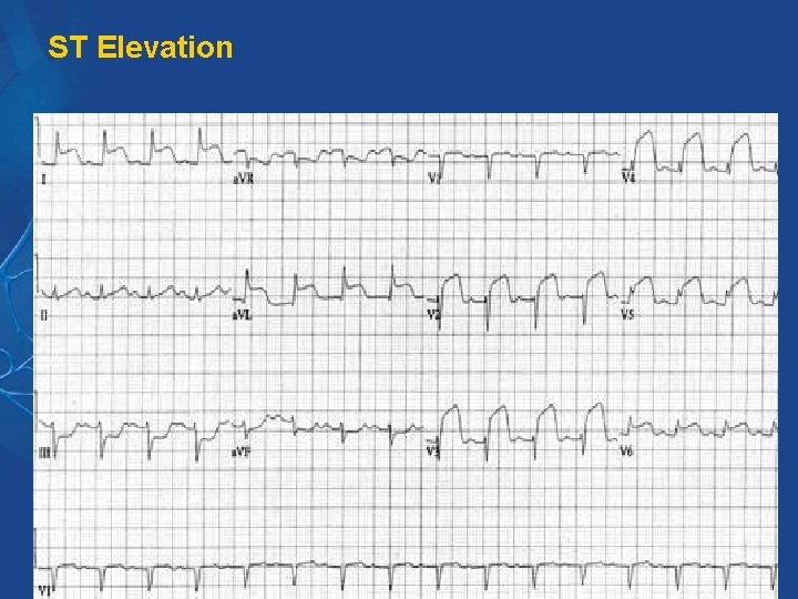
ST Elevation
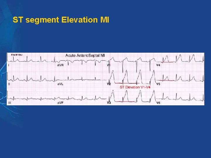
ST segment Elevation MI
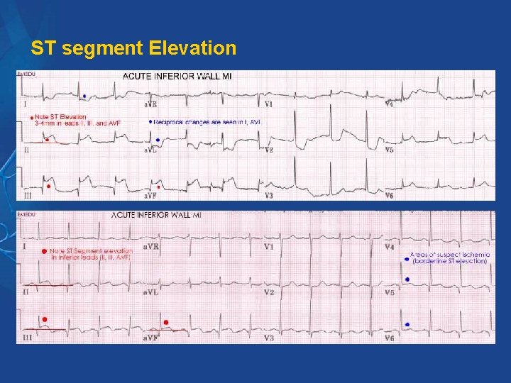
ST segment Elevation
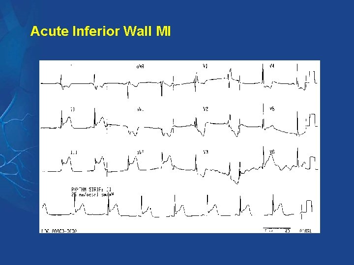
Acute Inferior Wall MI
Acute Posterior Wall MI
Stable angina. Diagnosis § § § § § History : angina pectoris is clinical diagnosis Physical exam Electrocardiogram: 12 ECG, 24 ECG Stress ECG : diagnostic and prognostic information Radioactive studies: thalium scan, . . Echocardiography CT Coronary angiography Serum lipid( LDL, HDL, TG), FBG, CBC Coronary angiography
Imaging Techniques Used to Assess Atherosclerosis Invasive techniques § Coronary angiography § Intravascular ultrasound (IVUS) Non-invasive techniques § Magnetic resonance imaging (MRI) § Computed tomography (CT) § Ultrasound (B-mode)
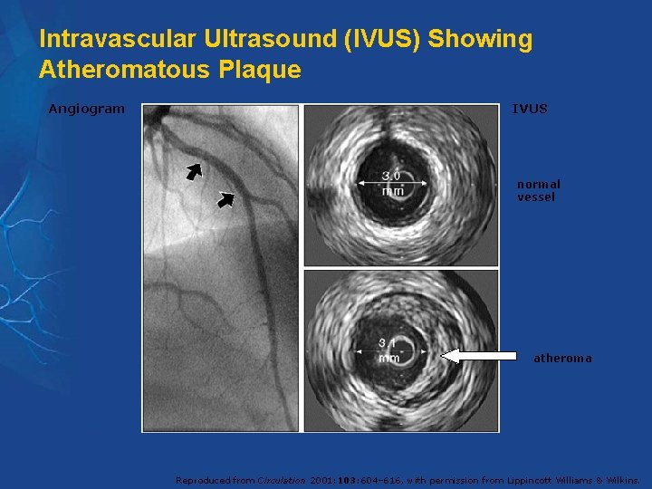
Intravascular Ultrasound (IVUS) Showing Atheromatous Plaque Angiogram IVUS normal vessel atheroma Reproduced from Circulation 2001; 103: 604– 616, with permission from Lippincott Williams & Wilkins.
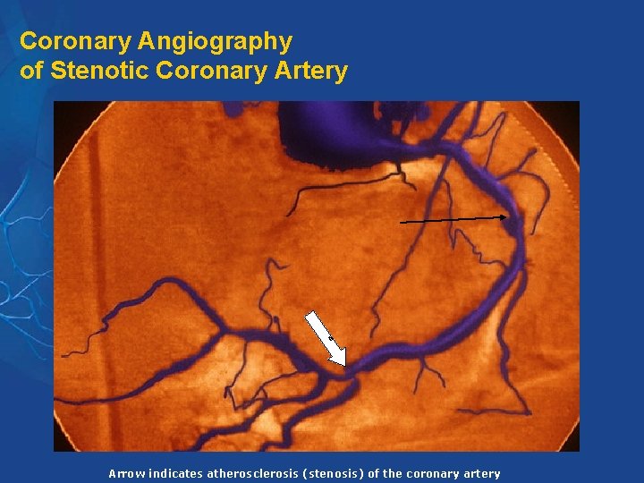
Coronary Angiography of Stenotic Coronary Artery 6 Arrow indicates atherosclerosis (stenosis) of the coronary artery
Management goals of stable angina To improve prognosis (mortality reduction) § Modification of risk factors § Aspirin § Lipid-lowering therapy § ACE-Inhibitor § Revascularization procedures (PTCA, CABG) To decrease anginal symptoms § Medical treatment ACC/AHA Guidelines. J Am Cardiol. 1999; 33: 2092 -2197. ESC Guidelines. Eur Heart J. 1997; 18: 394 -413.
Treatment of stable angina 1 - General measures 2 - Medical therapy: Increase O 2 supply Decrease O 2 demand 3 -Revasularization: PCI (percutaneous coronary intervension) CABG (coronary artery bypass grafting)
TREATMENT OF STABLE ANGINA General Measures 4. § Correction of established risk factors( reversible) § weight reduction (ideal body weight) § Areobic exercise: improve functional capacity, well-being sensation § Treatment of: anemia, thyrotoxicosis, arrhythmias, . .
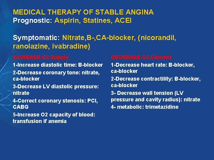
MEDICAL THERAPY OF STABLE ANGINA Prognostic: Aspirin, Statines, ACEI Symptomatic: Nitrate, B-, CA-blocker, (nicorandil, ranolazine, ivabradine) INCREASE O 2 Supply 1 -Increase diastolic time: B-blocker 2 -Decrease coronary tone: nitrate, ca-blocker 3 -Decrease LV diastolic pressure: nitrate 4 -Correct coronary stenosis: PCI, CABG 5 -Increase O 2 capacity of blood: transfusion if anemia DECREASE O 2 Demand 1 -Decrease heart rate: B-blocker, ca-blocker 2 -Decrease contractility: B-blocker, ca-blocker 3 - Decrease wall tension (LV pressure and cavity radius): nitrate 4 - metabolic: trimetazidine
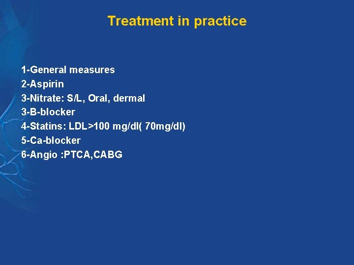
Treatment in practice 1 -General measures 2 -Aspirin 3 -Nitrate: S/L, Oral, dermal 3 -B-blocker 4 -Statins: LDL>100 mg/dl( 70 mg/dl) 5 -Ca-blocker 6 -Angio : PTCA, CABG
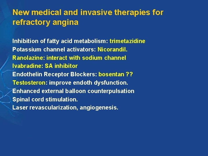
New medical and invasive therapies for refractory angina Inhibition of fatty acid metabolism: trimetazidine Potassium channel activators: Nicorandil. Ranolazine: interact with sodium channel Ivabradine: SA inhibitor Endothelin Receptor Blockers: bosentan ? ? Testosteron: improve endoth dysfunction. Enhanced external balloon counterpulsation Spinal cord stimulation. Laser revascularization, angiogenesis.
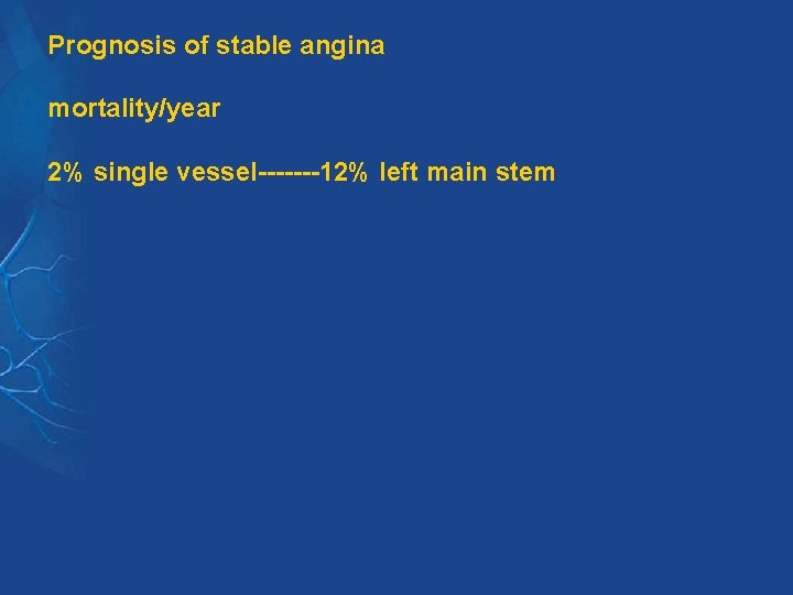
Prognosis of stable angina mortality/year 2% single vessel-------12% left main stem
VARIANT ANGINA-PRINZMETAL ANGINA Chest pain with ST-Segment elevation Usually at rest, Troponin: negative Female > male Spasm of large epicardial coronary vessel during the attack Vasospastic symptpms in other organs Can cause arrhythmias and death Treatment: CA-blocker, Nitrate B-blocker is contraindicated Prognosis: 5 year mortality < 5%