
The Arteries SHANDONG UNIVERSITY Liu Zhiyu The Arteries
- Slides: 43
The Arteries SHANDONG UNIVERSITY Liu Zhiyu
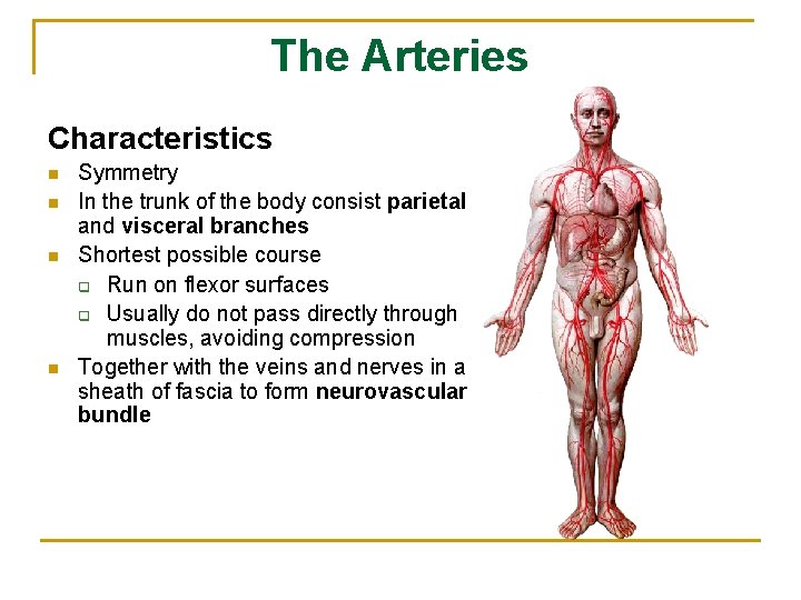
The Arteries Characteristics n n Symmetry In the trunk of the body consist parietal and visceral branches Shortest possible course q Run on flexor surfaces q Usually do not pass directly through muscles, avoiding compression Together with the veins and nerves in a sheath of fascia to form neurovascular bundle
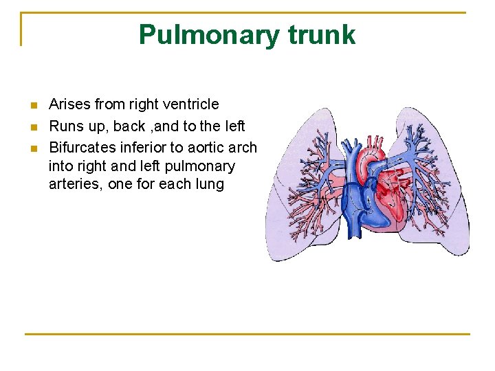
Pulmonary trunk n n n Arises from right ventricle Runs up, back , and to the left Bifurcates inferior to aortic arch into right and left pulmonary arteries, one for each lung
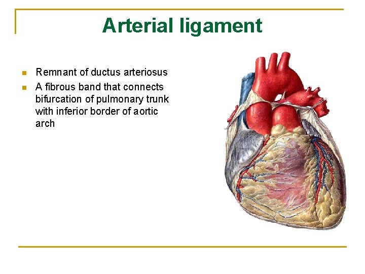
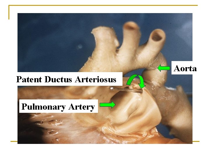
Arterial ligament n n Remnant of ductus arteriosus A fibrous band that connects bifurcation of pulmonary trunk with inferior border of aortic arch
Aorta Patent Ductus Arteriosus Pulmonary Artery
Arteries of systemic circulation
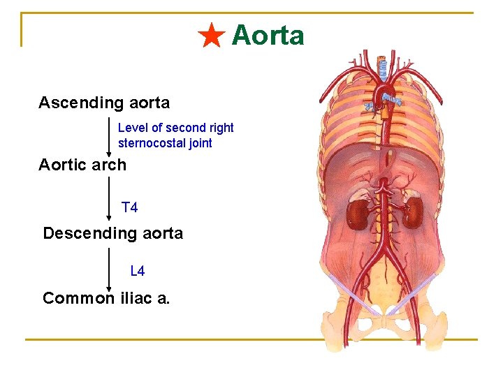
★ Aorta Ascending aorta Level of second right sternocostal joint Aortic arch T 4 Descending aorta L 4 Common iliac a.
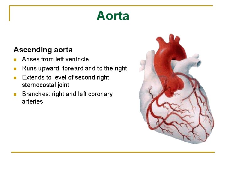
Aorta Ascending aorta n n Arises from left ventricle Runs upward, forward and to the right Extends to level of second right sternocostal joint Branches: right and left coronary arteries
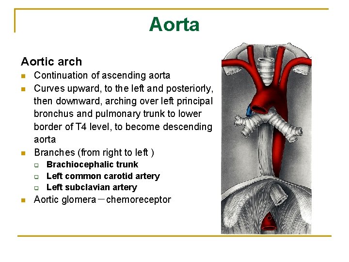
Aorta Aortic arch n n n Continuation of ascending aorta Curves upward, to the left and posteriorly, then downward, arching over left principal bronchus and pulmonary trunk to lower border of T 4 level, to become descending aorta Branches (from right to left ) q q q n Brachiocephalic trunk Left common carotid artery Left subclavian artery Aortic glomera-chemoreceptor
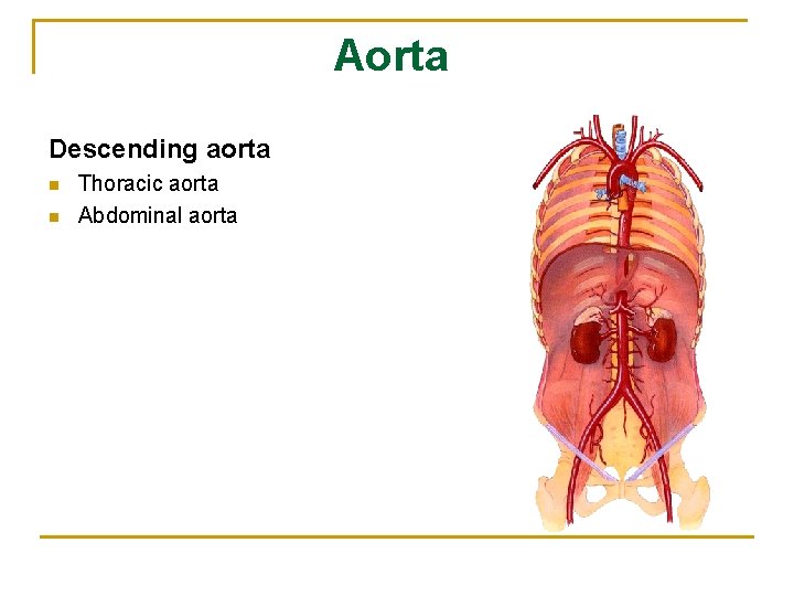
Aorta Descending aorta n n Thoracic aorta Abdominal aorta
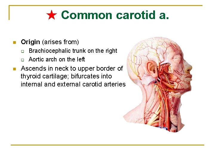
★ Common carotid a. n Origin (arises from) q q n Brachiocephalic trunk on the right Aortic arch on the left Ascends in neck to upper border of thyroid cartilage; bifurcates into internal and external carotid arteries
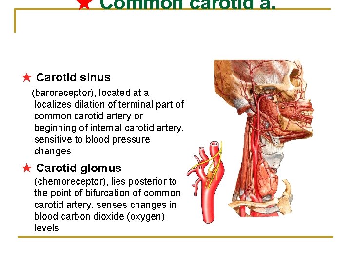
★ Common carotid a. ★ Carotid sinus (baroreceptor), located at a localizes dilation of terminal part of common carotid artery or beginning of internal carotid artery, sensitive to blood pressure changes ★ Carotid glomus (chemoreceptor), lies posterior to the point of bifurcation of common carotid artery, senses changes in blood carbon dioxide (oxygen) levels
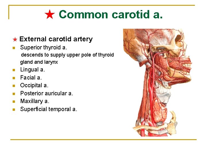
★ Common carotid a. ★ External n carotid artery Superior thyroid a. descends to supply upper pole of thyroid gland larynx n n n Lingual a. Facial a. Occipital a. Posterior auricular a. Maxillary a. Superficial temporal a.
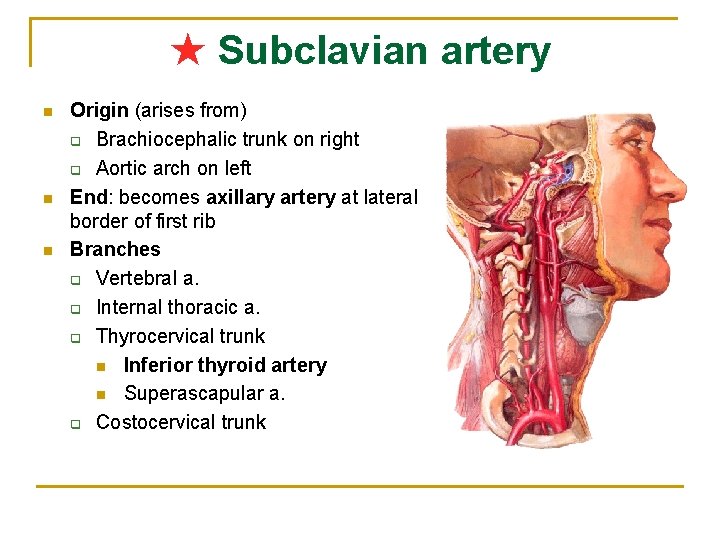
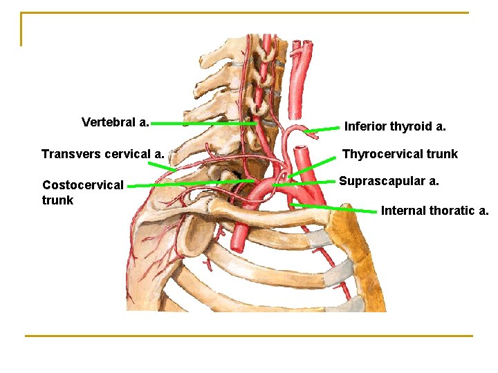
★ Subclavian artery n n n Origin (arises from) q Brachiocephalic trunk on right q Aortic arch on left End: becomes axillary artery at lateral border of first rib Branches q Vertebral a. q Internal thoracic a. q Thyrocervical trunk n Inferior thyroid artery n Superascapular a. q Costocervical trunk
Vertebral a. Inferior thyroid a. Transvers cervical a. Thyrocervical trunk Costocervical trunk Suprascapular a. Internal thoratic a.
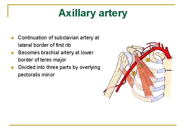
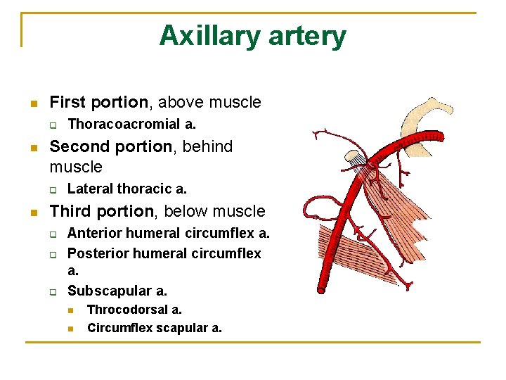
Axillary artery n n n Continuation of subclavian artery at lateral border of first rib Becomes brachial artery at lower border of teres major Divided into three parts by overlying pectoralis minor
Axillary artery n First portion, above muscle q n Second portion, behind muscle q n Thoracoacromial a. Lateral thoracic a. Third portion, below muscle q q q Anterior humeral circumflex a. Posterior humeral circumflex a. Subscapular a. n n Throcodorsal a. Circumflex scapular a.
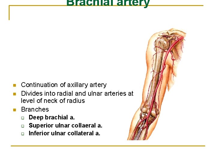
Brachial artery n n n Continuation of axillary artery Divides into radial and ulnar arteries at level of neck of radius Branches q q q Deep brachial a. Superior ulnar collaeral a. Inferior ulnar collateral a.
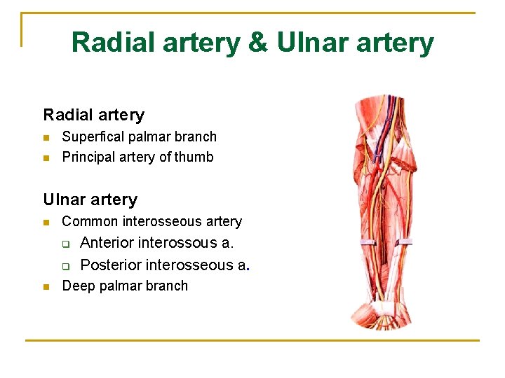
Radial artery & Ulnar artery Radial artery n n Superfical palmar branch Principal artery of thumb Ulnar artery n Common interosseous artery q q n Anterior interossous a. Posterior interosseous a. Deep palmar branch
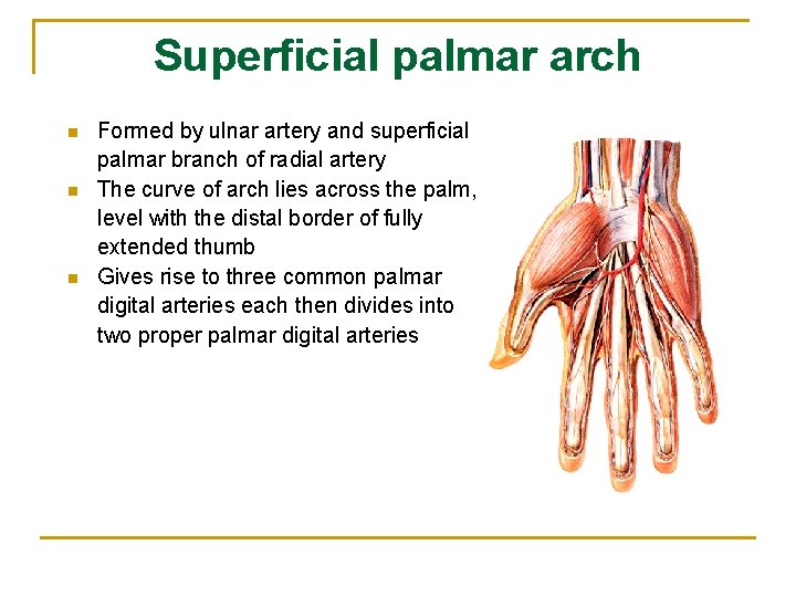
Superficial palmar arch n n n Formed by ulnar artery and superficial palmar branch of radial artery The curve of arch lies across the palm, level with the distal border of fully extended thumb Gives rise to three common palmar digital arteries each then divides into two proper palmar digital arteries
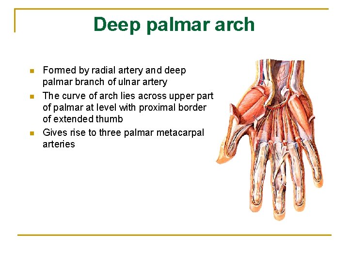
Deep palmar arch n n n Formed by radial artery and deep palmar branch of ulnar artery The curve of arch lies across upper part of palmar at level with proximal border of extended thumb Gives rise to three palmar metacarpal arteries
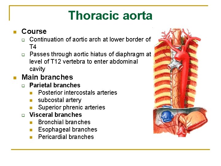
Thoracic aorta n Course q q n Continuation of aortic arch at lower border of T 4 Passes through aortic hiatus of diaphragm at level of T 12 vertebra to enter abdominal cavity Main branches q q Parietal branches n Posterior intercostals arteries n subcostal artery n Superior phrenic arteries Visceral branches n Bronchial branches n Esophageal branches n Pericardial branches
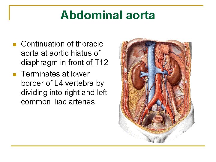
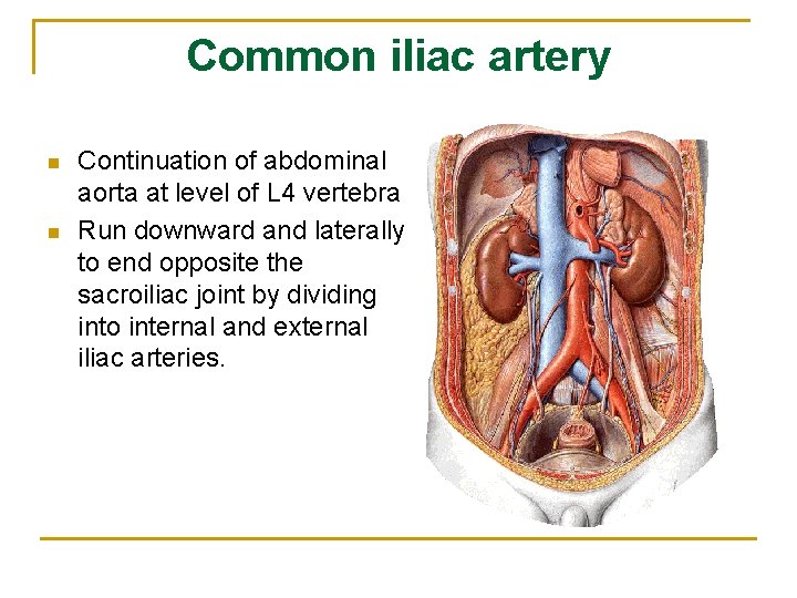
Abdominal aorta n n Continuation of thoracic aorta at aortic hiatus of diaphragm in front of T 12 Terminates at lower border of L 4 vertebra by dividing into right and left common iliac arteries
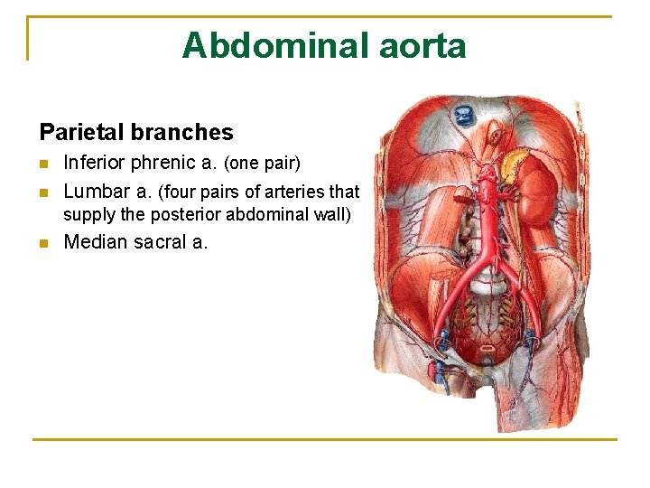
Abdominal aorta Parietal branches n n Inferior phrenic a. (one pair) Lumbar a. (four pairs of arteries that supply the posterior abdominal wall) n Median sacral a.
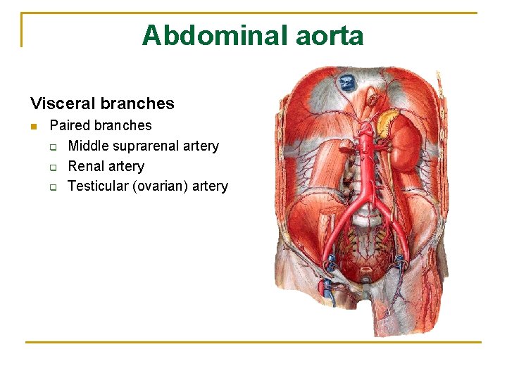
Abdominal aorta Visceral branches n Paired branches q Middle suprarenal artery q Renal artery q Testicular (ovarian) artery
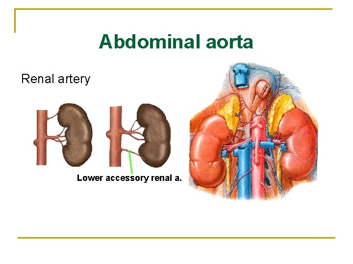
Abdominal aorta Renal artery Lower accessory renal a.
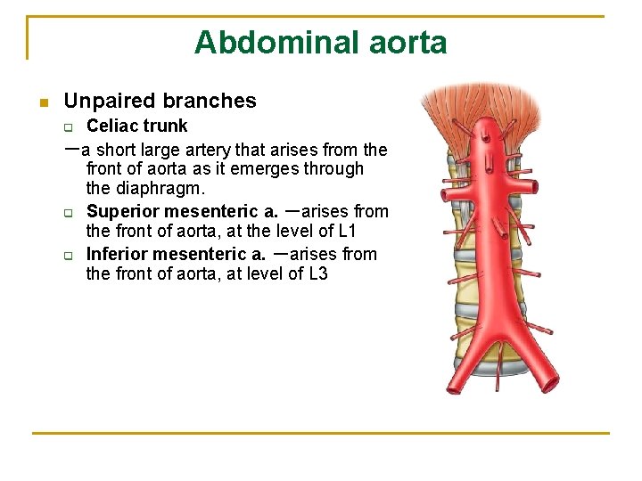
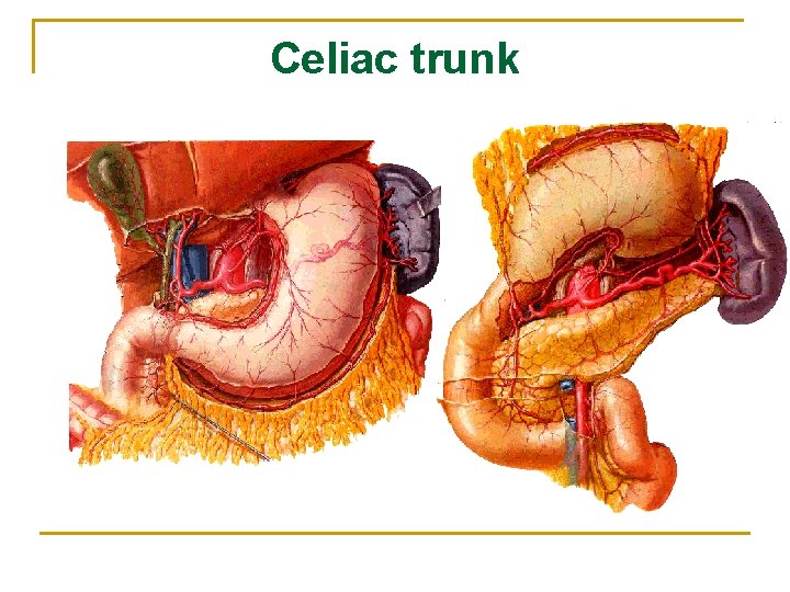
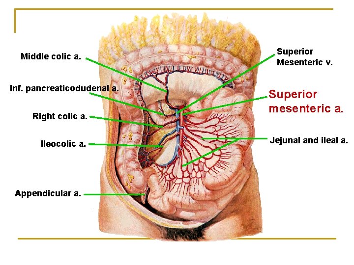
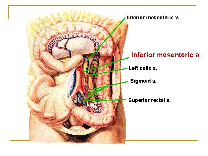
Abdominal aorta n Unpaired branches Celiac trunk -a short large artery that arises from the front of aorta as it emerges through the diaphragm. q Superior mesenteric a. -arises from the front of aorta, at the level of L 1 q Inferior mesenteric a. -arises from the front of aorta, at level of L 3 q
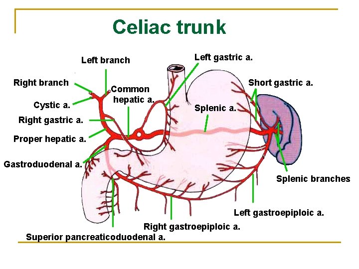
Celiac trunk Left branch Right branch Cystic a. Common hepatic a. Left gastric a. Short gastric a. Splenic a. Right gastric a. Proper hepatic a. Gastroduodenal a. Splenic branches Left gastroepiploic a. Right gastroepiploic a. Superior pancreaticoduodenal a.
Celiac trunk
Middle colic a. Inf. pancreaticodudenal a. Right colic a. Ileocolic a. Appendicular a. Superior Mesenteric v. Superior mesenteric a. Jejunal and ileal a.
Inferior mesenteric v. Inferior mesenteric a. Left colic a. Sigmoid a. Superior rectal a.
Common iliac artery n n Continuation of abdominal aorta at level of L 4 vertebra Run downward and laterally to end opposite the sacroiliac joint by dividing into internal and external iliac arteries.
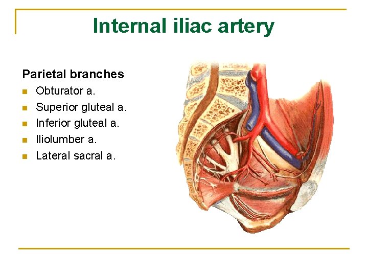
Internal iliac artery Parietal branches n n n Obturator a. Superior gluteal a. Inferior gluteal a. Iliolumber a. Lateral sacral a.
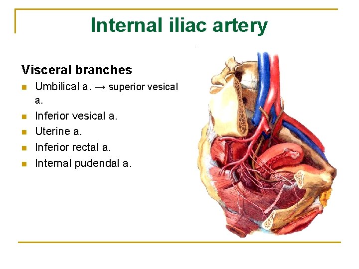
Internal iliac artery Visceral branches n Umbilical a. → superior vesical a. n n Inferior vesical a. Uterine a. Inferior rectal a. Internal pudendal a.
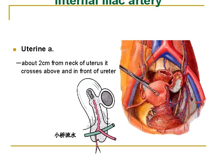
Internal iliac artery n Uterine a. -about 2 cm from neck of uterus it crosses above and in front of ureter 小桥流水
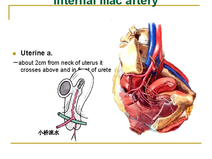
Internal iliac artery n Uterine a. -about 2 cm from neck of uterus it crosses above and in front of ureter 小桥流水
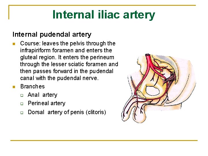
Internal iliac artery Internal pudendal artery n n Course: leaves the pelvis through the infrapiriform foramen and enters the gluteal region. It enters the perineum through the lesser sciatic foramen and then passes forward in the pudendal canal with the pudendal nerve. Branches q Anal artery q Perineal artery q Dorsal artery of penis (clitoris)
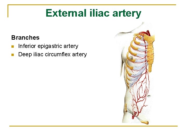
External iliac artery Branches n n Inferior epigastric artery Deep iliac circumflex artery
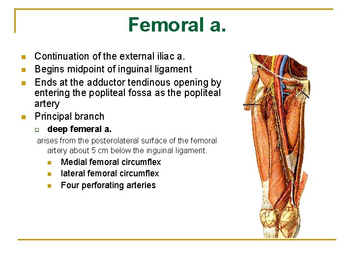
Femoral a. n n Continuation of the external iliac a. Begins midpoint of inguinal ligament Ends at the adductor tendinous opening by entering the popliteal fossa as the popliteal artery Principal branch q deep femeral a. arises from the posterolateral surface of the femoral artery about 5 cm below the inguinal ligament. n n n Medial femoral circumflex lateral femoral circumflex Four perforating arteries
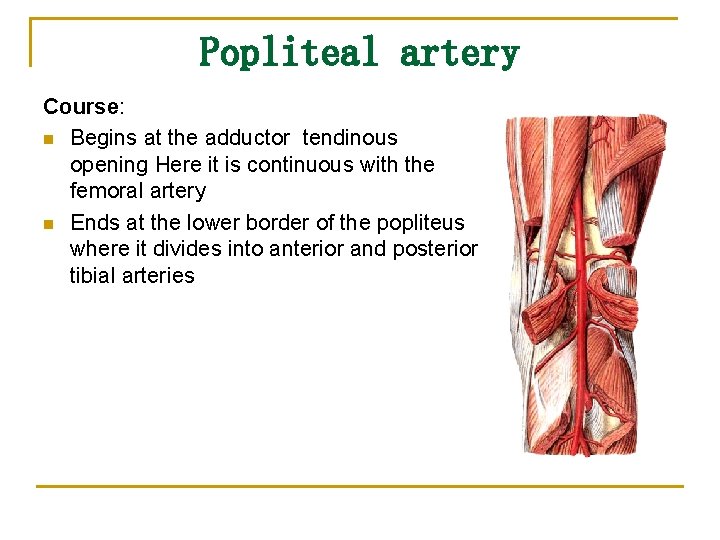
Popliteal artery Course: n Begins at the adductor tendinous opening Here it is continuous with the femoral artery n Ends at the lower border of the popliteus where it divides into anterior and posterior tibial arteries
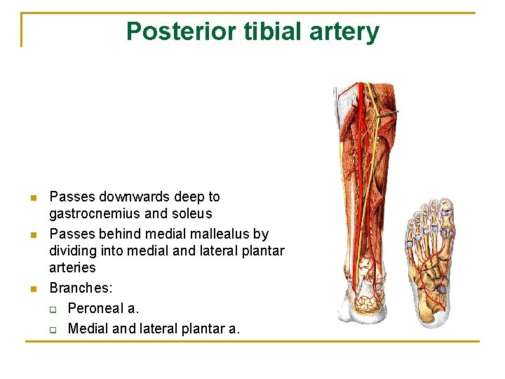
Posterior tibial artery n n n Passes downwards deep to gastrocnemius and soleus Passes behind medial mallealus by dividing into medial and lateral plantar arteries Branches: q Peroneal a. q Medial and lateral plantar a.
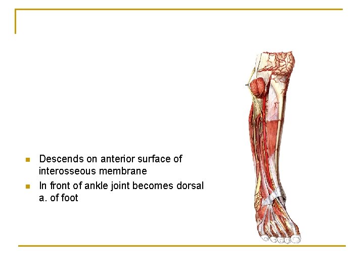
n n Descends on anterior surface of interosseous membrane In front of ankle joint becomes dorsal a. of foot
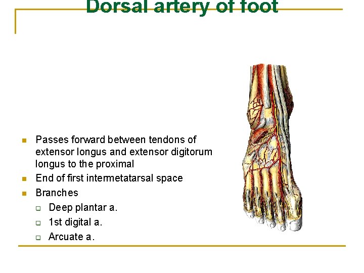
Dorsal artery of foot n n n Passes forward between tendons of extensor longus and extensor digitorum longus to the proximal End of first intermetatarsal space Branches q Deep plantar a. q 1 st digital a. q Arcuate a.