ATHEROSCLEROSIS Sufia Husain Pathology KSU Riyadh March 2015

 arteries • • aorta, common carotid, iliac lots of")


 SMCs are present in the vascular media SMCs are responsible")
 Atherosclerosis is characterized by intimal lesions called atheromas/ atheromatous/ fibrofatty plaques, which")

. Fatty streaks are the earliest lesion of")


: When too much LDL")


- Slides: 26
ATHEROSCLEROSIS Sufia Husain. Pathology. KSU. Riyadh. March 2015
Normal Blood Vessels Large (elastic) arteries • • aorta, common carotid, iliac lots of elastic fibers Medium (muscular) arteries • • coronary, renal arteries mostly smooth muscle cells Small arteries/arterioles • • all smooth muscle cells blood pressure controlled here Capillaries • • • diameter of RBC thin walls, slow flow great for exchanging oxygen, nutrients Venules/veins • large diameter, thin walls compressible, penetrable by tumor Have valves • • drain excess interstitial fluid pass through nodes • • Lymphatics
Artery www. full-health. com www. mananatomy. com
ENDOTHELIAL CELLS The endothelium is a single cell thick lining of endothelial cells and it is the inner lining of the entire cardiovascular system (arteries, veins and capillaries) and the lymphatic system. It is in direct contact with the blood/lymph and the cells circulating in it. Endothelial structural and functional integrity is fundamental to the maintenance of vessel wall homeostasis and normal circulatory function. www. udel. edu
Smooth muscle cells (SMC) SMCs are present in the vascular media SMCs are responsible for vasoconstriction and dilation in response to normal or pharmacologic stimuli. Any vascular injury or dysfunction stimulates SMCs. On stimulation they: 1. They migrate from the media to the intima. 2. In the intima they lose the capacity to contract and gain the capacity to divide. So they multiply/proliferate as intimal SMCs. 3. They synthesize collagen, elastin etc and deposit extracellular matrix (ECM).
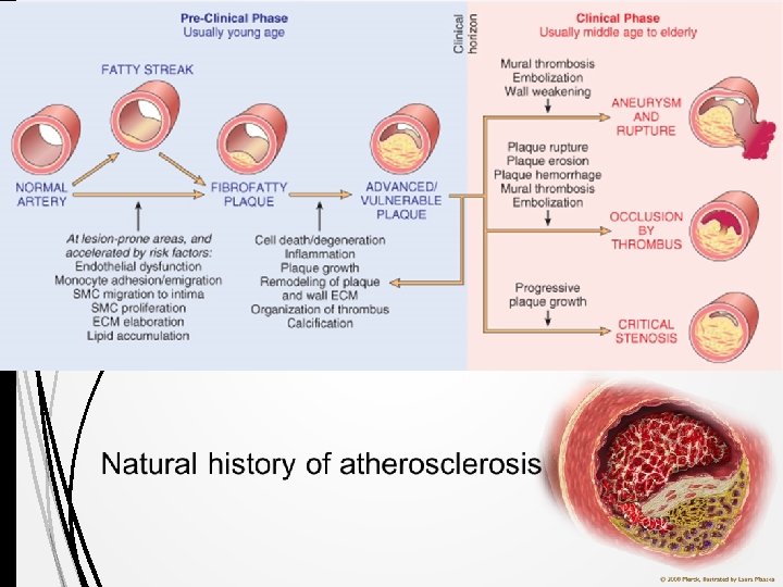
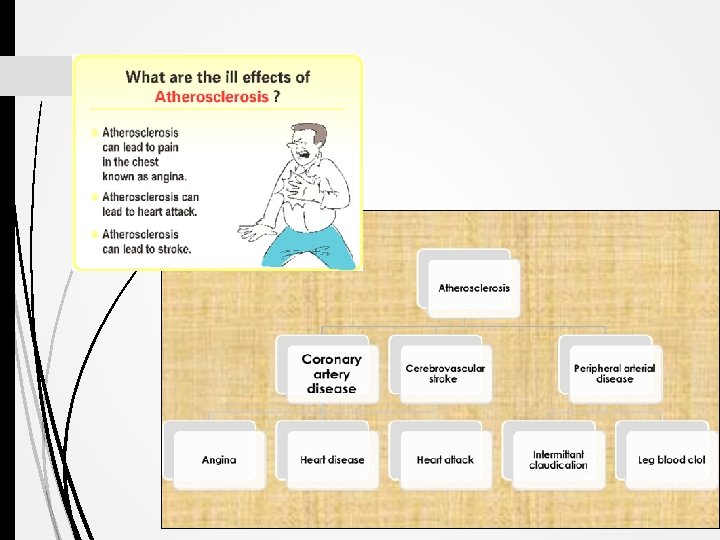
Atherosclerosis (AS) Atherosclerosis is characterized by intimal lesions called atheromas/ atheromatous/ fibrofatty plaques, which protrude into and obstruct vascular lumens and weaken the underlying media. They may lead to serious complications like Coronary artery disease (angina & MI) and Carotid atherosclerotic disease (stroke) The most heavily involved vessels are the abdominal aorta then coronary arteries, the popliteal arteries, the internal carotid arteries, and the vessels of the circle of Willis.
Gross morphology of atheroma/atheromatous plaque The key processes in AS is intimal thickening and lipid accumulation. The atheromatous plaques impinge on the lumen of the artery. They vary in size. atheromatous plaques usually involve only a partial circumference of the arterial wall ("eccentric" lesions) and are patchy and variable along the vessel length. www. med. uottawa. ca
Atherosclerosis: Aorta with fatty streaks ( arrows). Fatty streaks are the earliest lesion of atherosclerosis they are a collection of lipid laden foam cells in the intima. They do not cause any disturbance in blood flow. Fatty streaks begin as multiple yellow, flat spots less than 1 mm in diameter that coalesce into elongated streaks, 1 cm long or longer. They contain T lymphocytes and extracellular lipid in smaller amounts than in plaques. Photomicrograph of fatty streak in an experimental hypercholesterolemic rabbit, demonstrating intimal macrophage-derived foam cells ( arrow).
Atherosclerosis: Microscopic morphology An atheroma consists of a raised focal lesion in the intima, with a soft, yellow, grumous core of lipid (mainly cholesterol and cholesterol esters), covered by a firm, white fibrous cap. Atherosclerotic plaques have three principal components: 1. Cells: SMCs, macrophages, lymphocytes and foam cell 2. Extracellular matrix: including collagen, elastic fibers, and proteoglycans 3. Lipid: Typical atheromas contain relatively abundant lipid both intracellular and extracellular lipid. Foam cells are large, lipid-laden macrophages derived from blood monocytes, but SMCs can also imbibe lipid to become foam cells.
Atherosclerosis: microscopic morphology Typically, the superficial fibrous cap is composed of SMCs and extracellular matrix. With some macrophages and T lymphocytes. Below the fibrous cap is a necrotic core, containing a lipid deposits (primarily cholesterol and cholesterol esters), cholesterol clefts, debris from dead cells, foam cells, fibrin.
Gross views of atherosclerosis in the aorta. A. Mild atherosclerosis composed of fibrous plaques, one of which is denoted by the arrow. Slide 12. 7 B. Severe disease with diffuse and complicated lesions.
Overall architecture demonstrating an eccentric lesion with a fibrous cap and a central lipid core with typical cholesterol clefts. The lumen is moderately narrowed. http: //sphweb. bumc. bu. edu/otlt/MPHModules/PH/PH 709_Heart 3. html
PATHOLOGICAL COMPLICATIONS The advanced lesion of atherosclerosis is at risk for the following pathological changes that have clinical significance: 1) Focal rupture, ulceration, or erosion of the luminal surface of atheromatous plaques which may induce thrombus formation or discharge of debris into the bloodstream, producing microemboli composed of lesion contents (cholesterol emboli or atheroemboli). 2) Hemorrhage into a plaque (especially when atheroma in the coronary arteries) due to rupture of the overlying fibrous cap or the capillaries in the plaque. The hematoma may expand the plaque or induce plaque rupture 3) Superimposed thrombosis, the most feared complication, usually occurs on disrupted lesions (those with rupture, ulceration, erosion, or hemorrhage) the thrombus can lead to partial or completely occlusion of the lumen. The thrombus can also embolize. 4) Wall weakening with aneurysmal dilation. Atheroma can induced atrophy of the underlying media, with loss of elastic tissue, causing weakness, aneurysm and potential rupture 5) Calcifications: Atheromas often undergo calcification.
Illustration from Libby P: Inflammation in Atherosclerosis. Nature 202; 420: 868
Slide 12. 5 Natural history of atherosclerosis
Stroke/ cerebrovascular accident healthcaredir. com -
cardiologydoc. wordpress. com
Risk Factors for Atherosclerosis MINOR/UNCERTAIN/NONQUANTIT ATED RISK FACTORS Obesity MAJOR RISK FACTORS Physical inactivity NON-MODIFIABLE POTENTIALLY MODIFIABLE Stress ("type A" personality) Increasing age Hyperlipidemia Postmenopausal estrogen deficiency Male gender Hypertension High carbohydrate intake Family history Cigarette smoking Genetic abnormalities Diabetes Alcohol Lipoprotein Lp(a) Hardened (trans)unsaturated fat intake Chlamydia pneumoniae
Importance of types of lipoproteins in hypelipidemia Low-density lipoproteins (LDLs): When too much LDL (bad) cholesterol circulates in the blood, it promote atherosclerosis and therefore contributes to heart disease. Very-low-density lipoproteins (VLDLs): is also considered to be a type of bad cholesterol and it promote atherosclerosis Chylomicrons also promote atherosclerosis. High-density lipoproteins (HDLs): is known as “good” cholesterol, because high levels of HDL protects against heart attack. Low levels of HDL also increase the risk of heart disease. HDLs help to reverse the effects of high cholesterol.
PATHOGENESIS: response to injury hypothesis Central to this hypothesis are the following: Accumulation of lipoproteins (mainly LDL with its high cholesterol content) in the vessel wall Subtle Chronic endothelial injury Increased permeability, leukocyte adhesion, and thrombotic potential. Adhesion of blood monocytes (and other leukocytes) to the endothelium, followed by migration of monocytes into the intima and transformation into macrophages and foam cells Adhesion of platelets
PATHOGENESIS: response to injury hypothesis Release of factors from activated platelets, macrophages, or vascular cells that cause migration of SMCs from media into the intima Proliferation of smooth muscle cells in the intima, and elaboration of extracellular matrix, leading to the accumulation of collagen and proteoglycans Enhanced accumulation of lipids both within cells (macrophages and SMCs) and extracellularly.
SUMMARY / http: //www. pathophys. org
Angioplasty www. flickr. com