ASSESSMENT AND DIAGNOSIS Importance of Diagnosing Migraine Improved















- Slides: 54
ASSESSMENT AND DIAGNOSIS
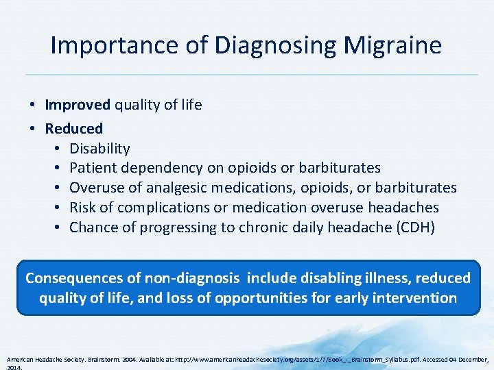
Importance of Diagnosing Migraine • Improved quality of life • Reduced • Disability • Patient dependency on opioids or barbiturates • Overuse of analgesic medications, opioids, or barbiturates • Risk of complications or medication overuse headaches • Chance of progressing to chronic daily headache (CDH) Consequences of non-diagnosis include disabling illness, reduced quality of life, and loss of opportunities for early intervention American Headache Society. Brainstorm. 2004. Available at: http: //www. americanheadachesociety. org/assets/1/7/Book_ _Brainstorm_Syllabus. pdf. Accessed 04 December,
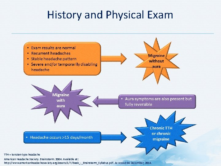
History and Physical Exam TTH = tension type headache American Headache Society. Brainstorm. 2004. Available at: http: //www. americanheadachesociety. org/assets/1/7/Book_ _Brainstorm_Syllabus. pdf. Accessed 04 December, 2014.
Red Flags in History Abrupt onset Very severe headache Progressive worsening of headache Headache precipitated by exertion Headache accompanied by generalized illness, fever, nausea, vomiting or stiff neck • Headache associated with neurological symptoms • Comorbidities • • •
Red Flags on Examination • • • Abnormal vital signs Papilledema Signs of meningeal irritation Presence of focal neurological signs Change in higher intellectual functions or cognition
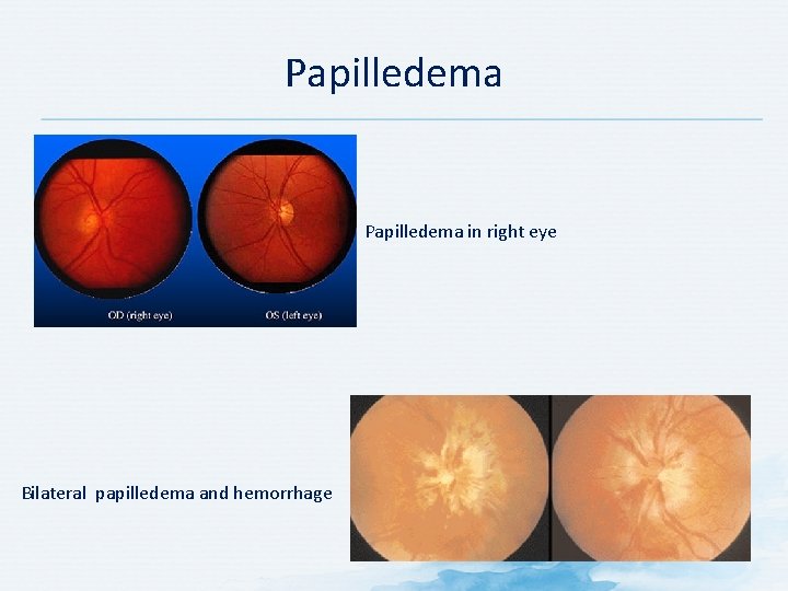
Papilledema in right eye Bilateral papilledema and hemorrhage
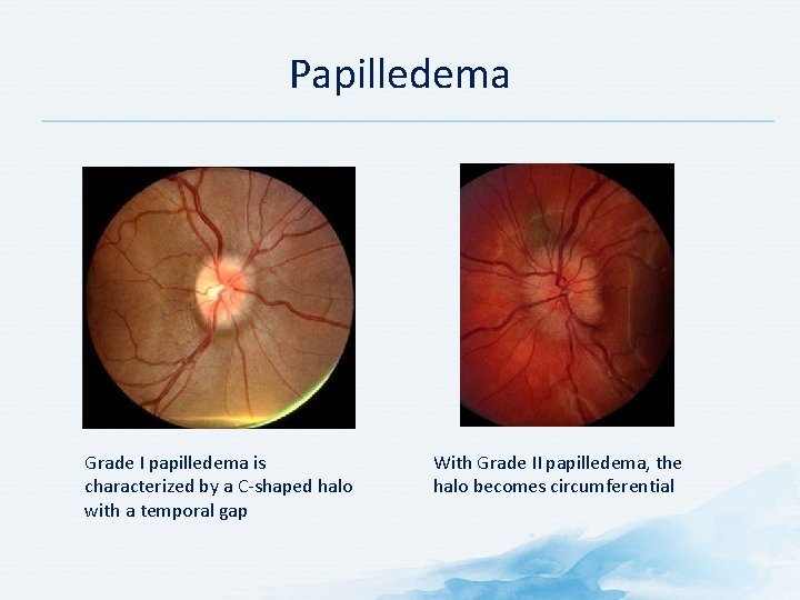
Papilledema Grade I papilledema is characterized by a C shaped halo with a temporal gap With Grade II papilledema, the halo becomes circumferential
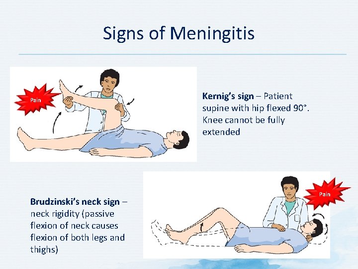
Signs of Meningitis Pain Brudzinski’s neck sign – neck rigidity (passive flexion of neck causes flexion of both legs and thighs) Kernig’s sign – Patient supine with hip flexed 90°. Knee cannot be fully extended Pain
Subarachnoid Hemorrhage
Brain Tumor
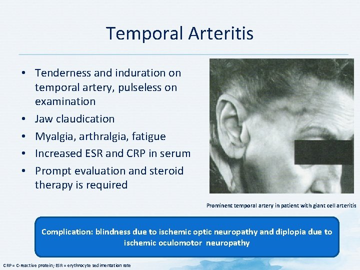
Temporal Arteritis • Tenderness and induration on temporal artery, pulseless on examination • Jaw claudication • Myalgia, arthralgia, fatigue • Increased ESR and CRP in serum • Prompt evaluation and steroid therapy is required Prominent temporal artery in patient with giant cell arteritis Complication: blindness due to ischemic optic neuropathy and diplopia due to ischemic oculomotor neuropathy CRP = C reactive protein; ESR = erythrocyte sedimentation rate
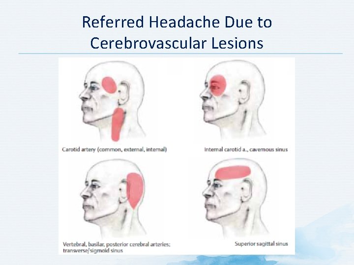
Referred Headache Due to Cerebrovascular Lesions
Patient History • Focus on most severe headache first • Ask standardized questions • Onset • Frequency/duration The goal of taking a patient’s • Location history and performing a • Severity physical exam is to rule out • Characteristics and other symptoms secondary headache and • Family history diagnose primary headache • What makes it better/worse • Medications taken • Recent changes in pattern • Other types of headaches • Neurologic symptoms (cognitive changes, changes in speech/language, loss of strength/sensation [including visual loss and diplopia], vertigo, faintness) • Disability – does the headache interfere with daily life? • Be alert for comorbid conditions complicating headache or diagnosis American Headache Society. Brainstorm. 2004. Available at: http: //www. americanheadachesociety. org/assets/1/7/Book_ _Brainstorm_Syllabus. pdf. Accessed 04 December,
Headache History: Useful Questions to Ask Patients 1. When do you think your worst headaches first started? 2. How often do you get headaches that if left untreated are so severe you find it difficult to function? 3. What is the pain like? 4. How long does the pain last? 5. Do you have other symptoms besides head pain with these headaches? 6. What makes your headaches better or worse? 7. How often do you take something for your headaches? 8. What do you take for your headaches? 9. Does anyone else in your family have similar headaches? 10. Do you get other kinds of headaches? 11. Has there been any recent change in your headaches? American Headache Society. Brainstorm. 2004. Available at: http: //www. americanheadachesociety. org/assets/1/7/Book_ _Brainstorm_Syllabus. pdf. Accessed 04 December,
Risk Factors for Chronic Migraine Modifiable Non-modifiable • Stressful life events • Frequent headache • Obesity • Duration of illness • Snoring • Medication overuse Granella F et al. Cephalalgia. 1998; 18(Suppl 2): 30 3; Scher AI et al. Curr Pain Headache Rep. 2002; 6: 486 91.
Vascular Risk Factors for Migraine • • • ↑ CRP levels ↑ interleukin levels ↑ TNF α and adhesion molecules (systemic inflammation markers) High homocysteine levels Oxidative stress and thrombosis Hypertension Hypercholesterolemia Impaired insulin sensitivity Stroke Coronary heart disease Increased body weight CRP = C reactive protein; TNF = tumor necrosis factor Chawla J. 2014. Available at http: //emedicine. medscape. com/article/1142556 overview. Accessed 05 January 2014.
Signs and Symptoms of Migraine
Typical Symptoms of Migraine • Throbbing or pulsatile headache • Moderate to severe pain; intensifies with movement or physical activity • Unilateral and localized pain in frontotemporal and ocular areas • Pain may be felt anywhere around head or neck • Pain builds up over 1 to 2 hours • Progresses posteriorly and becomes diffuse • Headache lasts 4 to 72 hours • Nausea (80%) and vomiting (50%), anorexia, food intolerance, and light headedness • Sensitivity to light and sound Chawla J. 2014. Available at http: //emedicine. medscape. com/article/1142556 overview. Accessed 05 January 2014.
Characteristics of Chronic Migraine • Onset usually between the ages of 20 and 301, 2 • By age 25 to 40 there is usually a gradual increase in headache frequency 1, 2 • Usually daily or almost daily mild to moderate head, neck, or face pain 1, 2 • Acute attacks resemble episodic migraine 3 • Day to day pain may resemble tension type headache or hemicrania continua 3 1. Saper JR. Headache Disorders. 1983; 2. Saper JR et al. Handbook of Headache Management. 2 nd edition. 1999; 3. Sanin LC et al. Cephalagia. 1994; 14(6): 443 6.
Migraine Aura Positive or negative focal neurological symptoms May start before or occur during the headache Usually lasts 5 20 minutes (<60 min) May present as visual, sensory, motor, speech or brainstem disorder • 80% of patients report headache after aura • •
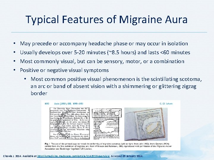
Typical Features of Migraine Aura • • May precede or accompany headache phase or may occur in isolation Usually develops over 5 20 minutes (~8. 5 hours) and lasts <60 minutes Most commonly visual, but can be sensory, motor, or a combination Positive or negative visual symptoms • Most common positive visual phenomenon is the scintillating scotoma, an arc or band of absent vision with a shimmering or glittering zigzag border Chawla J. 2014. Available at http: //emedicine. medscape. com/article/1142556 overview. Accessed 05 January 2014.
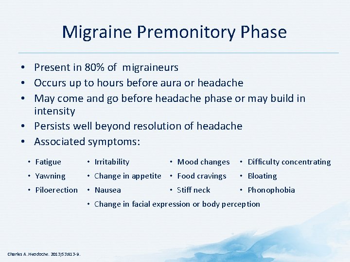
Migraine Premonitory Phase • Present in 80% of migraineurs • Occurs up to hours before aura or headache • May come and go before headache phase or may build in intensity • Persists well beyond resolution of headache • Associated symptoms: • Fatigue • Irritability • Yawning • Change in appetite • Food cravings • Piloerection • Nausea • Mood changes • Stiff neck • Difficulty concentrating • Bloating • Phonophobia • Change in facial expression or body perception Charles A. Headache. 2013; 53: 413 9.
Pathogenesis of the Premonitory Phase of Migraine • Role of dopamine? (yawning, nausea, drowsiness, lightheadedness) • Increased blood flow (shown by PET) • Increased activity of hypothalamus (mood, appetite, energy) • Orexin pathway in hypothalamus PET = positron emission tomography Charles A. Headache. 2013; 53: 413 9.
Postdrome Resolution Phase • Symptoms last for hours to days – Fatigue, weakness, cognitive difficulties, mood change, residual head pain, lightheadedness, gastrointestinal symptoms • Persistence of midbrain, dorsolateral pons, and hypothalamic activation, light induced activation of visual cortex Charles A. Headache. 2013; 53: 413 9.
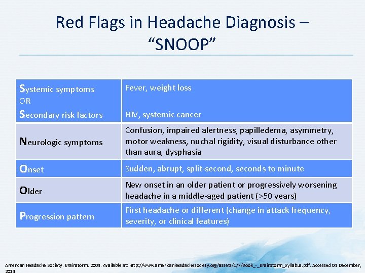
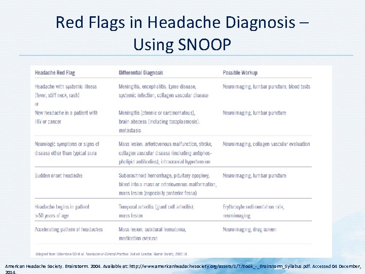
Red Flags in Headache Diagnosis – “SNOOP” Systemic symptoms Fever, weight loss Secondary risk factors HIV, systemic cancer Neurologic symptoms Confusion, impaired alertness, papilledema, asymmetry, motor weakness, nuchal rigidity, visual disturbance other than aura, dysphasia Onset Sudden, abrupt, split second, seconds to minute Older New onset in an older patient or progressively worsening headache in a middle aged patient (>50 years) Progression pattern First headache or different (change in attack frequency, severity, or clinical features) OR American Headache Society. Brainstorm. 2004. Available at: http: //www. americanheadachesociety. org/assets/1/7/Book_ _Brainstorm_Syllabus. pdf. Accessed 04 December,
Red Flags in Headache Diagnosis – Using SNOOP American Headache Society. Brainstorm. 2004. Available at: http: //www. americanheadachesociety. org/assets/1/7/Book_ _Brainstorm_Syllabus. pdf. Accessed 04 December,
Diagnosing Secondary Headaches • Secondary headaches are rare; lifetime prevalence is 0. 5% • Very few (0. 18%) patients with migraine symptoms and normal neurologic exam have significant intracranial pathology • Significantly changed headache should be regarded as a new headache • Abnormal neurologic exam in patients with headache require further evaluation Diagnosis of migraine does not preclude future development of secondary headache American Headache Society. Brainstorm. 2004. Available at: http: //www. americanheadachesociety. org/assets/1/7/Book_ _Brainstorm_Syllabus. pdf. Accessed 04 December,
Secondary Headaches: Warning Signs Warning signs in patients with no history of headaches or with changed headaches and a significantly different pattern: • Abrupt onset of a new type of severe headache • Is the worst headache the patient has ever had • Progressive worsening over of days or weeks • Headache is brought on by exertion (e. g. , coughing, sneezing, bending over, exercise, sexual arousal) • Headache is accompanied by generalized illness or fever, nausea, vomiting, or stiff neck American Headache Society. Brainstorm. 2004. Available at: http: //www. americanheadachesociety. org/assets/1/7/Book_ _Brainstorm_Syllabus. pdf. Accessed 04 December,
Secondary Headache: Diagnostic Testing • Lab testing is not routinely needed to evaluate headache • Patients with warning symptoms or signs of secondary headache must be evaluated • May be useful to establish a baseline lab screen before new drugs are prescribed • Cardiovascular factors should be reviewed before prescribing vasoconstrictors • Consider electrocardiogram on a case by case basis Electroencephalogram is not useful in headache evaluation American Headache Society. Brainstorm. 2004. Available at: http: //www. americanheadachesociety. org/assets/1/7/Book_ _Brainstorm_Syllabus. pdf. Accessed 04 December,
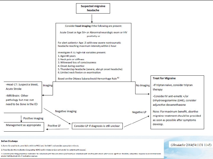
Secondary Headache: Neuroimaging • Abnormal findings on neurologic exam • No focal neurologic findings • Patient has progressively worsening headache • Patient has stable pattern of recurrent headache • Patient has new persistent headache • No history of seizures • Patient has a new, rapid onset headache (thunderclap headache)* • Headache does not respond to standard therapy MRI is more sensitive than CT for most CNS abnormalities *Sudden onset of a severe headache that reaches peak intensity in <1 minute CNS = central nervous system; CT = computed tomography; MRI = magnetic resonance imaging American Headache Society. Brainstorm. 2004. Available at: http: //www. americanheadachesociety. org/assets/1/7/Book_ _Brainstorm_Syllabus. pdf. Accessed 04 December,
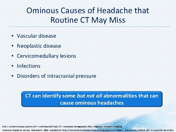
Ominous Causes of Headache that Routine CT May Miss • Vascular disease • Neoplastic disease • Cervicomedullary lesions • Infections • Disorders of intracranial pressure CT can identify some but not all abnormalities that can cause ominous headaches CNS = central nervous system; CSF = cerebrospinal fluid; CT = computed tomography; MRI = magnetic resonance imaging American Headache Society. Brainstorm. 2004. Available at: http: //www. americanheadachesociety. org/assets/1/7/Book_ _Brainstorm_Syllabus. pdf. Accessed 04 December,
Lab Testing for Migraine Investigate the atypical and red flags Lab testing is not routinely needed in the evaluation of a patient with headache Evans RE, Rozen TD, Adelman JU. Wolff’s Headache And Other Head Pain. 7 th ed. New York: Oxford University Press; 2001: 27 49. Campbell JK, Sakai F. The Headaches. 2 nd ed. Philadelphia, PA: Lippincott Williams & Wilkins; 2000: 359 63.
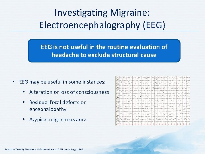
Investigating Migraine: Electroencephalography (EEG) EEG is not useful in the routine evaluation of headache to exclude structural cause • EEG may be useful in some instances: • Alteration or loss of consciousness • Residual focal defects or encephalopathy • Atypical migrainous aura Report of Quality Standards Subcommittee of AAN. Neurology. 1995.
Imaging for Migraine
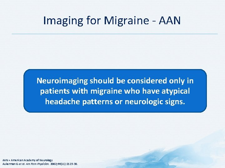
Imaging for Migraine AAN Neuroimaging should be considered only in patients with migraine who have atypical headache patterns or neurologic signs. AAN = American Academy of Neurology Aukerman G et al. Am Fam Physician. 2002; 66(11): 2123 30.
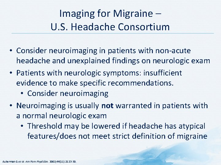
Imaging for Migraine – U. S. Headache Consortium • Consider neuroimaging in patients with non acute headache and unexplained findings on neurologic exam • Patients with neurologic symptoms: insufficient evidence to make specific recommendations. • Consider neuroimaging • Neuroimaging is usually not warranted in patients with a normal neurologic exam • Threshold may be lowered if headache has atypical features/does not meet strict definition of migraine Aukerman G et al. Am Fam Physician. 2002; 66(11): 2123 30.
37
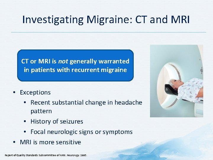
Investigating Migraine: CT and MRI CT or MRI is not generally warranted in patients with recurrent migraine • Exceptions • Recent substantial change in headache pattern • History of seizures • Focal neurologic signs or symptoms • MRI is more sensitive Report of Quality Standards Subcommittee of AAN. Neurology. 1995.
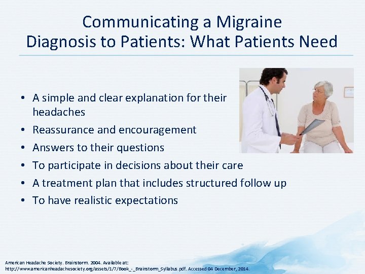
Communicating a Migraine Diagnosis to Patients: What Patients Need • A simple and clear explanation for their headaches • Reassurance and encouragement • Answers to their questions • To participate in decisions about their care • A treatment plan that includes structured follow up • To have realistic expectations American Headache Society. Brainstorm. 2004. Available at: http: //www. americanheadachesociety. org/assets/1/7/Book_ _Brainstorm_Syllabus. pdf. Accessed 04 December, 2014.
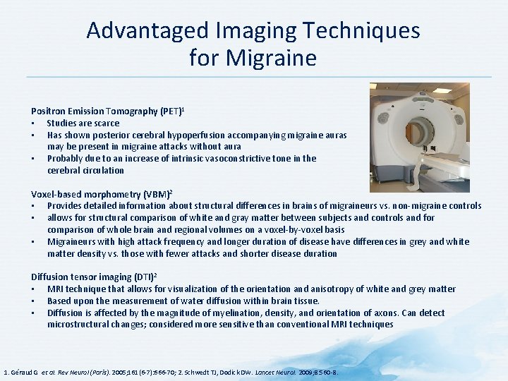
Advantaged Imaging Techniques for Migraine Positron Emission Tomography (PET)1 • Studies are scarce • Has shown posterior cerebral hypoperfusion accompanying migraine auras may be present in migraine attacks without aura • Probably due to an increase of intrinsic vasoconstrictive tone in the cerebral circulation Voxel-based morphometry (VBM)2 • Provides detailed information about structural differences in brains of migraineurs vs. non migraine controls • allows for structural comparison of white and gray matter between subjects and controls and for comparison of whole brain and regional volumes on a voxel by voxel basis • Migraineurs with high attack frequency and longer duration of disease have differences in grey and white matter density vs. those with fewer attacks and shorter disease duration Diffusion tensor imaging (DTI)2 • MRI technique that allows for visualization of the orientation and anisotropy of white and grey matter • Based upon the measurement of water diffusion within brain tissue. • Diffusion is affected by the magnitude of myelination, density, and orientation of axons. Can detect microstructural changes; considered more sensitive than conventional MRI techniques 1. Géraud G et al. Rev Neurol (Paris). 2005; 161(6 7): 666 70; 2. Schwedt TJ, Dodick DW. Lancet Neurol. 2009; 8: 560 8.
PET brainstem activation during acute migraine attack 41
International Headache Society Diagnostic Criteria
Diagnosis and Treatment of Headache – ICSI Main Algorithm ICSI = Institute for Clinical Systems Improvement Beithon J, Gallenberg M, Johnson K, Kildahl P, Krenik J, Liebow M, Linbo L, Myers C, Peterson S, Schmidt J, Swanson J. Institute for Clinical Systems Improvement. Diagnosis and Treatment of Headache. https: //www. icsi. org/_asset/qwrznq/Headache. pdf. Updated January 2013.
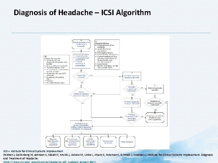
Diagnosis of Headache – ICSI Algorithm ICSI = Institute for Clinical Systems Improvement Beithon J, Gallenberg M, Johnson K, Kildahl P, Krenik J, Liebow M, Linbo L, Myers C, Peterson S, Schmidt J, Swanson J. Institute for Clinical Systems Improvement. Diagnosis and Treatment of Headache.
IHS Diagnostic Criteria for Cluster Headache A. At least five attacks fulfilling criteria B to D B. Severe or very severe unilateral orbital, supraorbital and/or temporal pain lasting 15 180 minutes when untreated C. Either or both of the following: 1. At least one of the following symptoms or signs a. Conjunctival injection and/or lacrimation b. Nasal congestion and/or rhinorrhea c. Eyelid edema d. Forehead and facial sweating e. Sensation of fullness in the ear f. Miosis and/or ptosis 2. A Sense of restlessness or agitation D. Attacks have a frequency between one every other day and eight per day for more than half of the time when the disorder is active E. Not better accounted for by another ICHD 3 diagnosis Link to IHS Diagnosis of Cluster Headache IHS = International Headache Society Headache Classification Committee of the International Headache Society (IHS). Cephalalgia. 2013; 33(9): 629 808.
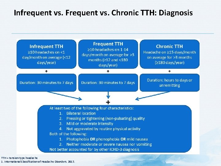
Infrequent vs. Frequent vs. Chronic TTH: Diagnosis TTH = tension type headache 1. International Classification of Headache Disorders. 2013.
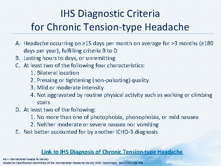
IHS Diagnostic Criteria for Chronic Tension type Headache A. Headache occurring on ≥ 15 days per month on average for >3 months (≥ 180 days per year), fulfilling criteria B to D B. Lasting hours to days, or unremitting C. At least two of the following four characteristics: 1. Bilateral location 2. Pressing or tightening (non pulsating) quality 3. Mild or moderate intensity 4. Not aggravated by routine physical activity such as walking or climbing stairs D. At least two of the following: 1. No more than one of photophobia, phonophobia, or mild nausea 2. Neither moderate or severe nausea nor vomiting E. Not better accounted for by another ICHD 3 diagnosis Link to IHS Diagnosis of Chronic Tension-type Headache IHS = International Headache Society Headache Classification Committee of the International Headache Society (IHS). Cephalalgia. 2013; 33(9): 629 808.
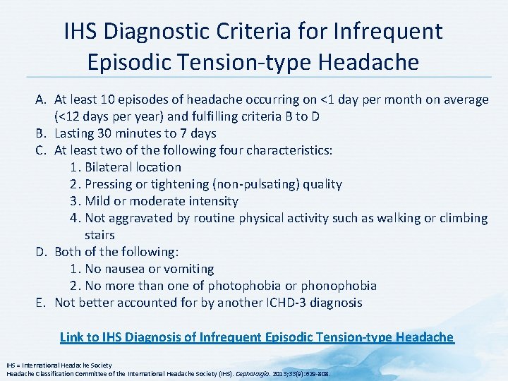
IHS Diagnostic Criteria for Infrequent Episodic Tension type Headache A. At least 10 episodes of headache occurring on <1 day per month on average (<12 days per year) and fulfilling criteria B to D B. Lasting 30 minutes to 7 days C. At least two of the following four characteristics: 1. Bilateral location 2. Pressing or tightening (non pulsating) quality 3. Mild or moderate intensity 4. Not aggravated by routine physical activity such as walking or climbing stairs D. Both of the following: 1. No nausea or vomiting 2. No more than one of photophobia or phonophobia E. Not better accounted for by another ICHD 3 diagnosis Link to IHS Diagnosis of Infrequent Episodic Tension-type Headache IHS = International Headache Society Headache Classification Committee of the International Headache Society (IHS). Cephalalgia. 2013; 33(9): 629 808.
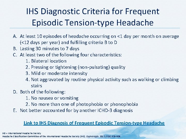
IHS Diagnostic Criteria for Frequent Episodic Tension type Headache A. At least 10 episodes of headache occurring on <1 day per month on average (<12 days per year) and fulfilling criteria B to D B. Lasting 30 minutes to 7 days C. At least two of the following four characteristics: 1. Bilateral location 2. Pressing or tightening (non pulsating) quality 3. Mild or moderate intensity 4. Not aggravated by routine physical activity such as walking or climbing stairs D. Both of the following: 1. No nausea or vomiting 2. No more than one of photophobia or phonophobia E. Not better accounted for by another ICHD 3 diagnosis Link to IHS Diagnosis of Frequent Episodic Tension-type Headache IHS = International Headache Society Headache Classification Committee of the International Headache Society (IHS). Cephalalgia. 2013; 33(9): 629 808.
IHS Diagnostic Criteria for Menstrual Migraine A. Attacks, in a menstruating woman, fulfilling criteria for migraine without aura B. Attacks occur exclusively on day 1+2 (i. e. , days 2 to +3)1 of menstruation in at least two out of three menstrual cycles and at no other times in the cycle Link to IHS Diagnosis of Menstrual Migraine 1 The first day of menstruation is day 1 and the preceding day is 1; there is no day 0 IHS = International Headache Society Headache Classification Committee of the International Headache Society (IHS). Cephalalgia. 2013; 33(9): 629 808.
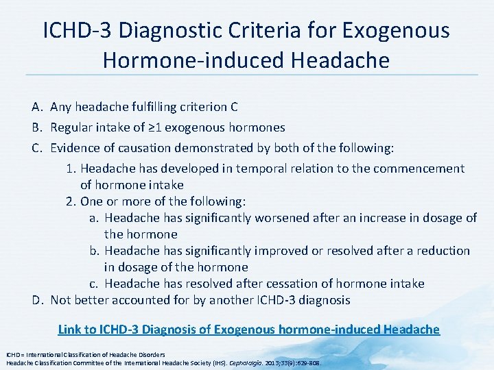
ICHD 3 Diagnostic Criteria for Exogenous Hormone induced Headache A. Any headache fulfilling criterion C B. Regular intake of ≥ 1 exogenous hormones C. Evidence of causation demonstrated by both of the following: 1. Headache has developed in temporal relation to the commencement of hormone intake 2. One or more of the following: a. Headache has significantly worsened after an increase in dosage of the hormone b. Headache has significantly improved or resolved after a reduction in dosage of the hormone c. Headache has resolved after cessation of hormone intake D. Not better accounted for by another ICHD 3 diagnosis Link to ICHD-3 Diagnosis of Exogenous hormone-induced Headache ICHD = International Classification of Headache Disorders Headache Classification Committee of the International Headache Society (IHS). Cephalalgia. 2013; 33(9): 629 808.
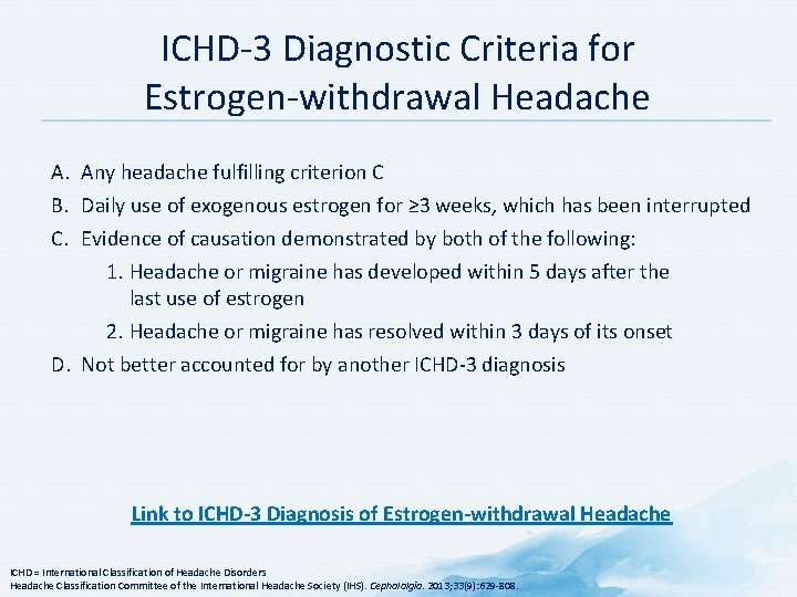
ICHD 3 Diagnostic Criteria for Estrogen withdrawal Headache A. Any headache fulfilling criterion C B. Daily use of exogenous estrogen for ≥ 3 weeks, which has been interrupted C. Evidence of causation demonstrated by both of the following: 1. Headache or migraine has developed within 5 days after the last use of estrogen 2. Headache or migraine has resolved within 3 days of its onset D. Not better accounted for by another ICHD 3 diagnosis Link to ICHD-3 Diagnosis of Estrogen-withdrawal Headache ICHD = International Classification of Headache Disorders Headache Classification Committee of the International Headache Society (IHS). Cephalalgia. 2013; 33(9): 629 808.
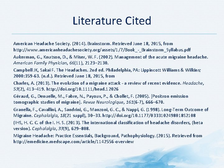
Literature Cited American Headache Society. (2014). Brainstorm. Retrieved June 18, 2015, from http: //www. americanheadachesociety. org/assets/1/7/Book_ _Brainstorm_Syllabus. pdf Aukerman, G. , Knutson, D. , & Miser, W. F. (2002). Management of the acute migraine headache. American Family Physician, 66(11), 2123– 2130. Campbell JK, Sakai F. The Headaches. 2 nd ed. Philadelphia, PA: Lippincott Williams & Wilkins; 2000: 359 63. (n. d. ). Retrieved June 18, 2015, from Charles, A. (2013). The evolution of a migraine attack a review of recent evidence. Headache, 53(2), 413– 419. http: //doi. org/10. 1111/head. 12026 Géraud, G. , Denuelle, M. , Fabre, N. , Payoux, P. , & Chollet, F. (2005). [Positron emission tomographic studies of migraine]. Revue Neurologique, 161(6 7), 666– 670. Granella, F. , Cavallini, A. , Sandrini, G. , Manzoni, G. C. , & Nappi, G. (1998). Long Term Outcome of Migraine. Cephalalgia, 18(21 suppl), 30– 33. http: //doi. org/10. 1177/0333102498018 S 2108 (IHS, H. C. C. of the I. H. S. (2013). The international classification of headache disorders, (beta version). Cephalalgia, 33(9), 629– 808. Migraine Headache: Practice Essentials, Background, Pathophysiology. (2015). Retrieved from http: //emedicine. medscape. com/article/1142556 overview
Literature Cited (cont) Minen, M. T. , Tanev, K. , & Friedman, B. W. (2014). Evaluation and Treatment of Migraine in the Emergency Department: A Review. Headache: The Journal of Head and Face Pain, 54(7), 1131– 1145. http: //doi. org/10. 1111/head. 12399 Neurology, A. A. of. (1995). Practice parameter: The electroencephalogram in the evaluation of headache (summary statement). Report of the Quality Standards Subcommittee of the American Academy of Neurology, 45(7), 1411– 1413. Pub. Med entry. (n. d. ). Retrieved from http: //www. ncbi. nlm. nih. gov/pubmed/16141953 Sanin, L. C. , Mathew, N. T. , Bellmeyer, L. R. , & Ali, S. (1994). The International Headache Society (IHS) headache classification as applied to a headache clinic population. Cephalalgia: An International Journal of Headache, 14(6), 443– 446. Saper, J. (n. d. ). Handbook of Headache Management 2 nd Edition. Saper, J. R. (1983). Headache disorders: current concepts and treatment strategies. J. Wright Psg Incorporated. Schwedt, T. J. , & Dodick, D. W. (2009). Advanced Neuroimaging of Migraine. Lancet Neurology, 8(6), 560– 568. http: //doi. org/10. 1016/S 1474 4422(09)70107 3