Urinary System Prefinals Urinary System Is the production







- Slides: 55
Urinary System Prefinals
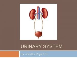
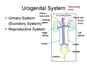
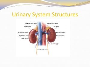
Urinary System �Is the production of urine and its elimination from the body. �Functions: �Remove nitrogenous wastes �Regulate water levels in the body. �Regulate acid-base balance and electrolyte levels of the blood.
Procedures: �Urography �Ivu or excretory urography �Hypertensive IVU �Percutaneous renal puncture �Retrograde urography �Retrograde cystography �Voiding cystourethrography �Retrograde urethrography
Urography �General term for radiologic investigations of the renal drainage. Or collecting system are performed by various procedures.
2 – methods routinely employed of filling the urinary canals with contrast medium 1. Excretory, or Intravenous Urography 2. Retrograde Pyelography
Excretory, or Intravenous Urography � most frequently employed method, in which the contrast agent is routinely administered intravenously.
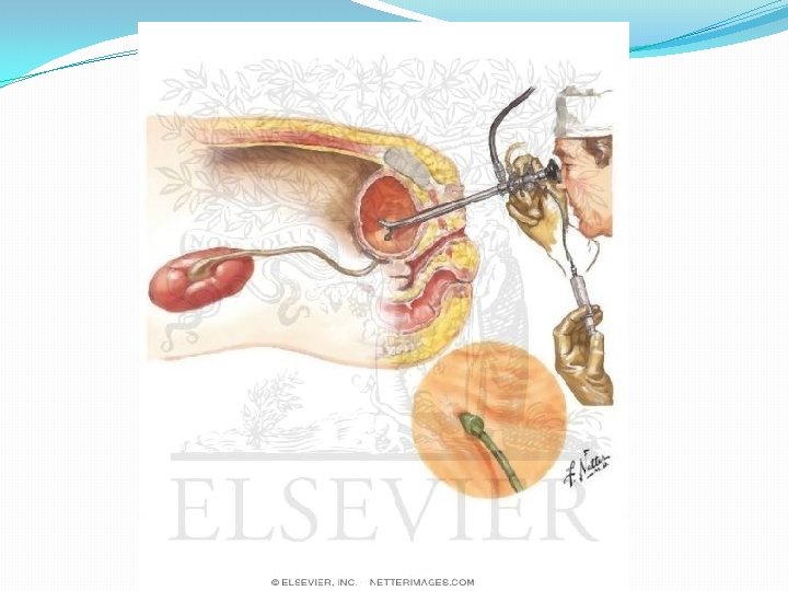
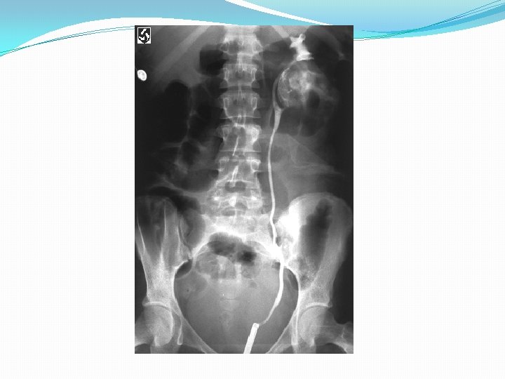
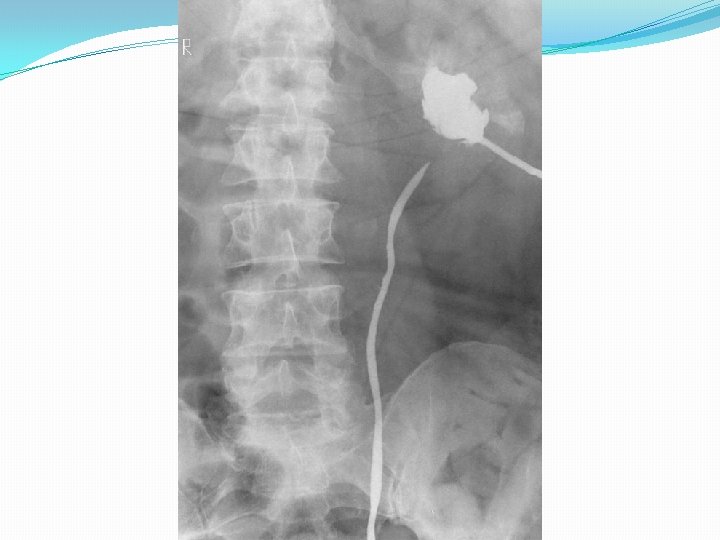
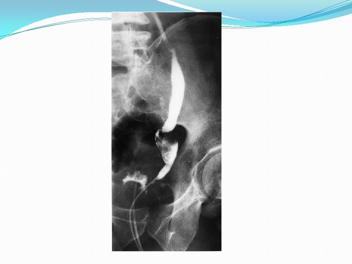
Retrograde Pyelography �contrast medium is introduced directly into the canals by means of catheterization – ureteral catherterization for contrast filling of the upper urinary tracts.
Percutaneous Antegrade Urography �Contrast solution is introduced directly into pelvicalyceal system by means of puncture of the renal pelvis. �It is seldom used method of introducing contrast media to the kidney
Preparation of Patient �General Use: 1. Low-residue diet for 1 – 2 days to prevent gas formation caused by excessive fermentation of the intestinal contents. 2. A light evening meal 3. Costive bowel action, a non-gas forming laxative the evening before the examination. 4. NPO after midnight on the day of examination. 5. For retrograge urography, patient is often requested to force water(4 -5 glassfuls) for severe hours before examination.
IVU or Excretory Urography (commonly known as IVP) �most common radiographic examination of urinary system. �Visualizes the minor and major calyces, renal pelves, ureters and urinary bladder – following an intravenous injection of contrast medium.
Purpose: � Visualize the collecting portion of the U. S. � Assess the functional ability of the kidneys.
Clinical Indications �Abdominal or pelvic mass �Renal or ureteral calculi �Kidney trauma �Flank pain �Hematuria or blood in the urine �Hypertension �Renal failure �Urinary tract infections
Contraindications �Hypersensitivity to contrast media �Anuria (non passage of urine) �multiple myeloma �diabetes �severe hepatic or renal disease �congestive heart failure �Pheochromocytoma (rare tumor that arise outside the adrenal gland) �sickle cell anemia (another type of anemia)
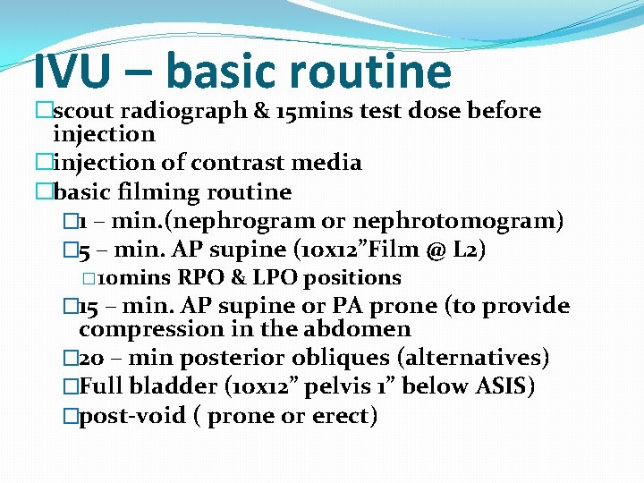
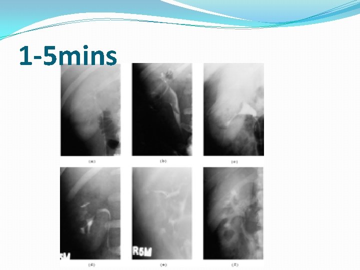
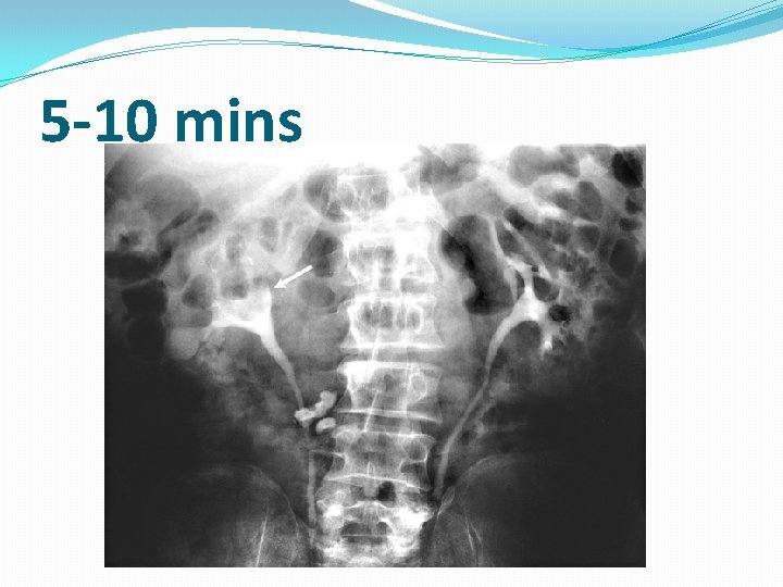
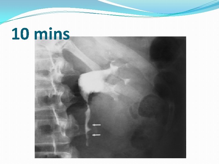
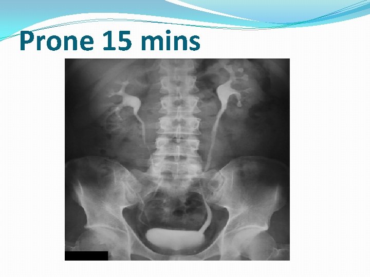
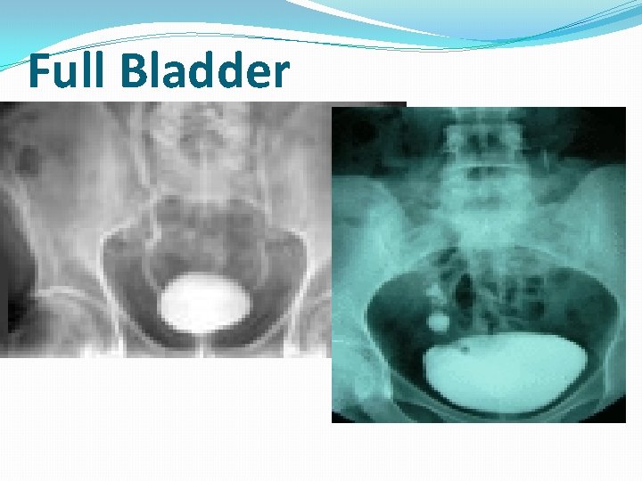
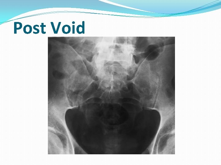
IVU – basic routine �scout radiograph & 15 mins test dose before injection �injection of contrast media �basic filming routine � 1 – min. (nephrogram or nephrotomogram) � 5 – min. AP supine (10 x 12”Film @ L 2) � 10 mins RPO & LPO positions � 15 – min. AP supine or PA prone (to provide compression in the abdomen � 20 – min posterior obliques (alternatives) �Full bladder (10 x 12” pelvis 1” below ASIS) �post-void ( prone or erect)
1 -5 mins
5 -10 mins
10 mins
Prone 15 mins
Full Bladder
Post Void
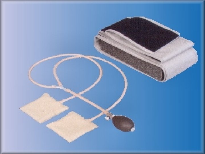
Ureteric Compression �Method utilized to enhance filling of the pelvicalyceal system and proximal ureters. �It allows the renal collecting system to retain the contrast medium longer for more complete study.
Ureteral Compression �The Ureteral Compression Device is used in excretory urography. �The belt fits around the waist of the patient so that he may be repositioned quickly for studies at any angle
Contraindications: �Possible ureteric stones �Abdominal mass �Aortic abdominal aneurysm �Recent abdominal surgery �Severe abdominal pain �Acute abdominal trauma
Hypertensive IVU �one special type of IVU. �This is done on patients with high blood pressure to determine if the kidneys are the caused of the hypertension. �Procedure: (sequence) � 1 min � 2 min � 30 seconds additional radiographs
�A hypertensive IVU was commonly requested to screen for Renovascular Hypertension. �This consisted of 30 -second and 1 -, 2 -, 3 -, and 5 -minute radiographs at the beginning of IVU, which were frequently referred to as “minute-sequence films. ” �The rationale for this study was that physiologic changes caused by the renal arterial stenosis would be demonstrated on early-phase excretion radiographs.
Percutaneous Renal Puncture �Radiologic procedure for the investigation of renal masses. �It is used to differentiate cysts and tumors of the renal parenchyma. �Introduced by Lindblom �It is replaced by the advent of ultrasounography.
Retrograde Urography �Is a nonfunctional radiographic examination of the urinary system which contrast medium is introduced directly into pelvicalyceal system via catheterization.
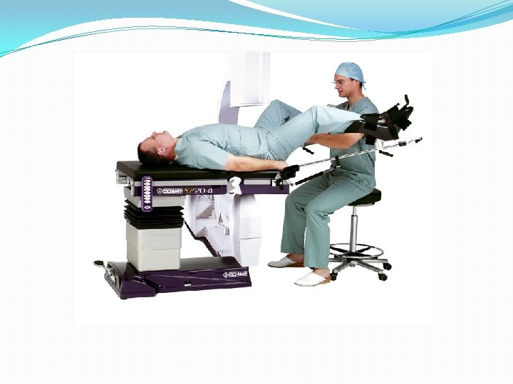
Procedure: � Modified Lithotomy Position � knees are flexed over stirrups of the adjustable leg supports.
* 3 – routine radiographs* � Preliminary radiographs ( showing the ureteral catheters in position) � The pyelogram � The ureterogram
Positioning �Routine �AP for Ureteral Catheters 14 x 17 film �AP for pyelogram 14 x 17 film �AP for Urography film �Additional �RPO �Lateral – for demonstration of anterior displacement of kidneys or ureters �Cross table – for demonstrating of ureteropelvic region with hydronephrosis patients.
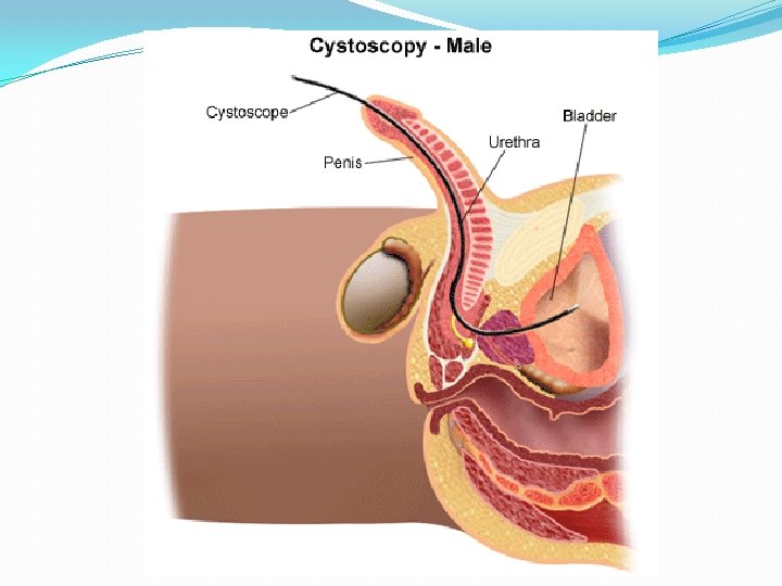
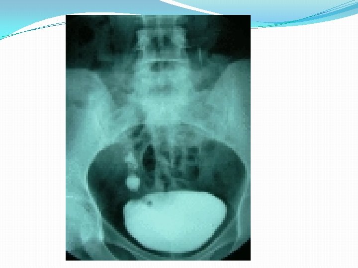
Retrograde Cystography �Another nonfunctional urinary system examination. �Radiographic examination of the urinary bladder following installation of an iodinated contrast medium via a urethral catheter.
Procedure: �contrast material is allowed to flow in by “gravity only” – using an asepto syringe or drip infusion. � 150 – 500 cc’s of contrast media to be instilled into the bladder.
Routine positions: � AP with 15 degrees caudal angulation � Both posterior obliques
Positioning �AP projections of the bladder and proximal part of the urethra with 15 degrees caudal angle. �Oblique projections of 40 -60 degrees, with perpendicular CR, or a 10 degrees angulations if needed. �AP projections with 20 -25 degree cephalic angulation to demonstrate, the shadow of the prostate above that of the pubic bones. �Lateral positions, to demonstrate the anterior and posterior bladder walls and base of the bladder.
�Chassard-Lapine method or “Squat shot”, is used to obtain an axial image of the posterior surface of the bladder and the lower end of the ureters. �AP projections, with a 15 -20 degrees tilt of the table to allow the filled bladders to stretch superiorly, where it will not superimpose the ureters.
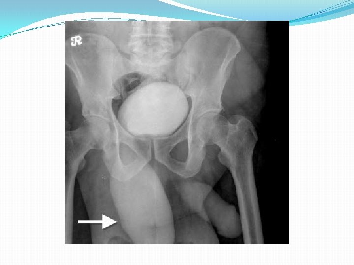
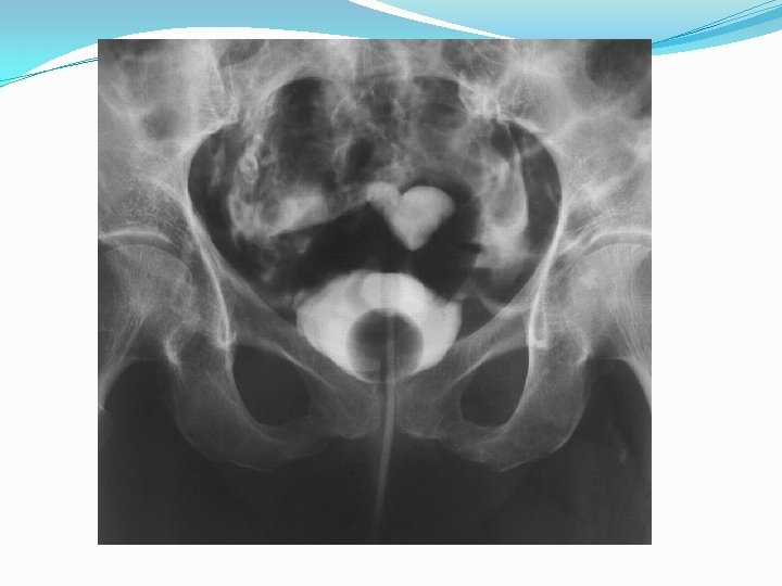
Voiding Cystourethrography �Study of the urethra and evaluate the patients ability to urinate. �Trauma or involuntary loss of urine are common clinical indiactions.
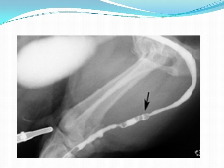
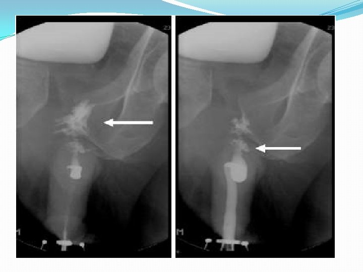
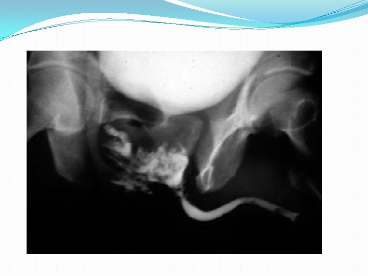
Retrograde Urethrography �Perform on the male patient to demonstrate the full length of the urethra. �Contrast medium is injected into distal urethra until the entire urethra is filled in retrograde fashion.
Procedure: �Brodney clamp – special device for injection of the c+ medium w/c is attached to distal penis. � 30 degs RPO – is the position of choice.
Summary �Urography �Ivu or excretory urography �Hypertensive IVU �Percutaneous renal puncture �Retrograde urography �Retrograde cystography �Voiding cystourethrography �Retrograde urethrography