Three Tasks in SICU Shock resuscitation 2 underlying






- Slides: 86
Three Tasks in SICU Shock ? resuscitation 2 underlying problems ? treatment 3 nutrition ? support 1 (metabolic support vs nutritional support)
Shock or Not?
Shock Inadequate tissue perfusion
1. What is the adequate tissue perfusion? 2. What is the optimal cardiac output for a patient at a specific time? For example: A patient with acute nercotizing pancreatitis?
Definition of Shock is a state of inadequate tissue perfusion 1 decreased tissue perfusion 2 inadequate tissue metabolism
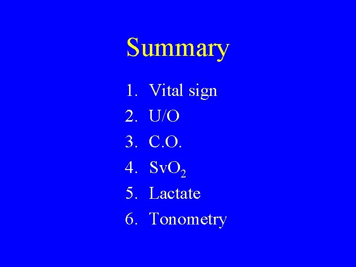
Diagnosis of Shock • • symptom & sign vital sign (BP, HR, RR) ABG urine output cardiac output Sv. O 2 (mixed venous oxygen saturation) serum lactate tonometry
Symptom & Sign Clinical finding: color, temp. , pulse, capillary refilling conscious level mental status: clear even in very low C. O. state but maybe confused in early sepsis
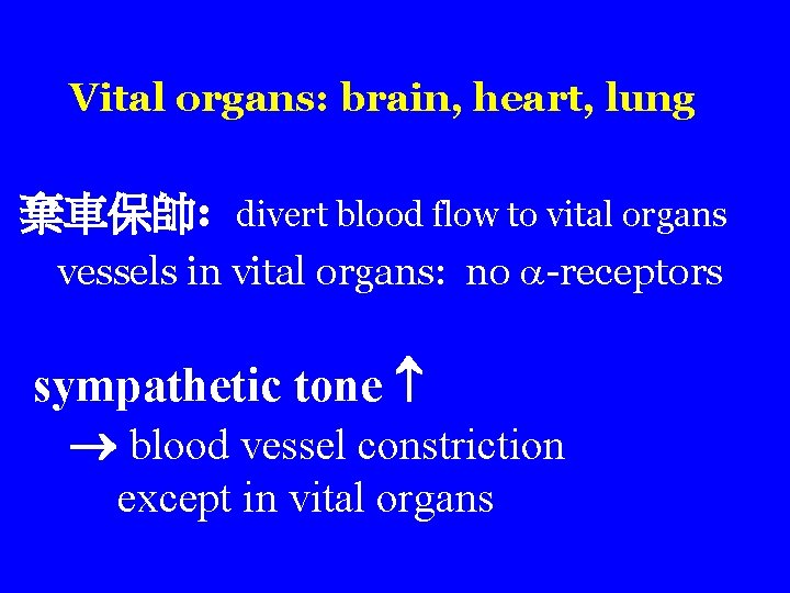
Vital organs: brain, heart, lung 棄車保帥: divert blood flow to vital organs vessels in vital organs: no -receptors sympathetic tone blood vessel constriction except in vital organs
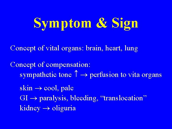
Symptom & Sign Concept of vital organs: brain, heart, lung Concept of compensation: sympathetic tone perfusion to vita organs skin cool, pale GI paralysis, bleeding, “translocation” kidney oliguria
Symptom & sign • Disadvantages: – Too late to be effective – Subjective – Not quantified – Unreliable in modern ICU
Vital Signs HR, BP, RR Disadvantages: • large overlap between normal & abnormal • when obvious (too late)
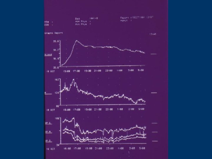
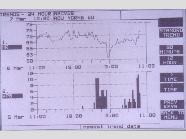
Vital Sign • Advantages: – Quantified, objective number – Automatic, computerized – Continuous --> trend analysis, not single value
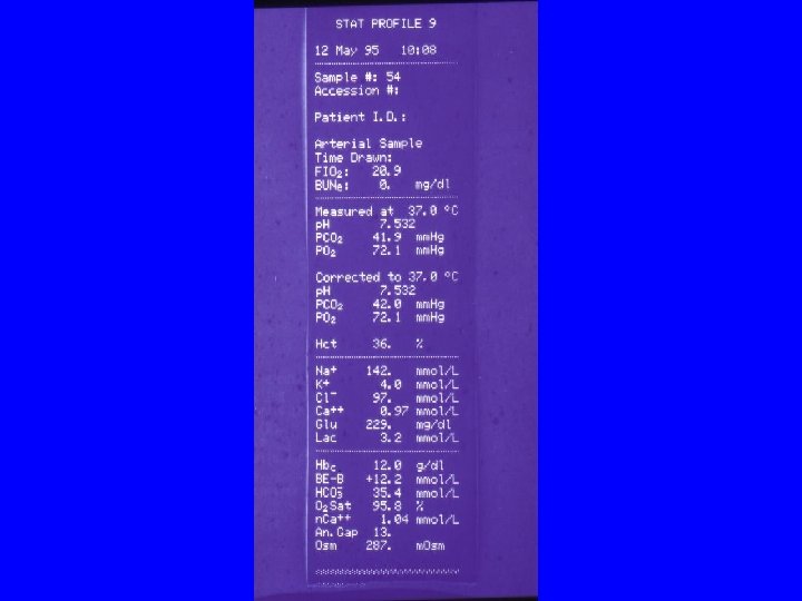
Blood Gas Analysis p. H, BE • intermittent measurement • p. H. : A real vital sign change only in decompensated state
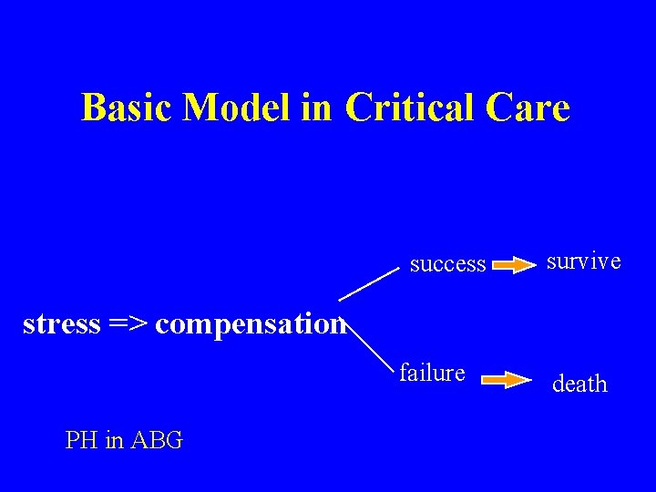
Basic Model in Critical Care success survive stress => compensation failure PH in ABG death
Urine Output • Advantages: – No instrument required (one Forley tube) – Simple and easy – Real time ( rapid reflection of renal perfusion) – Allow trend analysis
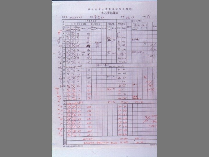
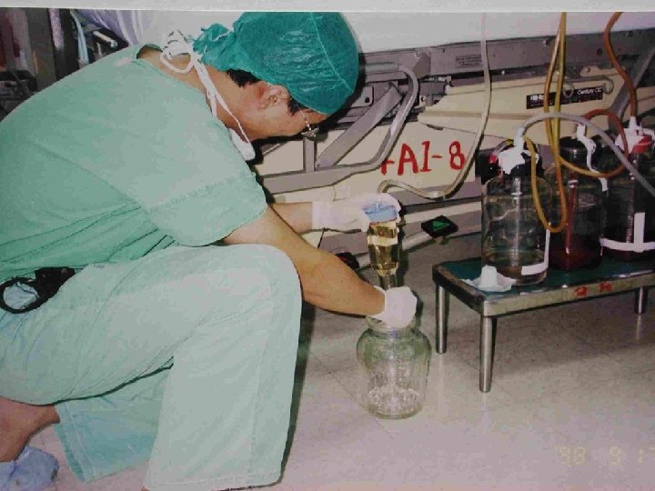
Urine Output NTUH SICU routine: U/O record q 1 h
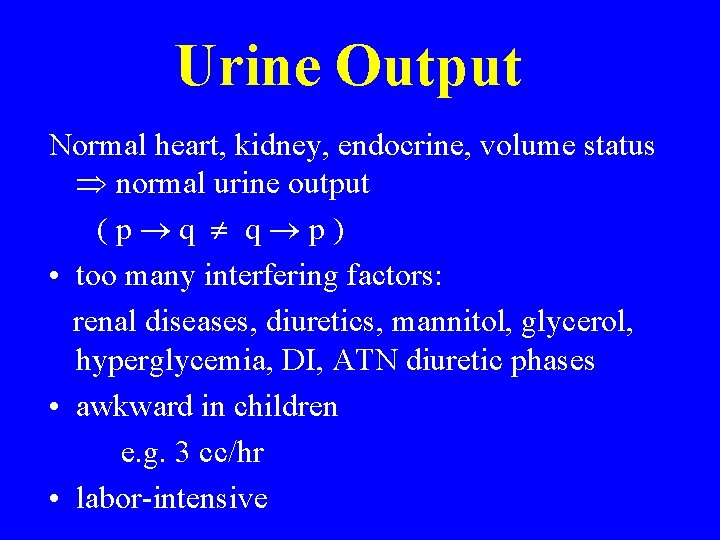
Urine Output Normal heart, kidney, endocrine, volume status normal urine output (p q q p) • too many interfering factors: renal diseases, diuretics, mannitol, glycerol, hyperglycemia, DI, ATN diuretic phases • awkward in children e. g. 3 cc/hr • labor-intensive
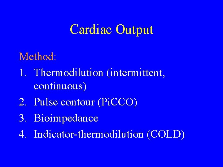
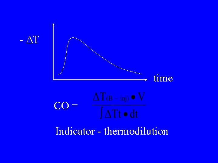
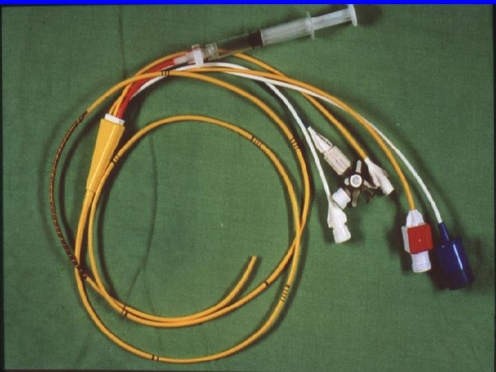
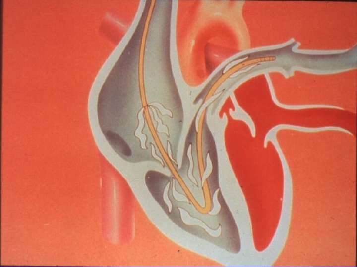
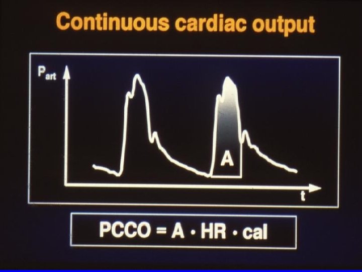
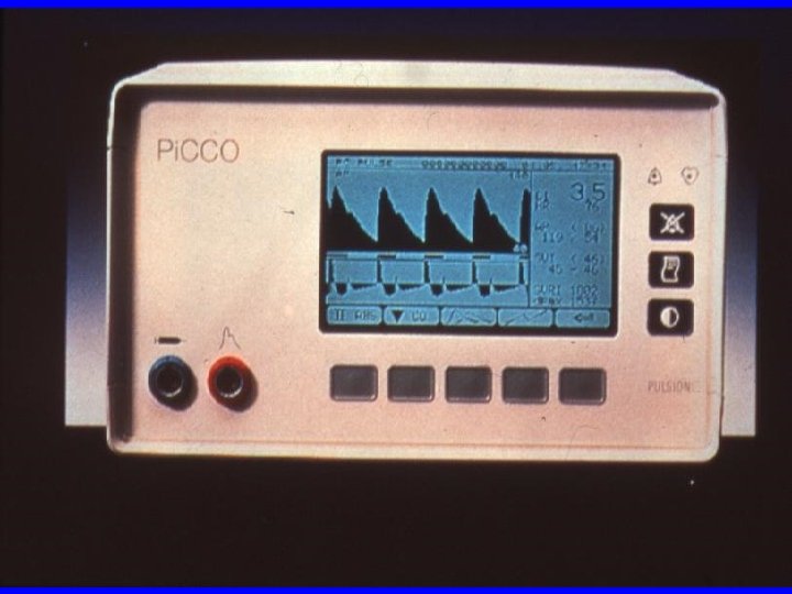
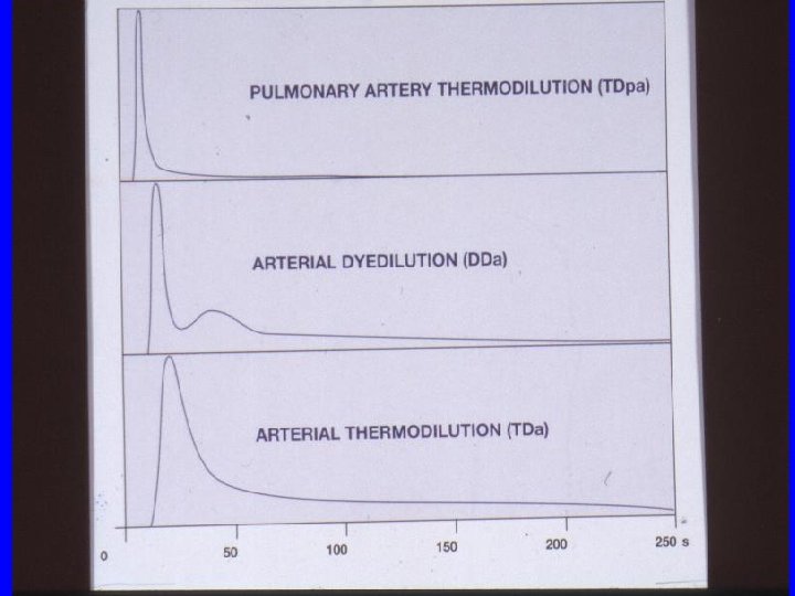
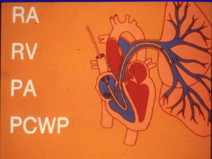
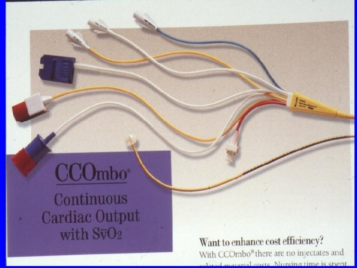
Cardiac Output Method: 1. Thermodilution (intermittent, continuous) 2. Pulse contour (Pi. CCO) 3. Bioimpedance 4. Indicator-thermodilution (COLD)
- T time CO = Indicator - thermodilution
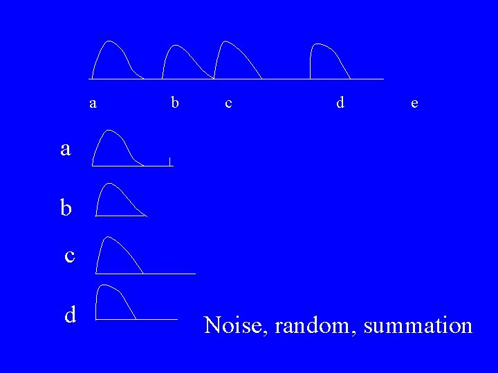
a b c d e a b c d Noise, random, summation
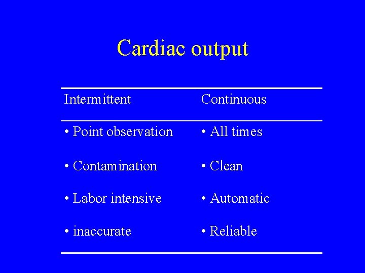
Cardiac output Intermittent Continuous • Point observation • All times • Contamination • Clean • Labor intensive • Automatic • inaccurate • Reliable
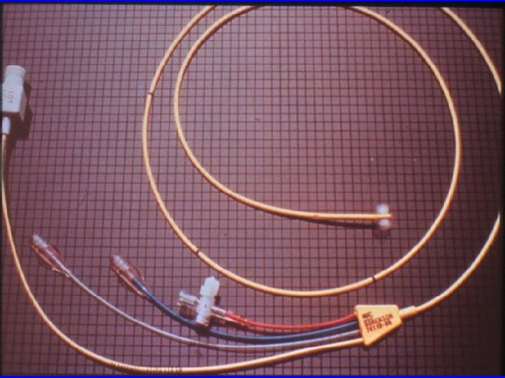
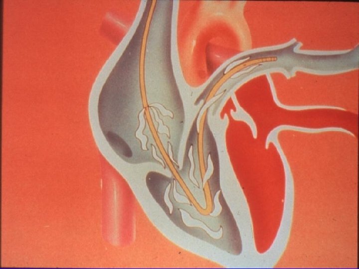
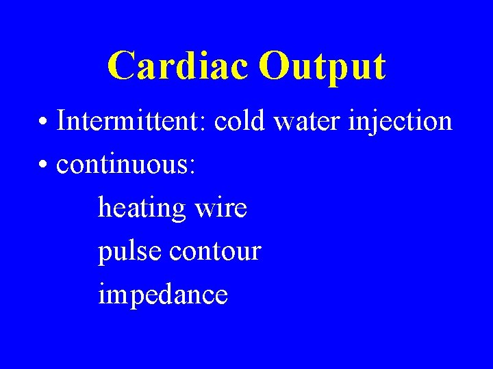
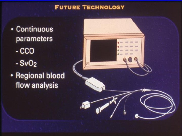
Cardiac Output • Intermittent: cold water injection • continuous: heating wire pulse contour impedance
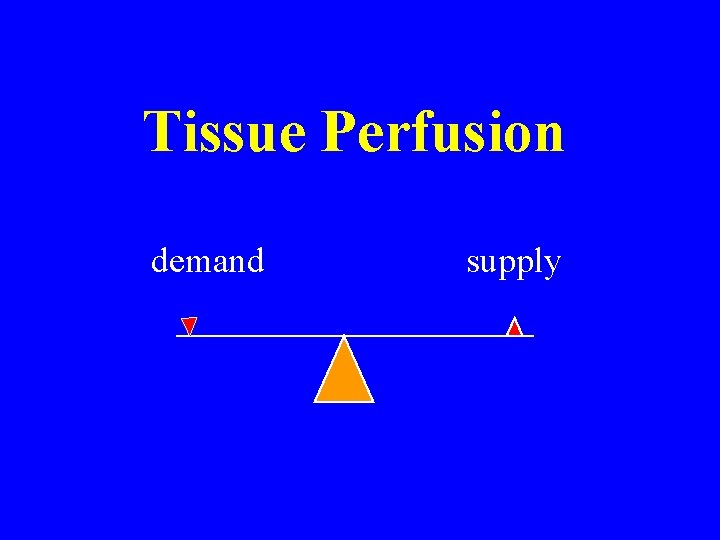
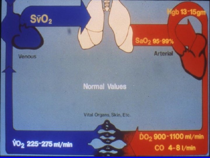
Tissue Perfusion demand supply
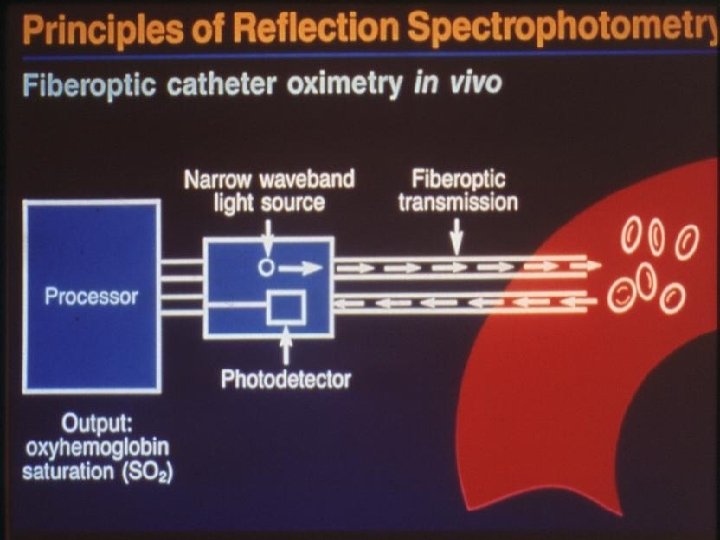
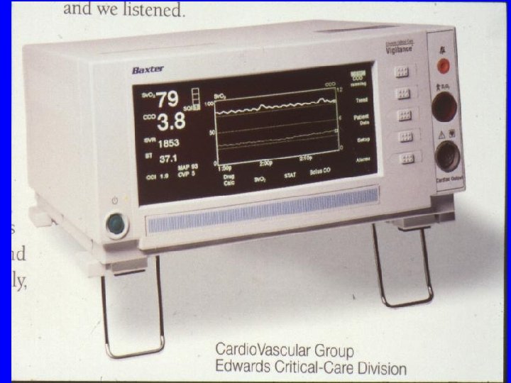
Sv. O 2 (mixed venous oxygen saturation)
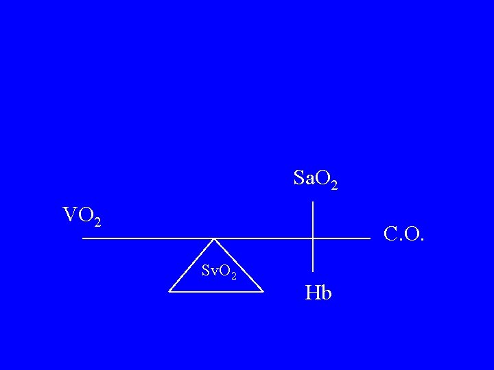
Sa. O 2 VO 2 C. O. Sv. O 2 Hb
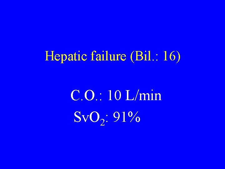
Hepatic failure (Bil. : 16) C. O. : 10 L/min Sv. O 2: 91%
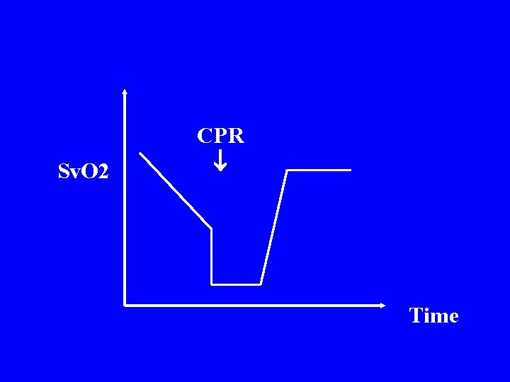
Sv. O 2 CPR Time
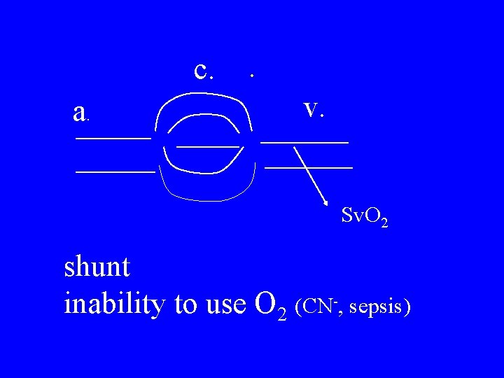
c. a. . v. Sv. O 2 shunt inability to use O 2 (CN-, sepsis)
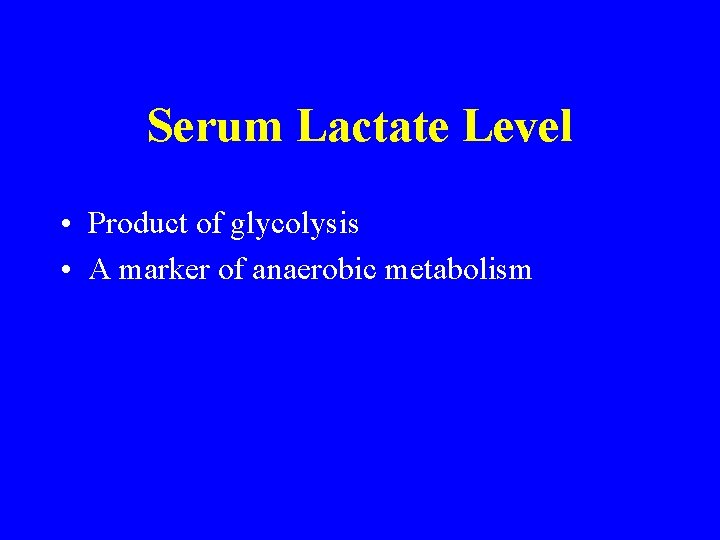
Serum Lactate Level • Product of glycolysis • A marker of anaerobic metabolism
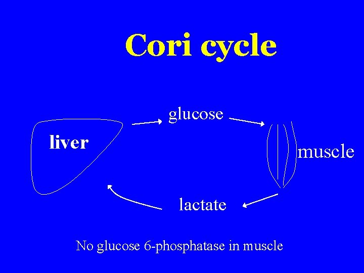
Cori cycle glucose liver muscle lactate No glucose 6 -phosphatase in muscle
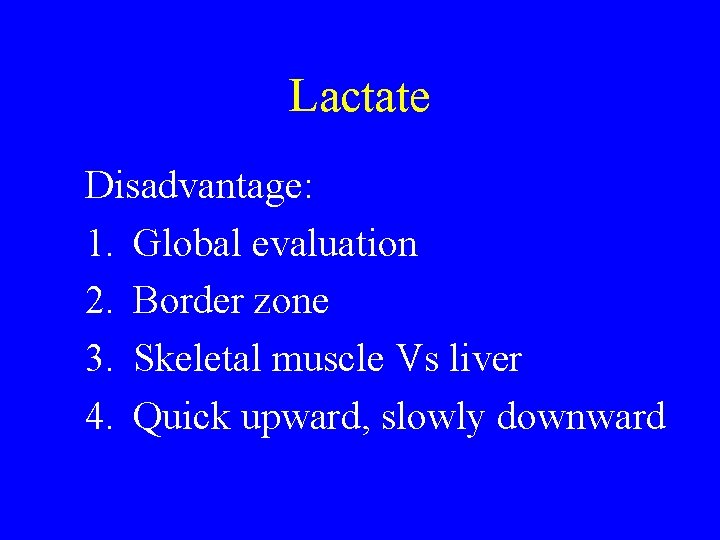
Lactate Disadvantage: 1. Global evaluation 2. Border zone 3. Skeletal muscle Vs liver 4. Quick upward, slowly downward
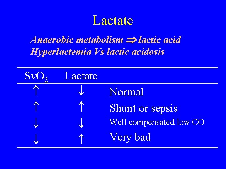
Lactate Anaerobic metabolism lactic acid Hyperlactemia Vs lactic acidosis Sv. O 2 Lactate Normal Shunt or sepsis Well compensated low CO Very bad
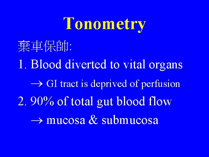
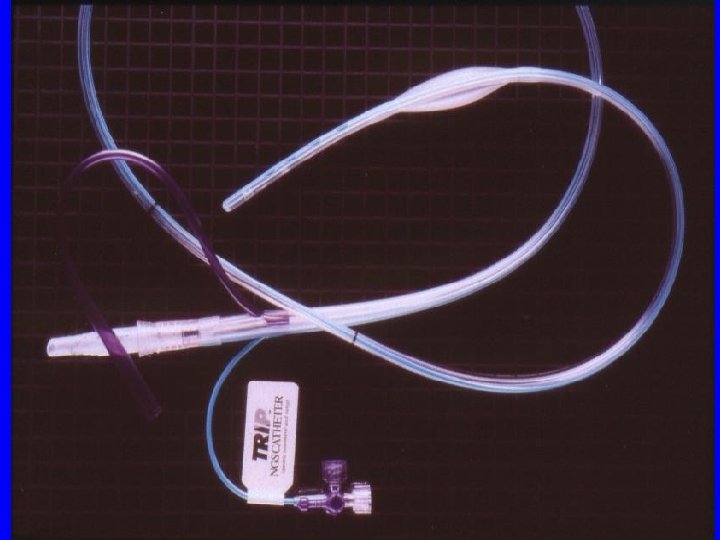
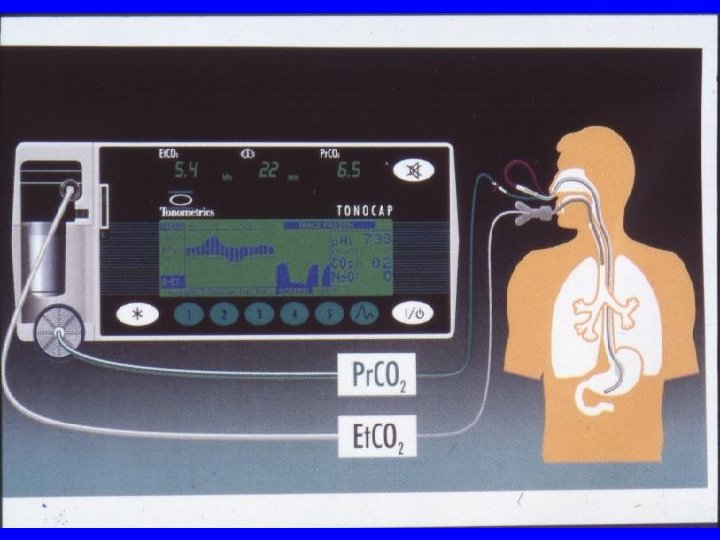
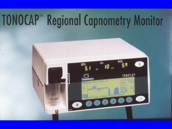
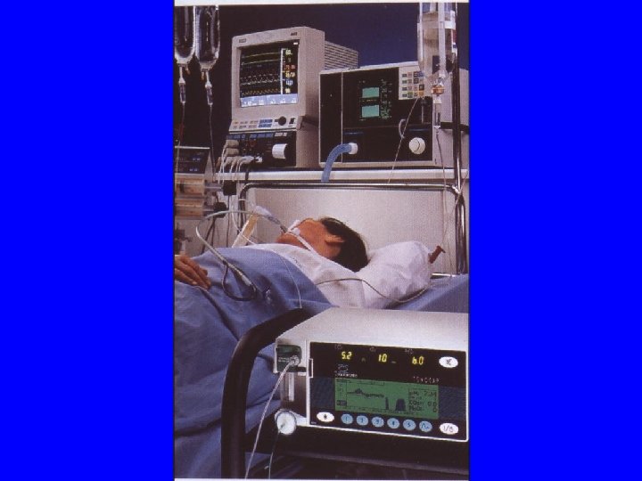
Tonometry 棄車保帥: 1. Blood diverted to vital organs GI tract is deprived of perfusion 2. 90% of total gut blood flow mucosa & submucosa
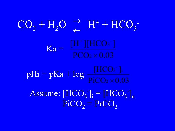
CO 2 + H 2 O H+ + HCO 3 - Ka = p. Hi = p. Ka + log Assume: [HCO 3 -]i = [HCO 3 -]a Pi. CO 2 = Pr. CO 2
Summary 1. 2. 3. 4. 5. 6. Vital sign U/O C. O. Sv. O 2 Lactate Tonometry
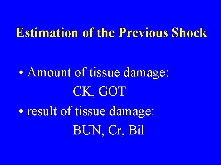
Estimation of the Previous Shock • Amount of tissue damage: CK, GOT • result of tissue damage: BUN, Cr, Bil
Treatment of Shock
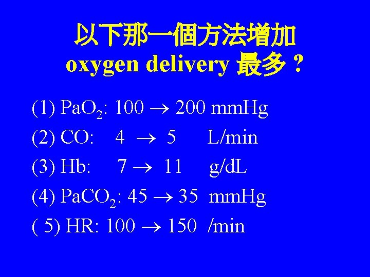
以下那一個方法增加 oxygen delivery 最多 ? (1) Pa. O 2: 100 200 mm. Hg (2) CO: 4 5 L/min (3) Hb: 7 11 g/d. L (4) Pa. CO 2: 45 35 mm. Hg ( 5) HR: 100 150 /min
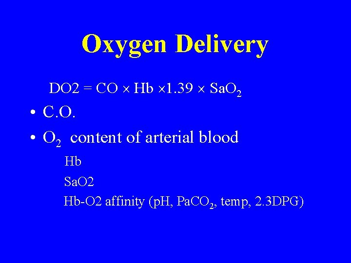
Oxygen Delivery DO 2 = CO Hb 1. 39 Sa. O 2 • C. O. • O 2 content of arterial blood Hb Sa. O 2 Hb-O 2 affinity (p. H, Pa. CO 2, temp, 2. 3 DPG)
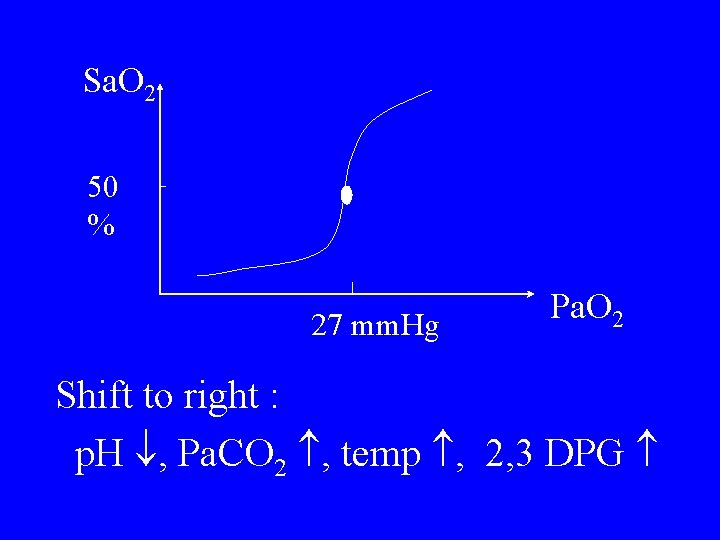
Sa. O 2 50 % 27 mm. Hg Pa. O 2 Shift to right : p. H , Pa. CO 2 , temp , 2, 3 DPG
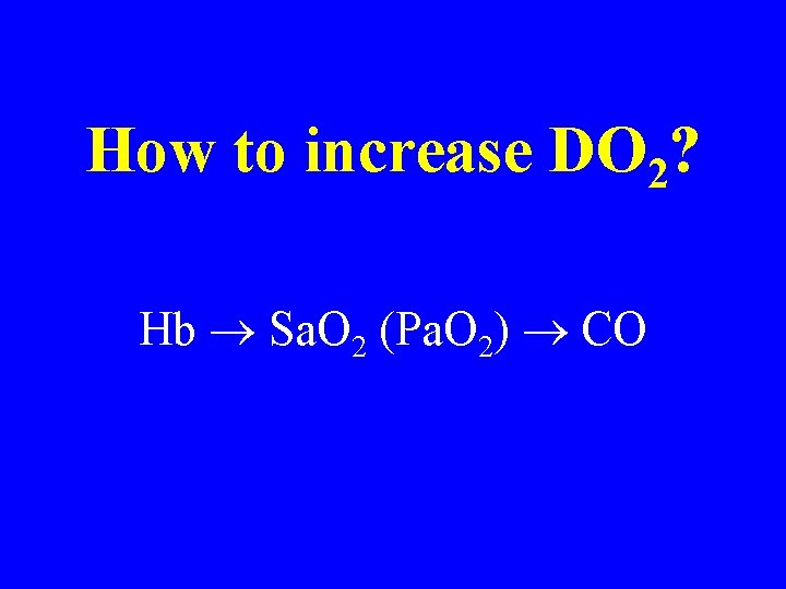
How to increase DO 2? Hb Sa. O 2 (Pa. O 2) CO
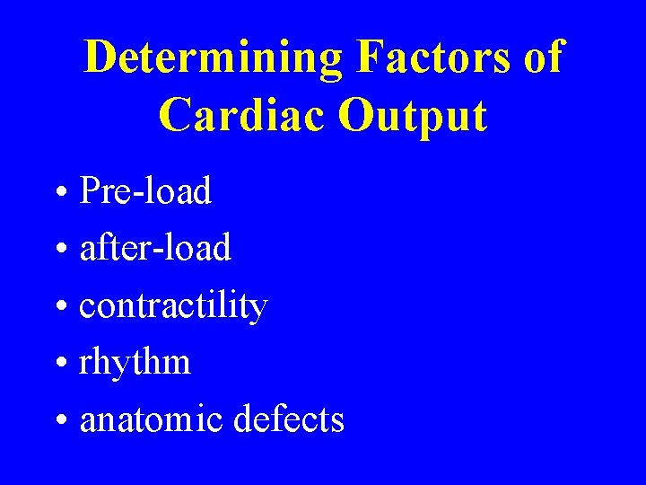
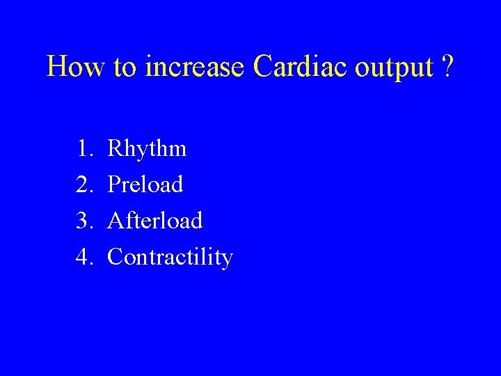
Determining Factors of Cardiac Output • Pre-load • after-load • contractility • rhythm • anatomic defects
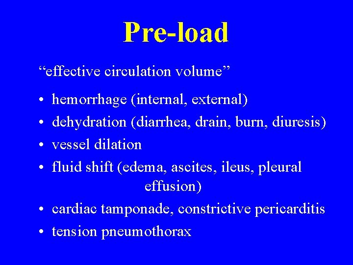
Pre-load “effective circulation volume” • • hemorrhage (internal, external) dehydration (diarrhea, drain, burn, diuresis) vessel dilation fluid shift (edema, ascites, ileus, pleural effusion) • cardiac tamponade, constrictive pericarditis • tension pneumothorax
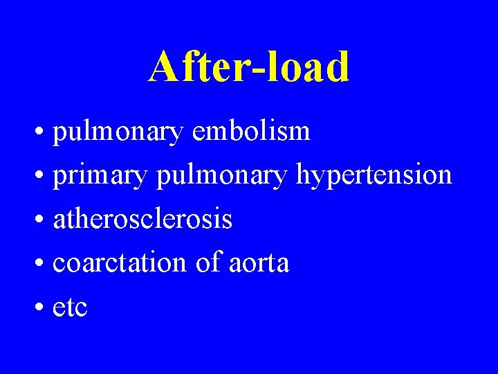
After-load • pulmonary embolism • primary pulmonary hypertension • atherosclerosis • coarctation of aorta • etc
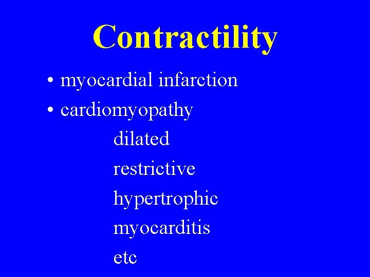
Contractility • myocardial infarction • cardiomyopathy dilated restrictive hypertrophic myocarditis etc
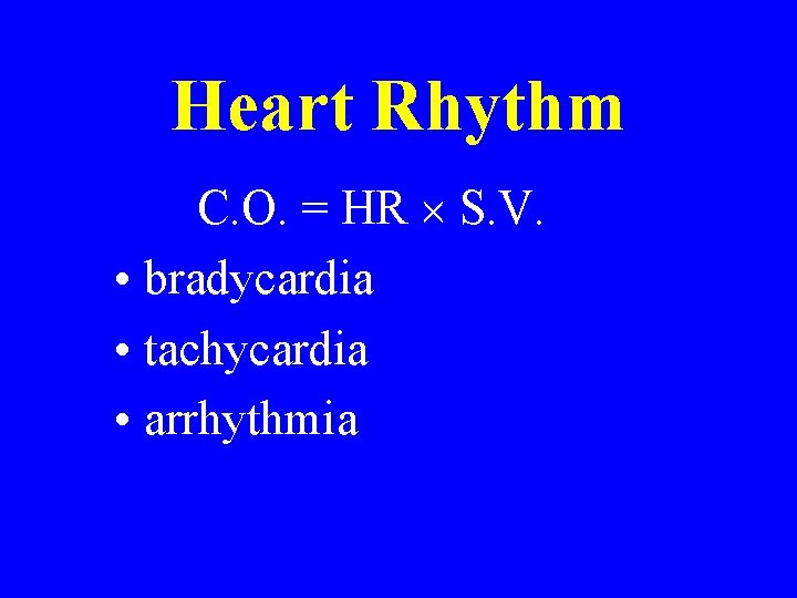
Heart Rhythm C. O. = HR S. V. • bradycardia • tachycardia • arrhythmia
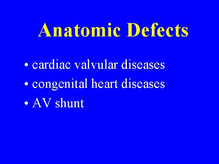
Anatomic Defects • cardiac valvular diseases • congenital heart diseases • AV shunt
How to increase Cardiac output ? 1. 2. 3. 4. Rhythm Preload Afterload Contractility
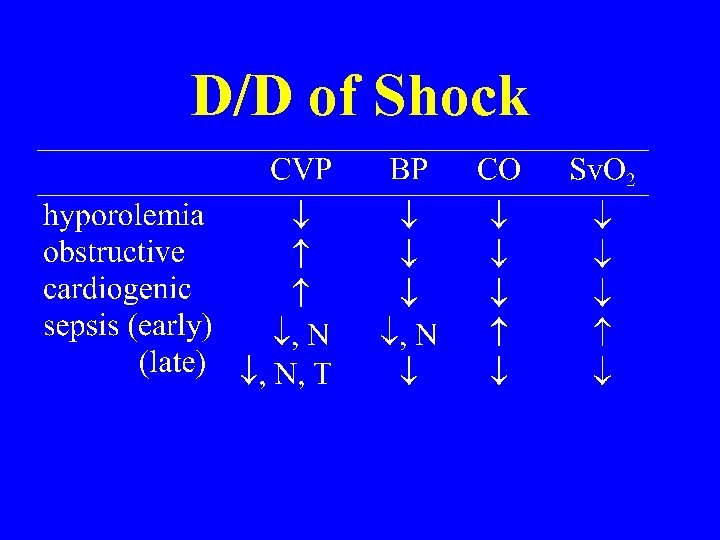
D/D of Shock
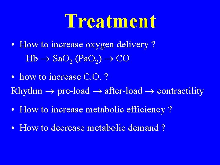
Treatment • How to increase oxygen delivery ? Hb Sa. O 2 (Pa. O 2) CO • how to increase C. O. ? Rhythm pre-load after-load contractility • How to increase metabolic efficiency ? • How to decrease metabolic demand ?
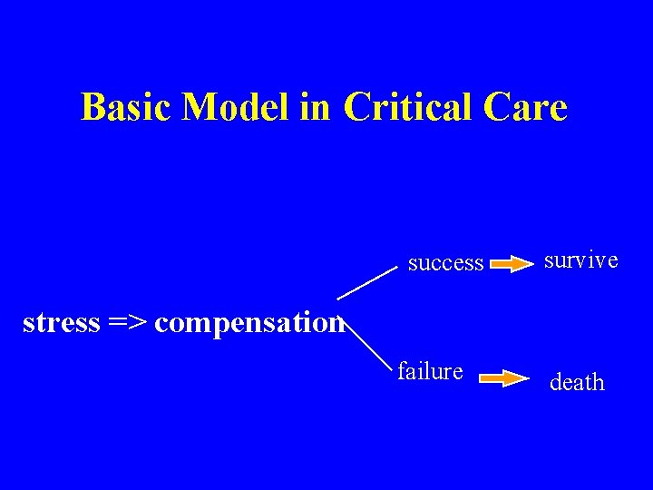
Basic Model in Critical Care success survive stress => compensation failure death