Hypersensitivity diseases Downloaded from Student Consult on 18


- Slides: 55
Hypersensitivity diseases
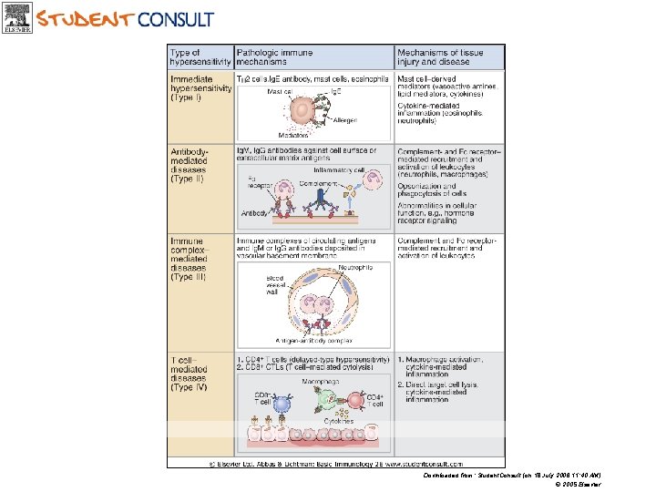
Downloaded from: Student. Consult (on 18 July 2006 11: 40 AM) © 2005 Elsevier
Type-I Hypersensitivity
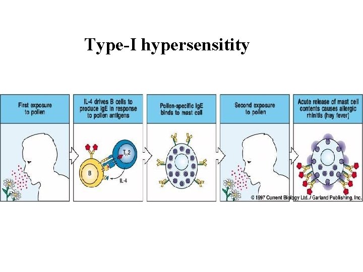
Basic terms • Type-I = Early= Ig. E-mediated = Atopic = Anaphylactic type of hypersensitivity • Atopy = genetic predisposition to type-I hypersensitivity diseases. It is a genetic predisposition to react by Ig. E production to various stimuli.
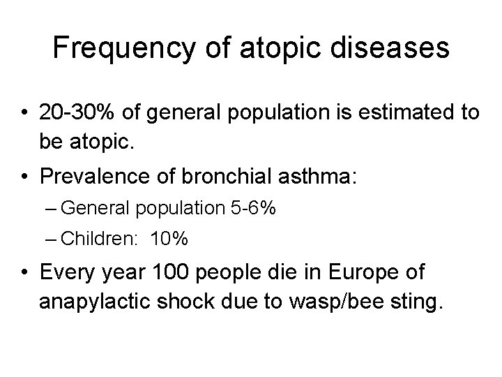
Frequency of atopic diseases • 20 -30% of general population is estimated to be atopic. • Prevalence of bronchial asthma: – General population 5 -6% – Children: 10% • Every year 100 people die in Europe of anapylactic shock due to wasp/bee sting.
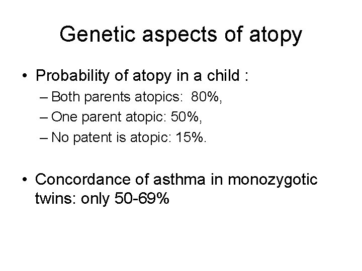
Genetic aspects of atopy • Probability of atopy in a child : – Both parents atopics: 80%, – One parent atopic: 50%, – No patent is atopic: 15%. • Concordance of asthma in monozygotic twins: only 50 -69%
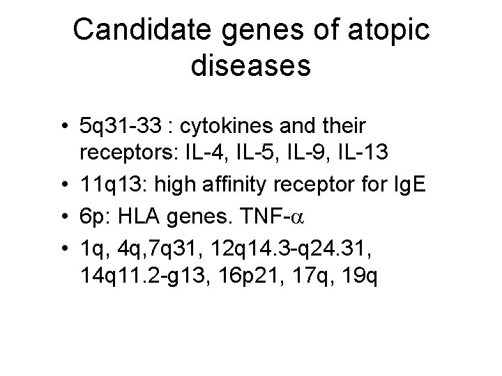
Candidate genes of atopic diseases • 5 q 31 -33 : cytokines and their receptors: IL-4, IL-5, IL-9, IL-13 • 11 q 13: high affinity receptor for Ig. E • 6 p: HLA genes. TNF-a • 1 q, 4 q, 7 q 31, 12 q 14. 3 -q 24. 31, 14 q 11. 2 -g 13, 16 p 21, 17 q, 19 q
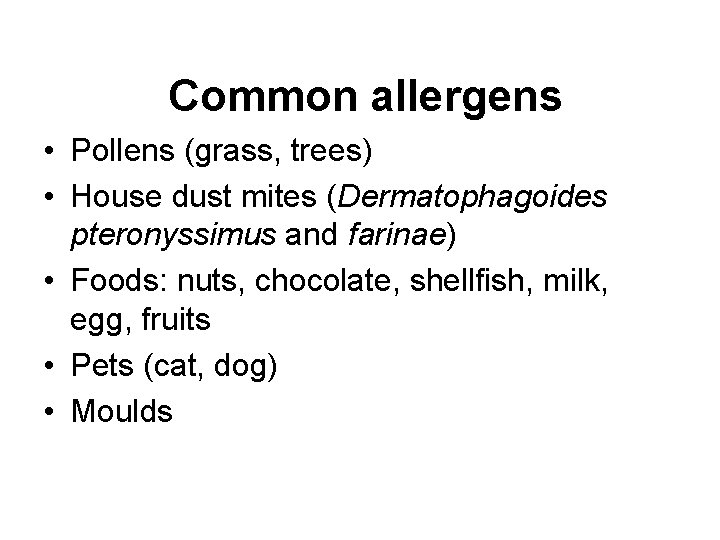
Common allergens • Pollens (grass, trees) • House dust mites (Dermatophagoides pteronyssimus and farinae) • Foods: nuts, chocolate, shellfish, milk, egg, fruits • Pets (cat, dog) • Moulds
Type-I hypersensitity
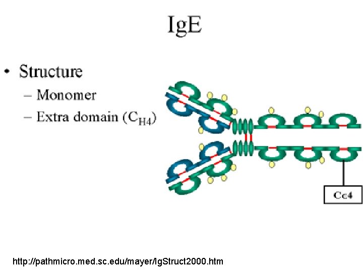
http: //pathmicro. med. sc. edu/mayer/Ig. Struct 2000. htm
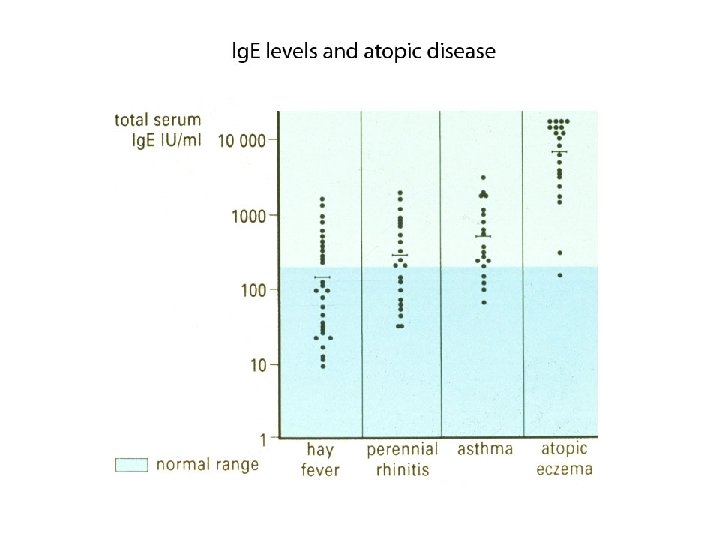
Serum Ig. E levels in atopic dirseses
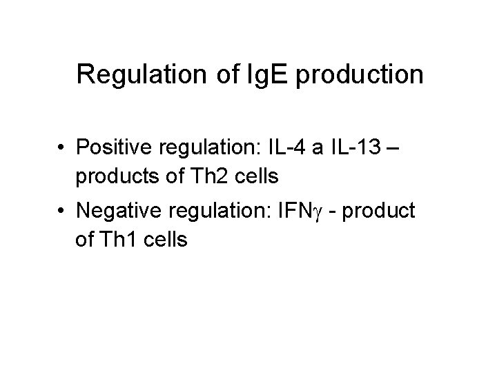
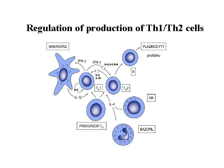
Regulation of Ig. E production • Positive regulation: IL-4 a IL-13 – products of Th 2 cells • Negative regulation: IFNg - product of Th 1 cells
Regulation of production of Th 1/Th 2 cells
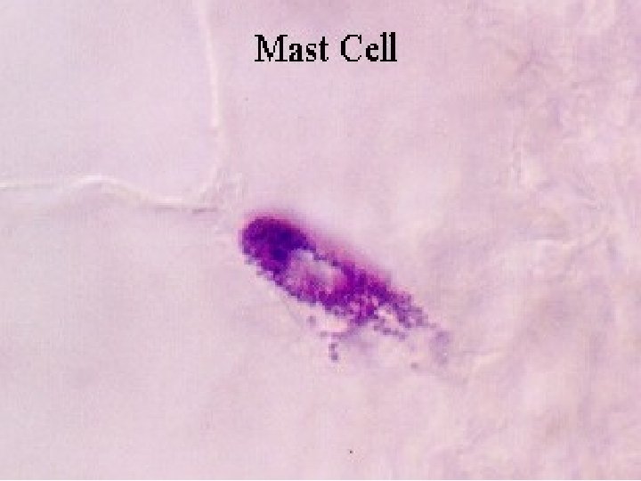
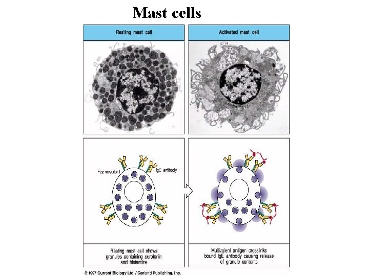
Mast cells
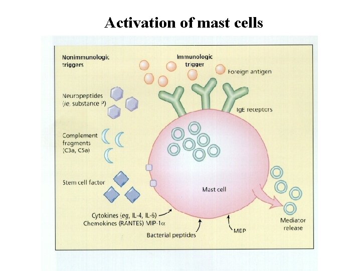
Activation of mast cells
Biological effects of histamin • H 1: Smooth muscle contraction, increased permeability of capillaries, vasodilatation, increased production of nasal and bronchial secretions, chemotaxis of leukocytes • H 2: increase in gastric juice production, increased production of secretions on respiratory tract • H 3: receptors present in CNS
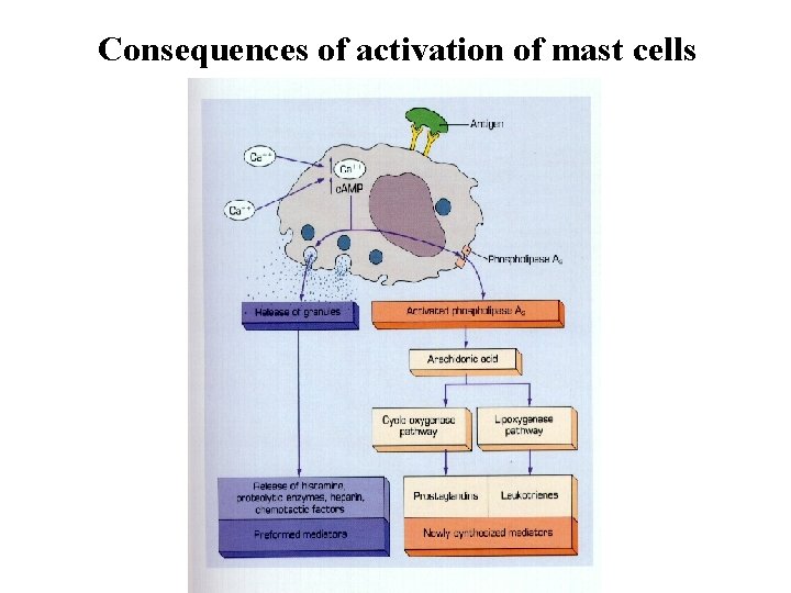
Consequences of activation of mast cells
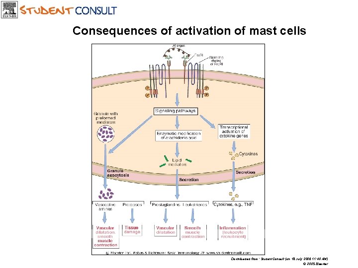
Consequences of activation of mast cells Downloaded from: Student. Consult (on 18 July 2006 11: 40 AM) © 2005 Elsevier
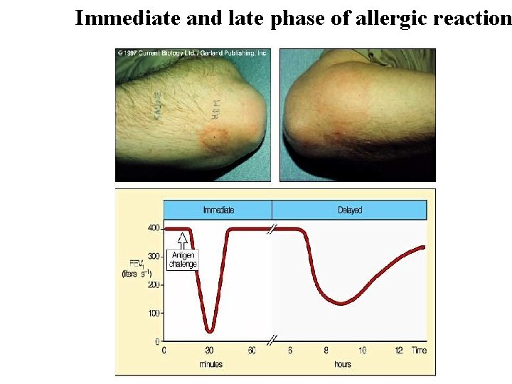
Immediate and late phase of allergic reaction
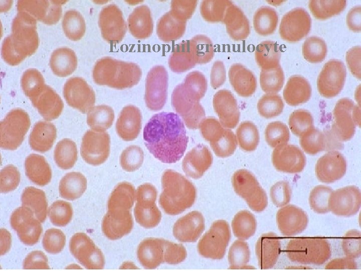
Phases of type-I hypersensitivity reaction • Immediate phase – clinical symptons evolve in several minutes. Mediated mainly by histamin. • Late phase – symptoms evolve after hours (6 -8). Mediated mainly by leukotriens. Presence of eosinophils plays an important role in allergic inflammation.
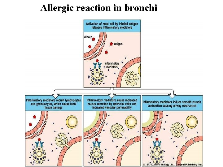
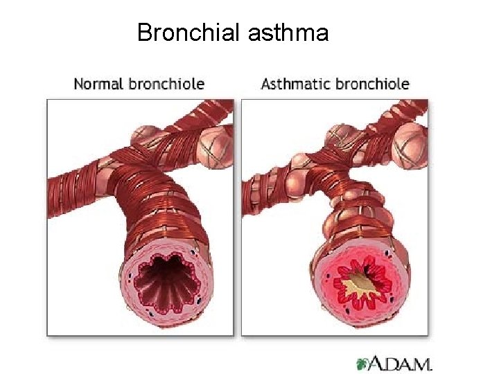
Allergic reaction in bronchi
Eozinophil granulocyte
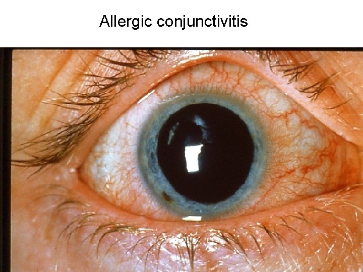
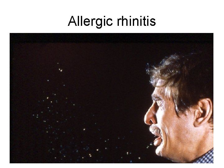
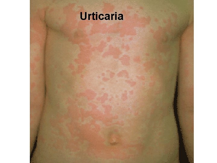
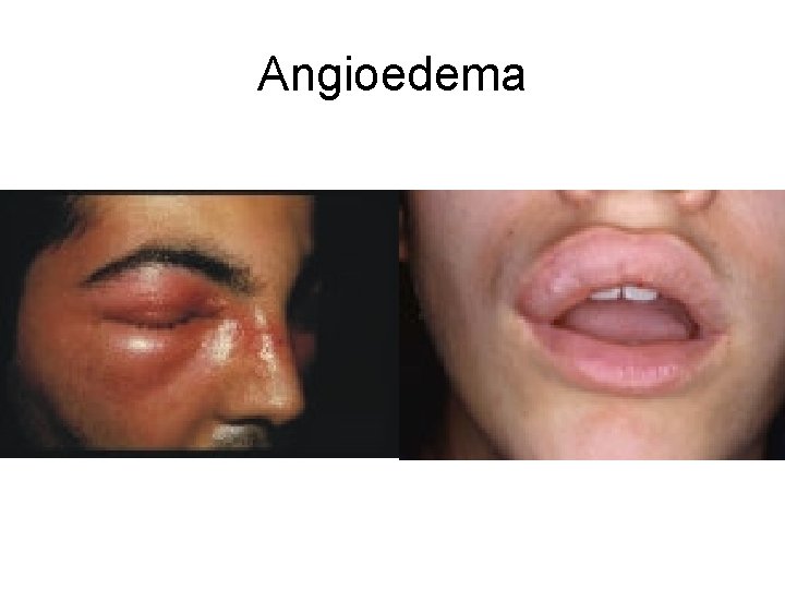
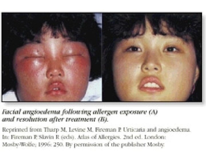
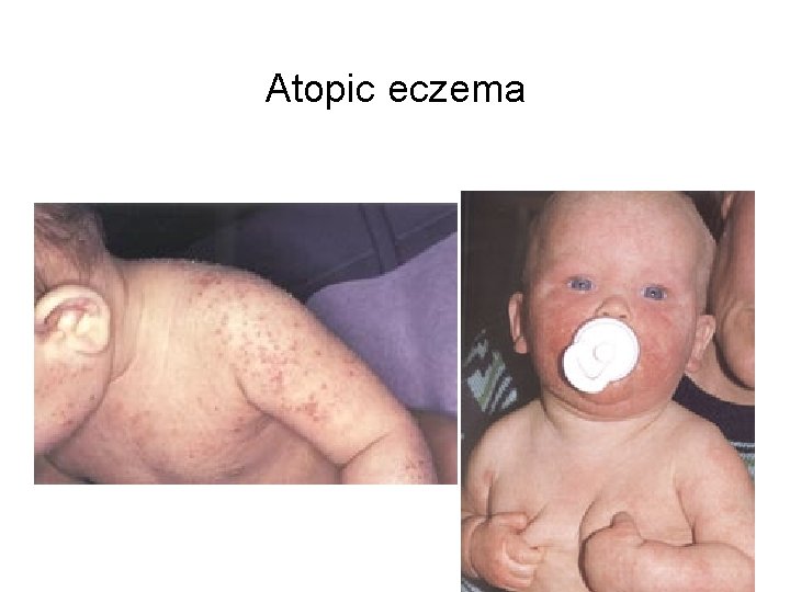
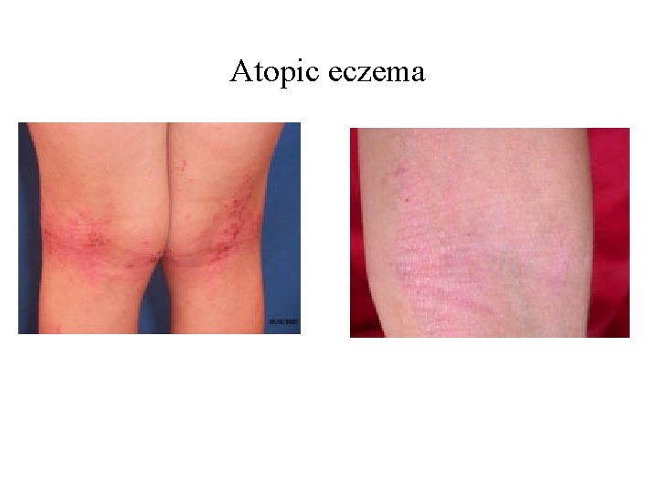
Clinical diseases caused by atopic hypersenitivity • • Allergic conjunctivitis Allergic rhinitis Bronchial asthma Allgergy of gastrointestinal tract Urticaria and angioedema Atopic eczema Anaphylactic shock
Allergic conjunctivitis
Allergic rhinitis
Bronchial asthma
Urticaria
Angioedema
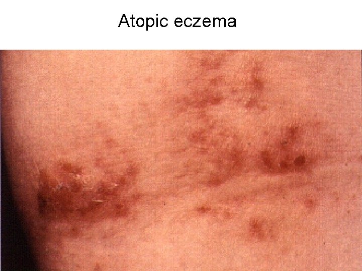
Atopic eczema
Atopic eczema
Atopic eczema
Treatment of allergic diseases • Allergen avoidance • Antihistaminics • Cromons (cromolyn sodium, nedocromil) stabilise membrane of the mast cells • Topical or systemic corticosteroids • Antilekotriens • In asthma: b-2 agonists, xantins • Allergen immunotherapy (desensitisation)
Diagnostic approaches in type-I hypersensitivity • • Past history Eosinophilia Skin tests Provocation and elimination tests
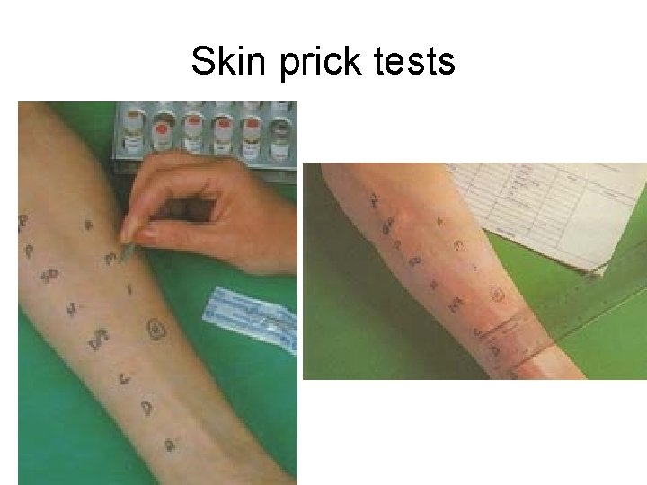
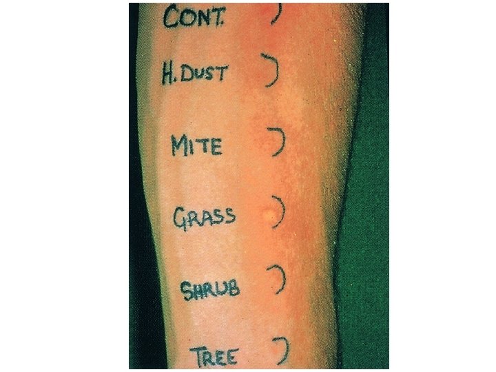
Skin prick tests
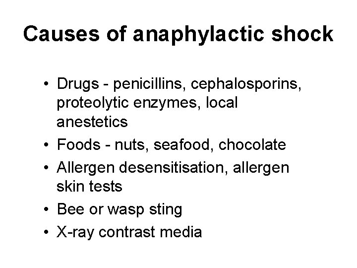
Causes of anaphylactic shock • Drugs - penicillins, cephalosporins, proteolytic enzymes, local anestetics • Foods - nuts, seafood, chocolate • Allergen desensitisation, allergen skin tests • Bee or wasp sting • X-ray contrast media
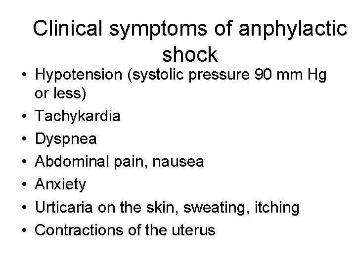
Clinical symptoms of anphylactic shock • Hypotension (systolic pressure 90 mm Hg or less) • Tachykardia • Dyspnea • Abdominal pain, nausea • Anxiety • Urticaria on the skin, sweating, itching • Contractions of the uterus
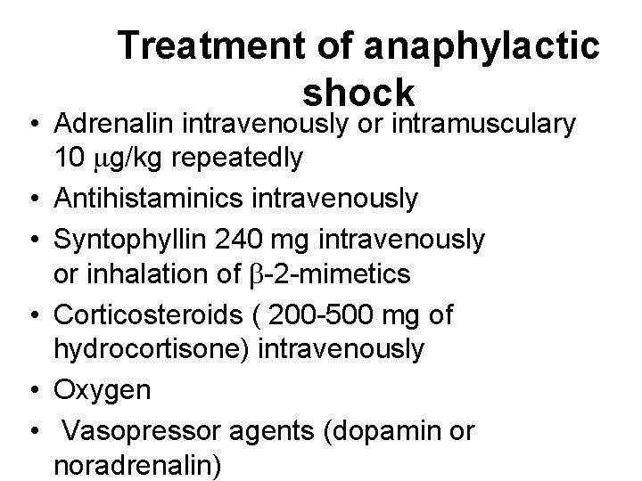
Treatment of anaphylactic shock • Adrenalin intravenously or intramusculary 10 mg/kg repeatedly • Antihistaminics intravenously • Syntophyllin 240 mg intravenously or inhalation of b-2 -mimetics • Corticosteroids ( 200 -500 mg of hydrocortisone) intravenously • Oxygen • Vasopressor agents (dopamin or noradrenalin)
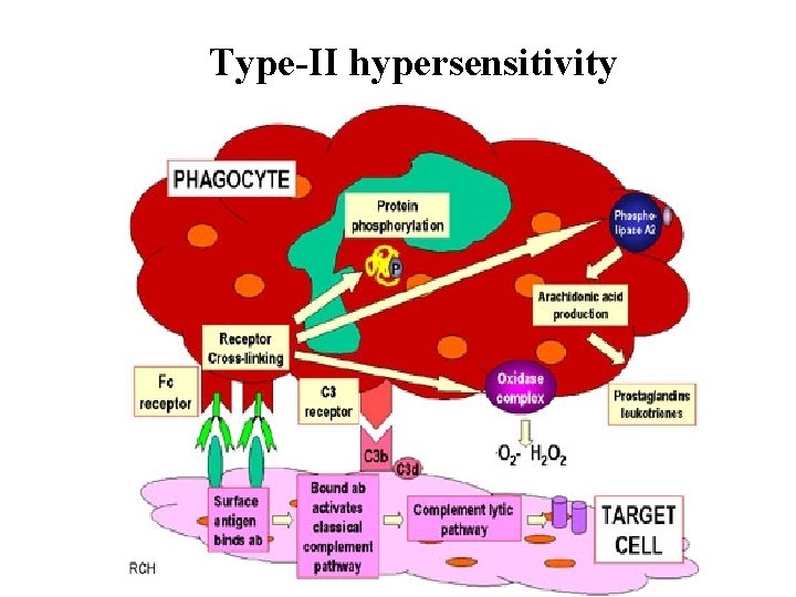
Type-II hypersensitivity
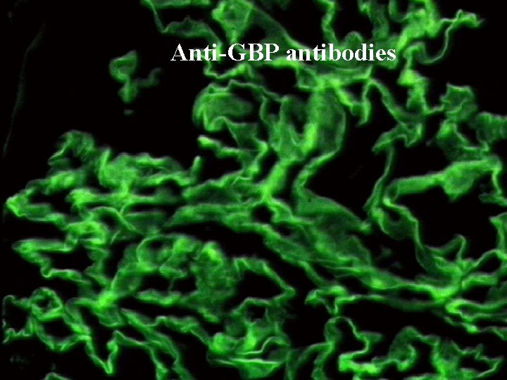
Anti-GBP antibodies
Diseased caused by immune complexes deposition • Caused by a disturbed transport or metabolism of immune complexes. • They usually deposit in the wall of vessels (causing vasculitis) or glomeruli (causing glomerulonephritis), less frequently in the place of their formation (extrinsic alveolitis). • The most important laboratory test is the direct immunofluorescence to detect the Ig. G part of the complexes.
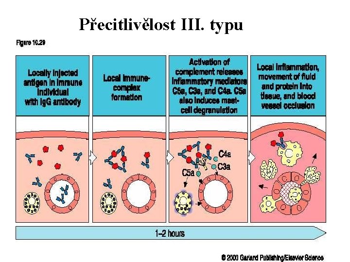
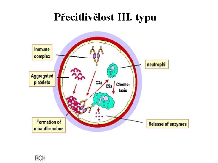
Immunocomplex diseases (type III immunopathological reaction • Caused by deposition of immune complexes in places different from their normal metabolism. • In case of circulating immune complexes (small, soluble complexes with excess of antigen), they deposit mainly in blood vessels walls and glomeruli leading to vasculitis and/or glomerulonephritis. • Less frequent is the situation when immune complexes deposit in the place of their formation (large complexes with excess of antibodies). They deposit in the place of their formation. • By activation of the complement system and phagocytioc cells they induce local inflammation.
Přecitlivělost III. typu
Přecitlivělost III. typu
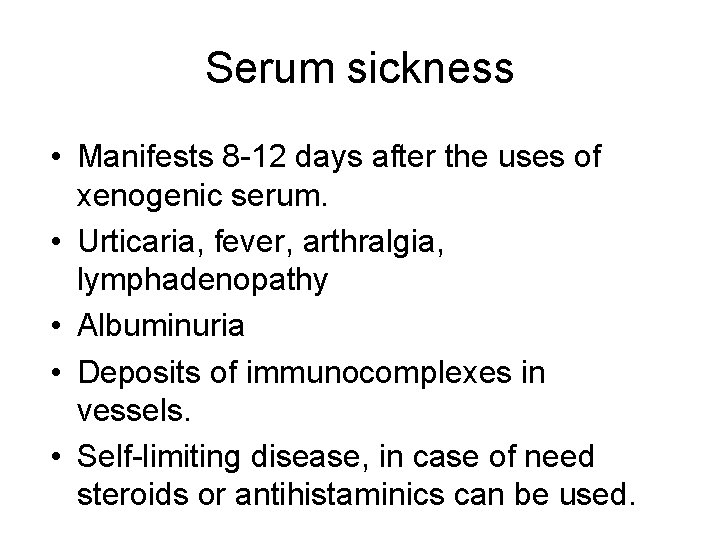
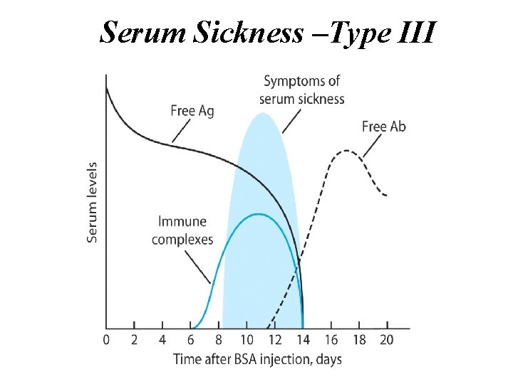
Serum sickness • Manifests 8 -12 days after the uses of xenogenic serum. • Urticaria, fever, arthralgia, lymphadenopathy • Albuminuria • Deposits of immunocomplexes in vessels. • Self-limiting disease, in case of need steroids or antihistaminics can be used.
Serum Sickness –Type III
Extrinsic alveolitis • Caused by deposition if insoluble immune complexes in the lung tissue. The complexes are formed from exogenous antigen and excess if antibodies of Ig. G class. • 6 -8 hours after exposition the patient suffers from dry cough, dyspnea, increased body temperature, lymphadenopathy. • Repeated expositions lead to lung fibrosis. . • Most frequently caused by bird antigens (pigeons – pigeon breeder´s disease, parrots), thermophil actinomycetes (farmers´s lungs disease).
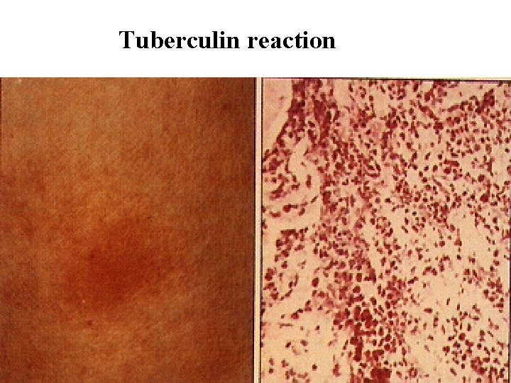
Tuberculin reaction
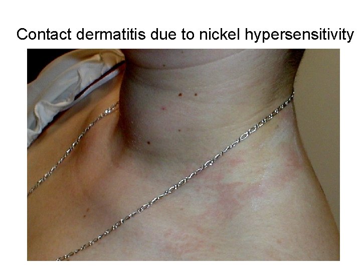
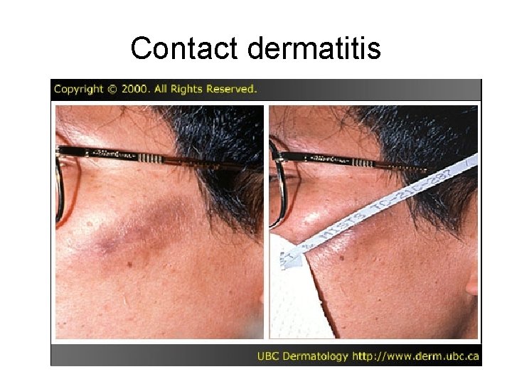
Examples of diseases where type-IV hypersensitivity plays a key role • • • Contact exzema Cavitation in tuberculosis Sarcoidosis Several types of vasculitis Autoimmune diseases where Tlymphocytes play a major role ( multiple sclerosis)
Contact dermatitis due to nickel hypersensitivity
Contact dermatitis