Hypersensitivity Reactions Allergies Refif alshawk Ph D Lec
 Lec 2&3 2018 -2019")


. Acts as a vasodilator and")

The Immediate Reaction")


 is a consequence of immediate hypersensitivity reactions to common")

 Causes shift to")


- Slides: 30
Hypersensitivity Reactions, Allergies Refif alshawk (Ph. D) Lec 2&3 2018 -2019
Learning objectives After reading this lecture, you should be able to: Distinguish between the four types of hypersensitivities, and understand the immunological mechanisms behind each of them. For each of the four types of hypersensitivities, recognize the harmful effects of these immune responses when they become inappropriately vigorous responses (hypersensitivity reactions). Discuss the roles of environmental factors and genetics in predisposition to allergies
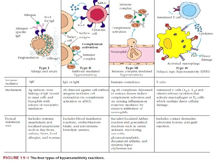
Hypersensitivity – responding inappropriately to an antigen Tissue injury Disease death Allergy: A Type I Hypersensitivity Reaction Antibody-Mediated (Type II) Hypersensitivity Reactions Immune Complex-Mediated (Type III) Hypersensitivity Delayed-Type (Type IV) Hypersensitivity (DTH
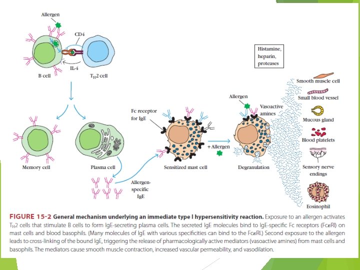
Types I : OVERVIEW OF Ig. EDEPENDENT ALLERGIC REACTIONS All allergic reactions share common feature: the production of Ig. E antibody, which is dependent on the activation of IL-4– producing helper T cells. allergic reactions differ greatly in the types of antigens that elicit these reactions and their clinical and pathologic manifestations. Antigens that elicit immediate hypersensitivity reactions called allergens.
The typical sequence of events in immediate hypersensitivity consists exposure to an antigen activation of lymphocytes (TH 2 cells, and B cells) specific for the antigen production of Ig. E antibody resulting in the release of mediators from the mast cells and triggering of the mast cells by re-exposure to the antigen binding of the antibody to Fc receptors of mast cells and the subsequent pathologic reaction.
Many allergens can elicit a type I response Antigens that elicit immediate hypersensitivity reactions (allergens) are proteins or chemicals bound to proteins. The important characteristics of allergens that, unlike microbes, they do not generally stimulate the innate immune responses that are associated with macrophage and dendritic cell secretion of TH 1 - and TH 17 -inducing cytokines. WHY? ? ? Chronic or repeated cell activation in the absence of strong innate immunity may drive CD 4+ T cells preferentially toward the TH 2 pathway. The chemical nature. include low to medium molecular weight, stability, glycosylation, and high solubility in body fluids. These structural features probably protect the antigens from denaturation and degradation in the gastrointestinal tract and allow them to be absorbed intact. Some nonprotein substances, such as penicillin, can elicit strong Ig. E responses, when react chemically with amino acid residues in self proteins to form hapten carrier conjugates, which induce IL-4–producing helper T cell responses and Ig. E production.
There is a genetic predisposition for the development of allergies. Atopic individuals produce high levels of Ig. E in response to environmental allergens, whereas normal individuals generally produce other Ig isotypes, such as Ig. M and Ig. G, and only small amounts of Ig. E.
� � Common components Allergens ○ Atopy – hereditary predisposition to development of immediate hypersensitivity reactions to common antigens - � Allows nonparasitic antigens to induce Ig. E response Ig. E ○ Normally lowest of all antibody classes in serum ○ Half-life is 2 -3 days but once bound to mast cells or basophils, can last for weeks � Mast cells and basophils � Ig. E binding receptors ○ High affinity FcεRI(mast cell, basophil, eosinophil, momncytes, platelets) ○ Low affinity. FcεRII CD 23(activated B-cell & various cell type) � Soluble � Atopic & membrane forme individuals have higher amount of soluble Ig. E receptor that has been shown to increase Ig. E production by B cells
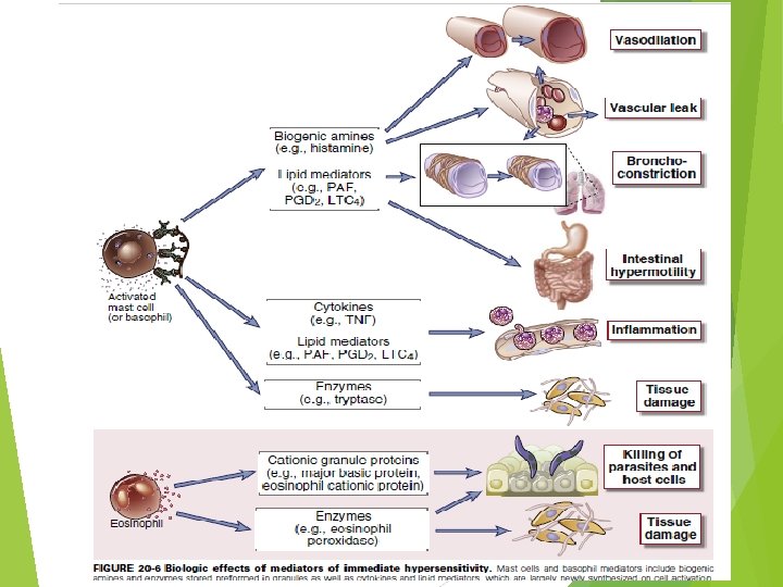
Mast cells, basophils, and eosinophils that are stimulated by FcεRI cross-linking release their granular contents (including histamine and proteases) in a process called degranulation) they also generate and secrete inflammatory cytokines and lipid inflammatory molecules (leukotrienes and prostaglandins) Mediators Derived from Mast Cells (primary & secondary mediators) Primary mediators: Biogenic Amines and proteolytic enzymes Histamine acts by binding to target cell receptors (e. g. , H 1, H 2, H 3) The actions of histamine are contraction of the endothelial cells, leading to increased inter endothelial spaces, increased vascular permeability, and leakage of plasma into the tissues and other effect cause vasodilation also causes contraction of intestinal and bronchial smooth muscle. proteolytic enzymes : tryptase - tissue damage
Secondary mediators Lipid Mediators prostaglandin D 2 (PGD 2). Acts as a vasodilator and a bronchoconstrictor. PGD 2 also promotes neutrophil chemotaxis and accumulation at inflammatory sites. leukotrienes its degradation products LTD 4 and LTE 4 which cause prolonged bronchoconstriction. (slow-reacting substance of anaphylaxis and are thought to be important mediators of asthmatic bronchoconstriction. platelet-activating factor (PAF) has direct bronchoconstricting actions.
Cytokines Mast cells produce many different cytokines that contribute to allergic inflammation (the late-phase reaction). These cytokines like TNF, IL-1, IL-4, IL-6, CCL 3…. . . and granulocytemonocyte colony-stimulating factor (GM-CSF). The cytokines that are released from activated mast cells and TH 2 cells are mainly responsible for the inflammation associated with the late-phase reaction. TNF activates endothelial expression of adhesion molecules and together with chemokines accounts for neutrophil and monocyte infiltrates.
Type I hyper. are characterized by both early and late responses 1)The Immediate Reaction The early vascular changes that occur during immediate hypersensitivity reactions are demonstrated by the wheal-and-flare reaction) within minutes of allergen exposure, including the release of histamine, leukotrienes, and prostaglandins. 2) The Late-Phase Reaction The immediate wheal-and-flare reaction is followed 2 to 4 hours later by a late-phase reaction consisting of the accumulation of inflammatory leukocytes, including eosinophils, which play a major role in this phase including recruitment of neutrophils, basophils. Degranulation of both cells induces tissue damage.
There are several categories of type I hypersensitivity reactions Systemic Anaphylaxis is a systemic immediate hypersensitivity reaction characterized by edema in many tissues and a decrease in blood pressure, secondary to vasodilation. These effects usually result from the systemic presence of antigen introduced by injection, an insect sting, or absorption across an epithelial surface such as gut mucosa. The allergen activates mast cells in many tissues, resulting in the release of mediators that gain access to vascular beds throughout the body. The decrease in vascular tone and leakage of plasma caused by mast cell mediators can lead to a significant decrease in blood pressure or shock, called anaphylactic shock, which is often fatal. The cardiovascular effects are accompanied by constriction of the upper and lower airways, laryngeal edema treatment is systemic epinephrine, which can be lifesaving by reversing the bronchoconstrictive and vasodilatory effects of mast cell mediators.
Localized hypersensitivity reactions Bronchial Asthma is an inflammatory disease caused by repeated immediate-type hypersensitivity and late-phase allergic reactions in the lung leading to the clinic-pathologic triad of intermittent and reversible airway obstruction, chronic bronchial inflammation with eosinophils, and bronchial smooth muscle cell hypertrophy and hyperreactivity to bronchoconstrictors
Asthma Inflammatory disease Induce expression of adhesion molecules on endothelial cells for eosinophils and neutrophils ○ Cause significant injury because of toxic enzymes, cytokines ○ Notice sloughing of the pseudostratified ciliated columnar epithelial cells lining the bronchiole
The pathophysiologic sequence in atopic asthma is probably initiated by mast cell activation in response to allergen binding to Ig. E as well as by TH 2 cells reacting to allergens. The lipid mediators and cytokines produced by the mast cells and T cells lead to the recruitment of eosinophils, basophils, and more TH 2 cells. The chronic inflammation in this disease may continue without mast cell activation. Smooth muscle cell hypertrophy and hyperreactivity are thought to result from leukocyte-derived mediators and cytokines. Mast cells, basophils, and eosinophils all produce mediators that constrict airway smooth muscle. The most important of the bronchoconstricting mediators are LTC 4, LTD 4, and LTE 4. Increased mucus secretion results from the action of cytokines, on bronchial epithelial cells. Corticosteroids may also be given systemically, especially once an attack is under way, to reduce inflammation and bronchial smooth muscle cell relaxants
Allergic rhinitis, (hay fever) is a consequence of immediate hypersensitivity reactions to common allergens such as plant pollen or house dust mites localized to the upper respiratory tract by inhalation. mucosal edema, leukocyte infiltration with abundant eosinophils, mucus secretion, coughing, sneezing, and difficulty in breathing. Food allergies are immediate hypersensitivity reactions to ingested foods that lead to the release of mediators from intestinal mucosal and submucosal mast cells of the GI tract, including the oropharynx. pruritis, tissue edema, enhanced peristalsis, increased epithelial fluid secretion, and associated symptoms of oropharyngeal swelling, vomiting and diarrhea. Allergic reactions to many different types of food most common are peanuts and shellfish. Common allergic reactions in the skin include urticaria and atopic dermatitis. Urticaria, or hives, is an acute wheal-and-flare reaction induced by mast cell mediators and occurs in response to direct local contact with an allergen or after an allergen enters the circulation. eczema. It is a common skin disorder that may be caused by a latephase reaction to an allergen in the skin. In the cutaneous late-phase reaction, TNF, IL-4, and other cytokines, probably derived from TH 2 cells and mast cells, act on endothelial cells to promote inflammation.
Clinical Methods to detect Type 1 Skin testing • small amounts of potential allergens are introduced at specific skin, • Thirty minutes later, the sites are reexamined, redness and swelling Checking serum level of Ig. E • using ELISA or Western blot technologies
Control of Type 1 Avoiding contact Immunotherapy Subcutaneous injections of allergens(Hyposensitization) Causes shift to Ig. G production instead of Ig. E Monoclonal anti-human Ig. E Though expensive and difficult administer. Drug therapies Can treated with pharmacological inhibitors of cellular and tissue responses and inflammation (antihistamines, leukotriene inhibitors and corticosteroids)
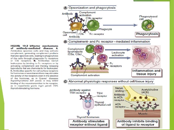
Type II – Antibody-Mediated Cytotoxic Hypersensitivity involves the binding of Ig. G antibodies to: [cell surface antigens or extracellular matrix molecules]. Antibody bound to a cell-surface antigen can induce death of the antibody-bound cell by three distinct mechanisms.
Type II – Antibody-Mediated Cytotoxic Hypersensitivity First, certain immunoglobulin subclasses can activate the complement system, creating pores in the membrane of a foreign cell. Secondly, antibodies can mediate cell destruction by antibody dependent cell-mediated cytotoxicity (ADCC), in which cytotoxic cells bearing Fc receptors bind to the Fc region of antibodies on target cells and promote killing of the cells. Finally, antibody bound to a foreign cell also can serve as an opsonin, enabling phagocytic cells with Fc or C 3 b receptors to bind and phagocytose the antibody-coated cell
Type II – Antibody-Mediated Cytotoxic Hypersensitivity Transfusion Reactions antigens that are associated with the blood types are identified as A, B, and H, Individuals have antibodies to blood types not their own and adults possess Ig. M antibodies to those Ag. Antibodies directed toward ABH antigens are termed isohemagglutinins Antibody attaches to RBC and initiates complement system to lyse RBC After lysis: ○ Hemoglobin detected in plasma, starts to filter through kidneys and found in urine (hemoglobinuria) ○ Hemoglobin converted to bilirubin – toxic at high levels ○ Fever, chills, blood clotting
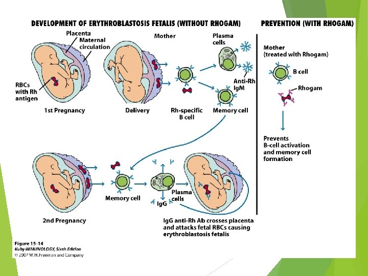
Type II – Antibody-Mediated Cytotoxic Hypersensitivity Hemolytic disease of newborn Rh+ fetus, Rh- mother Ig. G antibodies cross placenta(memory) Some of these antibodies may be anti-Rh antibodies - Can have severe consequences Antibodies against ABO blood groups produce less consequences, can be easily treated Rhogam shot ○ Given to mother ○ Anti-Rh antibodies bind to fetal cells that might have entered mother’s system during birthing process, facilitates clearing before there is a B cell response
Type II – Antibody-Mediated Cytotoxic Hypersensitivity Hemolytic Induced • • • Anemia Can Be Drug Certain antibiotics (e. g. , penicillin, cephalosporins, and streptomycin), as well as other well-known drugs adsorb nonspecifically to proteins on red blood cell membranes, forming a drug protein complex induce formation of antibodies inducing complement-mediated lysis progressive anemia
Refferences : v Immunology , Kuby, seventh edition 2013 v Immunology , Kuby, eighth edition 2019 v Medical microbiology, Jawetz, 26 th edition v Cellular and Molecular Immunology, Abul K. Abbas, 8 th edition.