Dentin Hypersensitivity The dentinal hypersensitivity DH is characterized
- Slides: 8
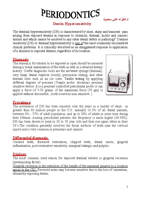
ﻋﺬﺭﺍﺀ ﻋﻠﻲ ﻣﺤﻤﻮﺩ. ﺩ Dentin Hypersensitivity The dentinal hypersensitivity (DH) is characterized by short, sharp and transient pain arising from exposed dentine in response to chemical, thermal, tactile and osmotic stimuli and which cannot be ascribed to any other dental defect or pathology” Dentine sensitivity (DS) or dentinal hypersensitivity is one of the most commonly encountered clinical problems. It is clinically described as an exaggerated response to application of a stimulus to exposed dentine, regardless of its location. Diagnosis The reason(s) for tubules to be exposed or open should be assessed during a visual examination of the teeth as well as a detailed dietary history. Useful diagnostic tools are the air/water syringe (thermal), very sharp dental explorer (touch), percussion testing, and othermal tests such as an ice cube. Tactile testing by applying different degrees of pressure (Yeaple probe: electronic pressure sensitive device. It is a pressure controlled periodontal probe. it can apply a force of 5 -70 grams. if the maximum force (70 gm) is applied without discomfort , tooth scored as non-sensitive. ) Prevalence The prevalence of DH has been reported over the years in a variety of ways: as greater than 40 million people in the U. S. annually 14. 3% of all dental patients, between 8% - 57% of adult population, and up to 30% of adults at some time during their lifetime. Among periodontal patients, the frequency is much higher (60 -98%). DH has been shown to peak in 20 to 30 year olds and then rise again when in their 50’s. The condition generally involves the facial surfaces of teeth near the cervical aspect and is very common in premolars and canines. Differential diagnosis: Cracked teeth, fractured restoration, chipped teeth, dental caries, gingival inflammation, post-restorative sensitivity, marginal leakage, and pulpitis. Etiology The most common cited reason for exposed dentinal tubules is gingival recession (predisposing factor). Gingival recession is the reduction of the height of the marginal gingiva to a location apical to the CEJ. Recessed areas may become sensitive due to the loss of cementum, ultimately exposing dentin. 1
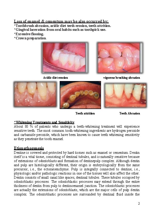
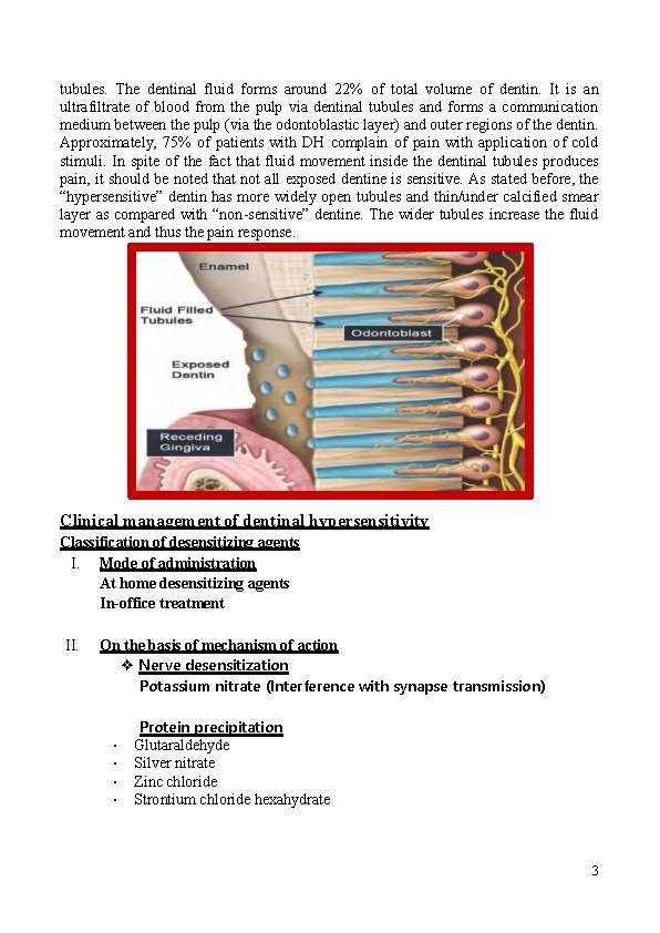
Loss of enamel & cementum may be also occurred by: *Toothbrush abrasion, acidic diet teeth erosion, teeth attrition. *Gingival laceration from oral habits such as toothpick use. *Excessive flossing. *Crown preparation. Acidic diet erosion vigorous brushing abrasion Teeth attrition Teeth Abrasion *Whitening Treatments and Sensitivity About 80 % of patients who undergo a teeth-whitening treatment will experience sensitive teeth. The most common tooth-whitening ingredients are hydrogen peroxide and carbamide peroxide, which have been known to cause teeth whitening sensitivity as they penetrate the tooth enamel. Etiopathogenesis Dentine is covered and protected by hard tissues such as enamel or cementum. Dentin itself is a vital tissue, consisting of dentinal tubules, and is naturally sensitive because of extensions of odontoblasts and formation of dentinepulp complex. Although dentin and pulp are histologically different, their origin is embryologically from the same precursor, i. e. , the ectomesenchyme. Pulp is integrally connected to dentine, i. e. , physiologic and/or pathologic reactions in one of the tissues will also affect the other. Dentin consists of small canal like spaces, dentinal tubules. These tubules occupied by odontoblastic processes. The odontoblastic processes may extend through the entire thickness of dentin from pulp to dentinoenamel junction. The odontoblastic processes are actually the extensions of odontoblasts, which are the major cells of pulp dentin complex. The odontoblastic processes are surrounded by dentinal fluid inside the 2
tubules. The dentinal fluid forms around 22% of total volume of dentin. It is an ultrafiltrate of blood from the pulp via dentinal tubules and forms a communication medium between the pulp (via the odontoblastic layer) and outer regions of the dentin. Approximately, 75% of patients with DH complain of pain with application of cold stimuli. In spite of the fact that fluid movement inside the dentinal tubules produces pain, it should be noted that not all exposed dentine is sensitive. As stated before, the “hypersensitive” dentin has more widely open tubules and thin/under calcified smear layer as compared with “non-sensitive” dentine. The wider tubules increase the fluid movement and thus the pain response. Clinical management of dentinal hypersensitivity Classification of desensitizing agents I. Mode of administration At home desensitizing agents In-office treatment II. On the basis of mechanism of action ❖ Nerve desensitization Potassium nitrate (Interference with synapse transmission) • • Protein precipitation Glutaraldehyde Silver nitrate Zinc chloride Strontium chloride hexahydrate 3
• • • • Plugging dentinal tubules Sodium fluoride Stannous fluoride Strontium chloride Potassium oxalate Calcium phosphate Calcium carbonate Bio active glasses (Si. O 2–P 2 O 5–Ca. O–Na 2 O) Dentine adhesive sealers Fluoride varnishes Oxalic acid and resin Glass ionomer cements Composites Dentin bonding agents Lasers Neodymium: yttrium aluminum garnet (Nd-YAG) laser. Ga. Al. As (gallium-aluminum-arsenide laser). Erbium-YAG laser. Diode laser. At home desensitizing therapy These “at home” desensitizing agents include: toothpastes, mouthwashes and chewing gums. Majority of the toothpastes contain *Potassium salts (potassium nitrate, potassium chloride or potassium citrate) *Sodium fluoride *strontium chloride *Dibasic sodium citrate *Formaldehyde *Sodium monofluorphosphate *Stannous fluoride. Potassium nitrate: Potassium salts act by diffusion along the dentinal tubules and decreasing the excitability of the intradental nerve fibers by blocking the axonic action. Various clinical studies have shown the efficacy of potassium salts in controlling the DH. The toothpaste (5% KNO 3) and a professionally applied sealant product (6% ferric oxalate) both produced some obliteration of the tubuli. It has been shown that toothpastes containing 5% potassium nitrate significantly reduced the DH. Also, toothpastes containing potassium nitrate and fluorides have been shown to reduce post-bleaching sensitivity. The desensitizing 4
toothpastes should be used with the help of a toothbrush with soft bristles. Patients should be advised to use minimal amount of water to prevent the dilution of the active agent. Along with the desensitizing toothpastes, mouthwashes and chewing gums containing potassium nitrate, sodium fluoride or potassium citrate are also recommended. The results of at-home desensitizing therapy should be reviewed after every 3– 4 weeks. If there is no relief in DH, in-office therapy should be initiated. In-office desensitizing agents Theoretically, the in-office desensitizing therapy should provide an immediate relief from the symptoms of DH. The in-office desensitizing agents can be classified as the materials which undergo a setting reaction (glass ionomer cement, composites) and which do not undergo a setting reaction (varnishes, oxalates). Fluorides Traditionally, fluorides have been used as a caries preventive material which can help in remineralization of enamel/dentin. Also, various clinical trials have shown that application of fluoride solution can decrease the DH. Fluorides decrease the dentinal permeability by precipitation of calcium fluoride crystals inside the dentinal tubules. These crystals are partially insoluble in saliva. SEM revealed granular precipitates in the peritubular dentin after application of fluorides. Various fluoride formulations are used to treat DH. These include sodium fluoride, stannous fluoride, sodium monofluorphosphate, fluorosilicates and fluoride combined with iontophoresis. Sodium fluoride has been used in dentifrices or may be professional applied in a concentration of 2%. The precipitates formed by sodium fluoride can be mechanically removed by the action of saliva or mechanical action. Therefore, an addition of acid formulation is recommended. The acidulated sodium fluoride can form precipitates deep inside the tubules. Also, some authors have recommended the use of iontophoresis along with sodium fluoride. The electric current is supposed to increase the ion diffusion. A clinical study has shown that 0. 4% stannous fluoride along with 0. 717% of fluoride can provide an immediate effect after a 5 minutes professional application. Stannous fluoride acts in a similar fashion as that of sodium fluoride, i. e. , formation of calcium fluoride precipitates inside tubules. Also, SEM studies have shown that stannous fluoride itself can form insoluble precipitates over the exposed dentine. (SEM) Treatment with non-anti-sensitive Fl (SEM) Treatment with 0. 4% Stannous Fl. 5
Fluorosilicates act by formation of precipitates of calcium phosphates from saliva. Ammonium hexafluorosilicate has been used as a desensitizing agent. It can present a continuous effect of dentinal tubule occlusion via precipitation of a mixture of calcium fluoride and fluoridated apatite. If the precipitate is predominantly composed of fluoridated apatite, it can form stable crystals deposited deep inside the dentinal tubules. These crystals are resistant to removal from the action of saliva, brushing or action of dietary substances. Oxalates can reduce dentinal permeability and occlude dentinal tubules. Thirty percent potassium oxalate had shown a 98% reduction in dentinal permeability. Also, topical application of 3% potassium oxalate reduced DH after periodontal therapy. The oxalate reacts with the calcium ions of dentine and forms calcium oxalate crystals inside the dentinal tubules as well as on the dentinal surface. This results in a better sealing as compared with an intact smear layer. It has been shown that the effect of oxalates on DH diminishes over a period of time. This can be attributed to the removal of the calcium oxalate crystals by brushing or dietary acids. The condition can be improved by acid etching of the dentinal surface, thus increasing the penetration of calcium oxalate crystals deep into the dentinal tubules. Many vegetables like rhubarb, spinach and mint contain oxalates. It has been shown that phytocomplexes obtained from these natural products can reduce the dentinal permeability. This can also be followed by covering the exposed surface with a dental adhesive. Potassium oxalate can lead to gastric irritation. Therefore, it should not be used with a tray with prolonged placement. Varnishes are commonly used in-office measures to treat DH. Copal varnish can be applied to cover the exposed dentinal surface. But its effect is for short term and is not recommended for long term management of DH. To improve its efficacy, removal of smear layer is advocated. Also, the varnishes can act as a vehicle for fluoride. The fluoride varnishes can be acidulated to increase the penetration of ions. Adhesive materials Resin-based dental adhesive systems can provide a more durable and long lasting dentine desensitizing effect. The adhesive resins can seal the dentinal tubules effectively by forming a hybrid layer. Various clinical studies have demonstrated the effectiveness of adhesives in management of DH. Traditionally, resin composites or dentin bonding agents are used as desensitizing agents. The conventional dentin bonding agents (DBA) removes the smear layer, etches the dentinal surface and forms deep dentinal resin tags inside the dentinal tubules. The combined dentin–resin layer (consisting of penetrating resinous tags) has been termed as hybrid layer. It effectively seals the dentinal tubules and prevents DH. 6
Gluma Its formula of (5%) glutaraldehyde & (35%) HEMA (hydroxyethylmethacrylate) in water is used to control DH Method of action • Gluma Dentin Bond is an adhesive system, where the primer contains 5% glutaraldehyde and 35% hydroxyethyl methacrylate. • Practitioners have reported a strong desensitizing effect of the Gluma system on dentin. Bioglass was developed to stimulate the formation of new bone. It has been used in dentistry to fill up the osseous defects during periodontal surgery. It has been reported that a formulation of bioglass can promote infiltration and remineralization of dentinal tubules. SEM analysis has shown that bioglass application forms an apatite layer, which occludes the dentinal tubules. Nova. Min: is the brand name of a particulate bioactiveglass. The active ingredient is Ca, Na and phsphosilicate. Nova. Min delivers silica and ionic calcium, phosphorus, & sodium which are necessary for bone and tooth mineralization. Nova. Min particles bind to the tooth surface and react with the water to release Ca and phosphate ions. These ions are protected by glass particles. A calcium phosphate layer then forms hydroxyapatite. The occlusion of dentinal tubules results from the hydroxyapatite layer & the residual Novamin particles. The clinical technique requires: • The tooth be clean and dry by using a blotting technique with a cotton roll. • Applied for 30 -60 sec. using a gentle rubbing motion then, using a very gentle air spray, blow off most of the excess but leave the surface wet. Pro-Argin Technology The essential components of this new technology are: Arginine is a naturally occurring amino acid positively charged at physiologic p. H (6. 57. 5). Bicarbonate, a p. H buffer. Insoluble Calcium carbonate, a source of calcium to form a deposit that seals the opened dentinal tubules. . An in-office product paste contains 8% arginine and calcium carbonate. Other products based on Pro-Argin technology include: 7
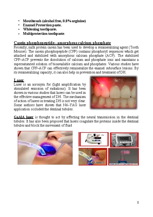
• • Mouthwash (alcohol free, 0. 8% arginine) Enamel Protection paste. Whitening toothpaste. Multiprotection toothpaste. Casein phosphopeptide– amorphous calcium phosphate Recently, milk protein casein has been used to develop a remineralizing agent (Tooth Mousse). The casein phosphopeptide (CPP) contains phosphoryl sequences which get attached and stabilized with amorphous calcium phosphate (ACP). The stabilized CPP–ACP prevents the dissolution of calcium and phosphate ions and maintains a supersaturated solution of bioavailable calcium and phosphates. Various studies have shown that CPP–ACP can effectively remineralize the enamel subsurface lesions. By its remineralizing capacity, it can also help in prevention and treatment of DH. Laser is an acronym for (light amplification by stimulated emission of radiations). It has been shown in various studies that lasers can be used in the effective management of DH. The mechanism of action of lasers in treating DH is not very clear. Some authors have shown that Nd–YAG laser application occluded the dentinal tubules. Ga. Al. A laser is thought to act by affecting the neural transmission in the dentinal tubules. It has also been proposed that lasers coagulate the proteins inside the dentinal tubules and block the movement of fluid 8