Shock OBJECTIVES Shock definitions Types of shock mainly

Shock OBJECTIVES Shock definitions Types of shock, mainly hypovolemia, septic, obstructive Diagnosis Management

Shock Definition Is an imbalance between oxygen delivery and oxygen demand this result in cell dysfunction and ultimately cell death and multi organ failure Fact Tissue oxygen delivery may be inadequate and the blood pressure and other vital signs are normal.

Shock Types Hypovolaemic shock The commonest Result from a reduction in intravascular volume secndary to loss of blood (e. g. trauma, gastrointestinal


Septic shock Results from complex disturbances in oxygen delivery and oxygen consumption. Gram negative (38%) Gram positive (52%) bacteria being the most frequently identified pathogens. The commonest sites Ølungs (50 70%) Øabdomen (20 25%) Øurinary tract (7 10%) Øskin

Septic shock Infection triggers a cytokine mediated pro inflammatory response results in: peripheral vasodilatation redistribution of blood flow endothelial cell activation increased vascular permeability formation of micro thrombi within the microcirculation.

Septic shock Cardiac output typically increases in septic shock to compensate for the peripheral vasodilation. However, despite a global increase in oxy gen delivery, microcirculatory dysfunction impairs oxygen delivery to the cells.

Cardiogenic shock A cardiac output insufficient to meet the metabolic requirements of the body (pump failure) and can be caused by myocardial infarction, arrhythmias, valve dysfunction, cardiac tamponade, massive pulmonary embolism, and tension pneumothorax.
 triggering the release")
Anaphylactic shock Systemic hypersensitivity reaction following exposure to an agent (allergen) triggering the release of vasoactive mediators (histamine, kinins and prostaglandins) from basophils and mast cells. Results from vasodilation, intravascular volume redistribution, capillary leak and a reduction in cardiac output.

Neurogenic shock Caused by a loss of sympathetic tone to vascular smooth muscle. This typically occurs following injury to the (thoracic or cervical) spinal cord and results in profound vasodilation, a fall in systemic vascular resistance and hypotension. It can also occur in 'high' spinal anaesthesia.

Pathophysiology In clinical practice there is often significant overlap between the causes of shock for example, patients with septic shock are frequently also hypovolaemic.
 is associated with increased sympathetic activity All share common")
Pathophysiology Most shock (exception neurogenic) is associated with increased sympathetic activity All share common pathophysiological features at the cellular level.
 can occur in the context of a low,")
Macrocirculation Shock (inadequate tissue oxygen delivery) can occur in the context of a low, normal or high cardiac output. In hypovolaemic shock there is catecholamine release from the adrenal medulla and sympathetic nerve endings, as well as the generation of Angiotesin II from the renin angiotensin system.

Macrocirculation The resulting tachycardia and increased myocardial contractility act to preserve cardiac output vasoconstriction acts to maintain arterial blood pressure and divert the available blood to vital organs (e. g. brain, heart and muscle) and away from non vital organs (e. g. skin and gut).

Macrocirculation Clinically this manifests as pale, clammy skin with collapsed peripheral veins and a prolonged capillary refill time. The resulting splanchnic hypoperfusion is implicated in many of the complications associated with prolonged or untreated shock.
 induce")
Macrocirculation In septic shock, circulating pro inflammatory cytokines (notably TNF interferon and IL) induce endothelial expression of the enzyme nitric oxide synthetase and the production of which leads to smooth muscle relaxation, vasodilation and a fall in systemic vascular resistance.

Macrocirculation The initial cardiovascular response is a reflex tachycardia and an increase in stroke volume resulting in an increased cardiac output. Clinically this manifests as warm, well perfused peripheries, a low diastolic blood pressure and raised pulse pressure. As septic shock progresses endothelial dysfunction results in significant extra vasation of fluid and a loss of intravascular volume.


Macrocirculation Ventricular dysfunction also impairs the com pensatory increase in cardiac output. As a result, peripheral perfusion falls and the clinical signs may become indistinguishable from those associated with the low output state described above.

Macrocirculation In neurogenic shock, traumatic disruption of sympathetic efferent nerve fibres results in loss of vasomotor tone peripheral vasodilation fall in systemic vascular resistance.
 conditions, glycolysis converts glucose to pyruvate is converted to")
Cellular function Under normal (aerobic) conditions, glycolysis converts glucose to pyruvate is converted to acetyl coenzyme A (acetyl Co. A) and enters the Krebs cycle.

Cellular function 0 xidation of acetyl Co. A in the TCA cycle generates nicotin amide adenine di nucleotide ( ADH) and flanine adenine dinucleo tide (FADH, which enter the electron transport chain and a re oxidized to ADP oxidative phosphorylation of adenosine diphosphate (ADP) to ATP.

Cellular function Oxidative metabolism of glucose is energy efficient, yielding up to 38 moles of ATP for each mole of glucose, but requires a continuous supply of oxygen to the cell. Hypoxaemia blocks mitochondrial oxidative phosphorylation, inhibiting ATP synthesis. and an accumulation of pyruvate that is unable to enter the TCA cycle.

Cellular function Cytosolic conversion of pyruvate to lactate allows the regeneration of some ADP, enabling the limited production of ATP by anaerobic glycolysis. However, anaerobic glycolysis is significantly less efficient, generating only 2 moles of ATP per mole of glucose and predisposing cells to ATP depletion. Anaerobic metabolism leads to a rise in lactic acid in the systemic circulation.

Cellular function Under normal conditions, the tissues globally extract about 25% of the oxygen delivered to them, with the normal oxygen saturation of mixed venous blood being 70 75%.

Cellular function In absence of significant renal or liver disease, serum lactate concentration may is a useful marker of global cellular hypoxia. Similarly, a fall in mixed venous oxygen saturations may reflect increased oxygen extraction by the tissues and an imbalance between oxygen delivery and oxygen demand.

Cellular function, Cell Necrosis In sepsis Mitochondrial dysfunction + Failure of ATP dependent cell functions + Reduction of intracellular p. H associated with high lactic acid Disruption of protein synthesis Damage lysosomal & mitochondrial membranes cell necrosis


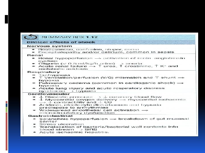
Shock & Organ Systems: compensatory mechanisms Shock leads to increased sympathetic activity. leads to Rise CO, SVR and MAP. Preservation and redistribution of cardiac output, coupled. organ autoregulation, helps to maintain adequate perfusion and oxygen delivery to vital organs (brain, heart, skeletal muscle).

Shock & Organ Systems: These compensatory mechanisms have limits. In case of severe, pro longed and / or uncorrected shock ('decompensated' shock), the clinical manifestations of organ hypoperfusion become apparent. Shock also leads to the up regulation of pro inflammatory cytokines (TNF a, IL l. P and IL 6)

Shock & Organ Systems: Cardiovascular Cardiogenic shock leads to a fall in CO Neurogenic shock leads to vasodilation and reduced SVR. Significant myocardial and vascular dys function frequently occur in other causes of shock.
hypotension results in an imbalance between myocardial oxygen")
Shock & Organ Systems: Cardiovascular Severe (diastolic)hypotension results in an imbalance between myocardial oxygen supply and demand ischaemia result in endocardium. This impairs myocardial contraction Hypoxaemia and acidosis deplete myocardial stores and diminish the cardiac response to both endogenous and exogenous catecholmines. Acid base and electrolyte abnormalities predispose to both atrial and ventricular dysrhythmias.

Shock & Organ Systems: Cardiovascular Inflammatory mediators in sepsis: ØDepress myocardial contractility and ventricular function. ØIncrease indothelial permeability (volume depletion). ØWidespread activation of both coagulation and fibrinolysis (Dl. C).

Shock & Organ Systems: Respiratory Tachypnoea Driven by pain, pyrexia, local lung patholgy, pulmonary oedema, metabolic acidosis or cytokines One of the earliest features of shock.

Shock & Organ Systems: Respiratory Increased minute volume results in reduced PC 02 and respiratory alkalosis Initially this is to compensate for the metabolic acidosis of shock but eventually this mechanism is overwhelmed and blood p. H falls.

Shock & Organ Systems: Respiratory In hypovolaemic states Reduction in pulmonary blood flow underperfusion of ventilated alveoli increasing ventilation perfusion (Y /Q) mismatch

Respiratory In cardiogenic shock Left ventricular failure and pulmonary oedema often compromises the ventilation of perfused alveolar units increasing the shunt fraction within the lung.

Shock & Organ Systems: Respiratory Increased V / Q mismatch and shunt fraction also occur in sepsis. Net result is hypoxaemia that may be refractory to increases in inspired oxygen concentration.

Shock & Organ Systems: Respiratory Sepsis and hypovolaemic shock are both recognized causes of acute lung injury and its more severe variant, the acute respiratory distress syndrome (ARDS).

Shock & Organ Systems: Renal Reduced renal blood flow results in: low urine volume (< O. 5 ml / kg/ h) high urine osmolality low urine sodium content urine. If shock is not reversed, hypoxia leads to acute tubular necrosis (ATN) characterized by oligouria or anurine with a high sodium concentration and an osmolality close to that of plasma.

Shock & Organ Systems: Renal With a fall in glomerular filtration High blood urea and creatinine Hyperkalaernia and a metabolic acidosis Renal failure occurs in about 30 50% of patients with septic shock.

Shock & Organ Systems: Nervous system Due to the increased sympathetic activity, patients appear anxious. Cerebral hypoperfusion and hypoxia cause increasing restlessness, confusion, stupor and coma. Unless cerebral hypoxia has been prolonged, effective resuscitation will usually correct the depressed conscious level rapidly.

Shock & Organ Systems: Gastrointestinal Redistribution of cardiac output observed in shock leads to a marked reduction in splanchnic blood flow.

Shock & Organ Systems: Gastrointestinal In the stomach, the resulting mucosal hypo perfusion and hypoxia predispose to stress ulceration and haemorrhage. In the intestine, movement (translocation) of bacteria and/or bacterial endotoxin from the lumen to the portal vein and cisterna chili; from here it goes into the systemic circulation.

Shock & Organ Systems: Hepatobiliary Ischaemic hepatic injury is frequently seen following hypovolaemic or cardiogenic shock. Reversible transaminase levels elevation indicates hepatocellular injury & occurs 1 3 days after ischaemic insult. Increases in prothrombin time and/or hypoglycaemia are markers of severe injury.

Shock Management General principles: Identification and treatment of the underlying cause. Maintenance of adequate tissue oxygen delivery. CAB Treatment and diagnosis should occur simultaneously. The early recognition and treatment of potentially reversible causes (e. g. bleeding, intra abdominal sepsis, myocardial ischaemia, pulmonary embolus, cardiac tamponade). Most patients with shock will require admission to a high dependency (HDU) or intensive care unit (ICU).

Shock Management Airway and breathing Hypoxaemia Must be prevented If present, rapidly corrected by maintaining a clear airway (e. g. head tilt if no trauma, chin lift) and high flow oxygen (e. g. 10 15 litres/min). The adequacy of this therapy can be estimated continuously using pulse oximetry (Sp. O 2 Severe hypoxaemia need intubation.
. Blood for unresponsive shock or")
Shock Management Circulation Stop bleeding and providing fluid (crystalloid). Blood for unresponsive shock or low haemoglobin. If remain hypoperfused low vasopressors and/ or inotropes may be required.
 cause peripheral vasoconstriction and an increased SVR")
Shock Management Circulation Vasopressors (e. g. noradrenaline) cause peripheral vasoconstriction and an increased SVR Inotropes (e. g. dobutamine) increase myocardial contractility, stroke volume and cardiac output.

Shock Management

Shock Management Hypovolaemic shock The commonest cause of acute hypovolaemic shock in surgical practice is bleeding followed by trauma, ruptured aortic aneurysm, gastrointestinal and obstetric haemorrhage.

Shock Management

Shock Management

Shock Management Hypovolaemic shock It remains unclear whether permissive hypotension is appro priate for all cases of haemorrhagic shock but it appears to improve outcomes following penetrating trauma and ruptured aortic aneurysm.

Shock Management Hypovolaemic shock Rapid fluid resuscitation requires secure vascular access and this is best achieved through two wide bore (14 or 16 gauge) peripheral IV cannulae Cannulation of a central vein provides an alternative means.

Shock Management Hypovolaemic shock In the case of Iife threatening or continued haemorrhage, blood will be required early in the resuscitation. ldeally, fully cross matched packed red blood cells (PRBCs) should be administered, but type specific or O Rhesus negative blood may be used until it becomes available.

Shock Management Hypovolaemic shock Resuscitation strategies aggressively targeting the 'lethal triad' of hypthermia, acidosis and coagulopathy appear to significantly improve outcome following military trauma and observational studies support the immediate use of measures to prevent hypothermia, early correction of severe metabolic acidosis (p. H < 7. 1), maintenance of ionized calcium> l. o mmol and the earl y empirical use of clotting factors and platelets.

Shock Management Hypovolaemic shock Where possible, correction of coagulopathy should be guided by laboratory results (platelet count, prothrombin tme, activated partial thromboplastin time and fibrinogen concentration).
 or rotational thrombo elastometry (ROTEM) Provide near")
Shock Management Hypovolaemic shock Thrombo elastography (TEG) or rotational thrombo elastometry (ROTEM) Provide near patient functional assays of clot formation, platelet function and fibrinolysis. Used to guide the management of coagulopathy.

Shock Management Hypovolaemic shock The antifibrinolytic, tranexamic acid, can be used to inhibit fibrinolysis and has been shown to reduce mortality from bleeding when used early (< 3 hours) and following major trauma.

Shock Management Hypovolaemic shock ln the case of rapid haemorrhage, it is often not possible to use traditional laboratory results to guide the correction of coagulopathy because of the time delay in obtaining these results.

Shock Management Hypovolaemic shock Formula driven approach to the use of PRBC, FFP and platelets 1: 1: 1 targeting the early empirical treatment of coagulopathy. Need further studies

Shock Management Septic shock The Surviving Sepsis Campaign evidence based guidelines on the management of severe sepsis and septic shock: http: / www. survivingsepsis. org.

Shock Management Septic shock Early recognition of severe sepsis and septic shock is critical. Both crystalloid and colloid can be used HES solutions should probably be avoided concerns about inducing acute renal failure. because of Persistent hypotension (MAP < 65 mm. Hg) following restoration of circulating volume is best treated with a vasopressor such as noradrenaline.

Shock Management Septic shock If lactate remain high , oxygen delivery can be increased by transfusion of PRBC to achieve Hg concentration of about 10 g/dl (haematocrit around 0. 3) and/or increasing cardiac output using an inotrope such as dobutamine. Adequate infectiouse source control and the administration of appropriate antibiotics. Blood cultures should be taken prior to the administration of antibiotics but this must not delay therapy.

Shock Management Septic shock Hospital acquired infection should always be considered as a cause of clinical deterioration in surgical patients. Source control includes the removal of infected devices, abscess drainage, the debridement of infected tissue and interventions to prevent ongoing microbial contamination such as repair of a perforated viscus or biliary drainage.

Shock Management Septic shock usually we use empirical broad spectrum antibiotics in the first instance, to cover all bacteria which can be changed to reduce the spectrum of cover once the results of microbiological investigations become available.

Shock Management Septic shock Resuscitation goals for the first 6 hours are: • Central venous pressure (CVP) of 8 12 mm. Hg • Mean arterial blood pressure (MAP)< 65 mm. Hg Urine output < 0. 5 ml/kg/h 2 • Central venous (superior vena cava) 02 sat. 70%

Shock Management Septic shock Culture of urine, cerebrospinal fluid, faeces and bronchoalveolar lavage fluid may also be indicated. Targeted imaging (CXR, ultrasound, computed tomography) may also help identify the source of infection.

Shock Management

Shock Management Cardiogenic shock The commonest cause of cardiogenic shock is myocardial infarction. Treatment based upon the identification and treatment of reversible causes and supportive management to maintain adequate tissue oxygen delivery

Shock Management Cardiogenic shock Routine investigations to identify the cause of car diogenicshock include Serial 12 lead ECGs Troponin or creatinine kinase MB (CK MB) levels and a CXR Echo to identify the cause

Shock Management Cardiogenic shock High concentrations of inspired oxygenation correction of hypovolaemia, needs carful titration. Vasoactive drugs if remain hypoperfused. Intra aortic balloon pump (IABP) as an adjunct in the supportive management of cardiogenic shock (bridge to PCI or CABG)

Obstructive shock Cause Tension Pneoumothorax Cardiac Temponade Massive Pulmonary Embolism Treatments Needle decompression then chest tube pericardiocentisis Tpa Thrombolytic(Fibrinolytic) if in shock

Anaphylactic shock
- Slides: 78