
PEMCOR TACHYCARDIA Frank P Carnevale M D Department

- Slides: 40
PEM|COR: TACHYCARDIA Frank P. Carnevale, M. D. Department of Pediatrics Division of Pediatric Emergency Medicine State University of New York at Buffalo November 13, 2013
PEM|Co. R: 2013 -2014 � � � � 09 -18 -13: Hypovolemic & Distributive Shock 10 -30 -13: Cardiogenic & Obstructive Shock 11 -13 -13: Tachycardia 01 -29 -14: Bradycardia 02 -12 -14: Fever Work-up 03 -19 -14: ATLS & RSI Issues 04 -30 -14: Cardiac Arrest 05 -07 -14: Neonatal Resuscitation
Learning Objectives � � � Differentiate SVT from ST Recognize and manage pediatric tachycardia with a pulse and adequate vs. poor perfusion Describe how and when to use vagal maneuvers, adenosine, and synchronized cardioversion for the treatment of SVT
The sequel to the Halloween Candy Monster (2013)… � http: //youtu. be/RK-o. Qf. FTo. Vg
Learning Objectives � � � Differentiate SVT from ST Recognize and manage pediatric tachycardia with a pulse and adequate vs. poor perfusion Describe how and when to use vagal maneuvers, adenosine, and synchronized cardioversion for the treatment of SVT
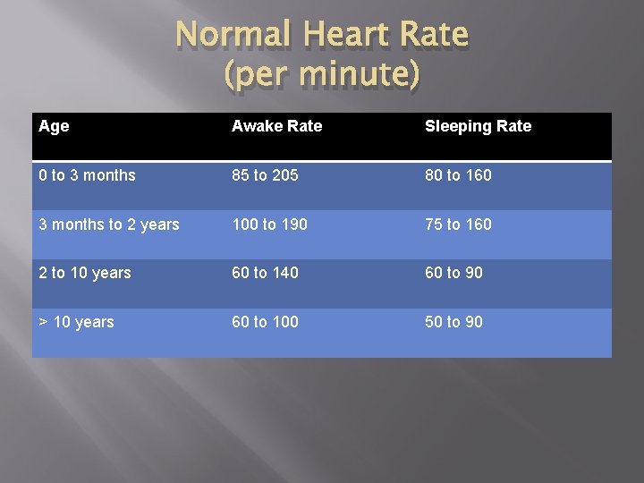
Normal Heart Rate (per minute) Age Awake Rate Sleeping Rate 0 to 3 months 85 to 205 80 to 160 3 months to 2 years 100 to 190 75 to 160 2 to 10 years 60 to 140 60 to 90 > 10 years 60 to 100 50 to 90
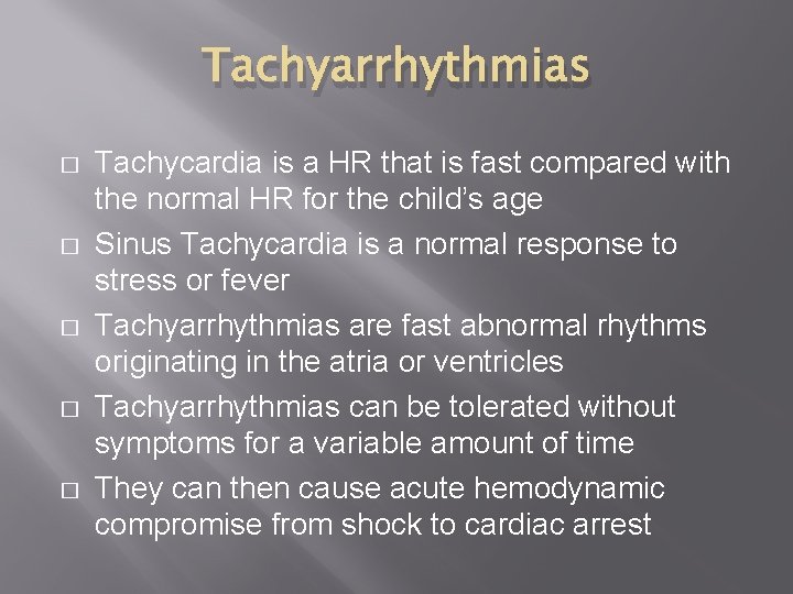
Tachyarrhythmias � � � Tachycardia is a HR that is fast compared with the normal HR for the child’s age Sinus Tachycardia is a normal response to stress or fever Tachyarrhythmias are fast abnormal rhythms originating in the atria or ventricles Tachyarrhythmias can be tolerated without symptoms for a variable amount of time They can then cause acute hemodynamic compromise from shock to cardiac arrest
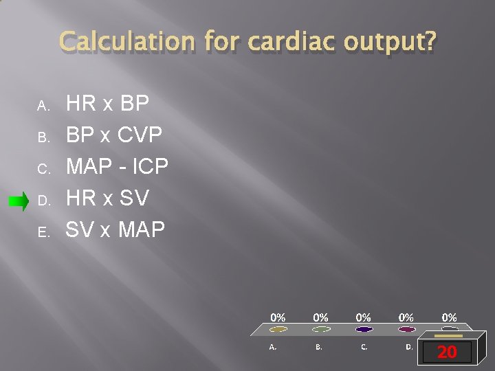
Calculation for cardiac output? A. B. C. D. E. HR x BP BP x CVP MAP - ICP HR x SV SV x MAP 20
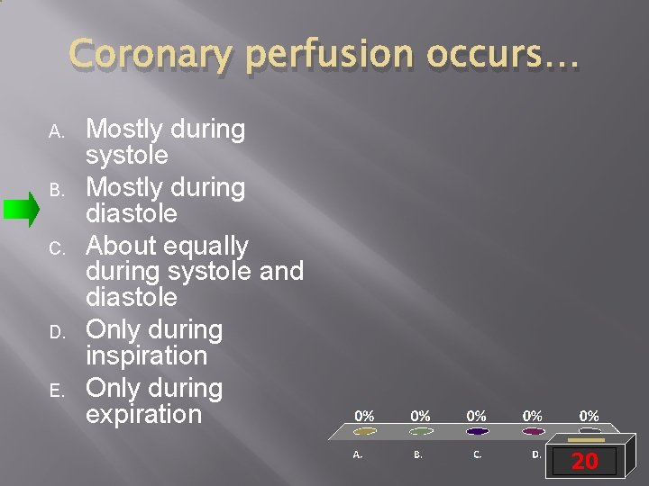
Coronary perfusion occurs… A. B. C. D. E. Mostly during systole Mostly during diastole About equally during systole and diastole Only during inspiration Only during expiration 20
Effect on Cardiac Output � � � CO = HR x SV Increase HR and you increase CO (to a point) You reach a point when diastole is so short that the heart doesn’t have time to fill When end-diastolic filling time decreases, SV decreases and therefore CO decreases Also, coronary perfusion occurs during diastole, so this can be compromised This, along with increased metabolic demand from tachycardia can lead to cardiogenic shock
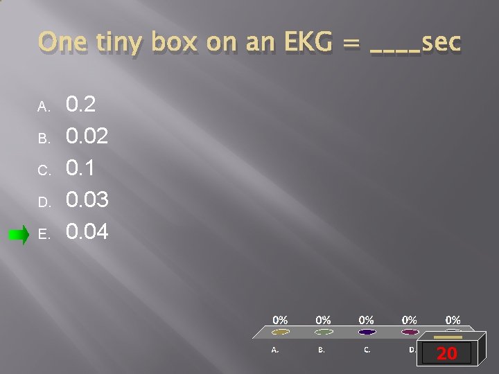
One tiny box on an EKG = ____sec A. B. C. D. E. 0. 2 0. 02 0. 1 0. 03 0. 04 20
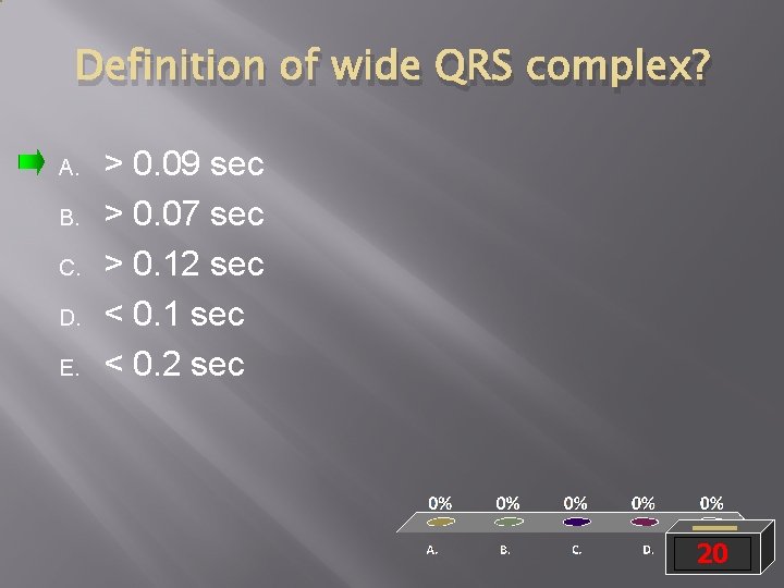
Definition of wide QRS complex? A. B. C. D. E. > 0. 09 sec > 0. 07 sec > 0. 12 sec < 0. 1 sec < 0. 2 sec 20
Wide QRS seen in: A. B. C. D. E. Sinus Tach SVT VT Atrial flutter A fib 20
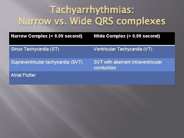
Tachyarrhythmias: Narrow vs. Wide QRS complexes Narrow Complex (< 0. 09 second) Wide Complex (> 0. 09 second) Sinus Tachycardia (ST) Ventricular Tachycardia (VT) Supraventricular tachycardia (SVT) SVT with aberrant intraventricular conduction Atrial Flutter
Sinus Tachycardia � � � A sinus node discharge rate faster than normal for a child’s age Typically develops in response to body’s need for increased cardiac output Common causes: exercise, pain, anxiety, tissue hypoxia, hypovolemia, shock, fever, metabolic stress, injury, toxins, and anemia
ST has beat-to-beat variability. A. B. True False 20
In ST, P waves are present and normal in appearance. A. B. True False 20
ECG characteristics of ST: A. B. C. D. PR normal; R-R constant PR normal; R-R variable PR variable; R-R constant PR variable; R-R normal 20
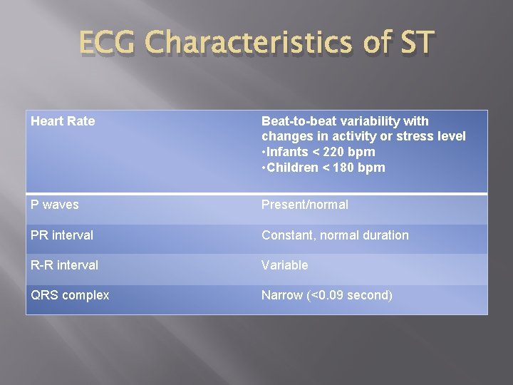
ECG Characteristics of ST Heart Rate Beat-to-beat variability with changes in activity or stress level • Infants < 220 bpm • Children < 180 bpm P waves Present/normal PR interval Constant, normal duration R-R interval Variable QRS complex Narrow (<0. 09 second)
Sinus Tachycardia on monitor � http: //youtu. be/0 Uy 8 TVGo. Njo
Supraventricular Tachycardia (SVT) � � � An abnormally fast rhythm originating above the ventricles Most commonly caused by a reentry mechanism that involves an accessory pathway The most common tachyarrhythmia that causes cardiovascular compromise during infancy
SVT: Clinical Presentation � � A rapid, regular rhythm that appears abruptly and may be episodic In infants, often diagnosed when symptoms of CHF develop Infants: irritability, poor feeding, rapid breathing, unusual sleepiness, vomiting, and pale, mottled, gray, or cyanotic skin Older children: palpitations, SOB, chest pain, dizziness, light-headedness, syncope
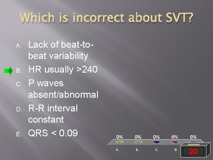
Which is incorrect about SVT? A. B. C. D. E. Lack of beat-tobeat variability HR usually >240 P waves absent/abnormal R-R interval constant QRS < 0. 09 20
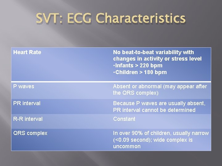
SVT: ECG Characteristics Heart Rate No beat-to-beat variability with changes in activity or stress level • Infants > 220 bpm • Children > 180 bpm P waves Absent or abnormal (may appear after the QRS complex) PR interval Because P waves are usually absent, PR interval cannot be determined R-R interval Constant QRS complex In over 90% of children, usually narrow (<0. 09 second); wide complex is uncommon
SVT on monitor � http: //youtu. be/Re. Jo 4 acl. Ow 8
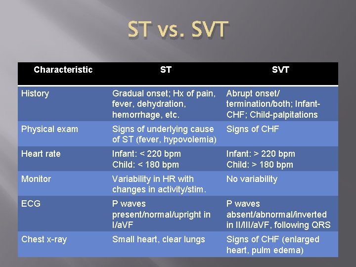
ST vs. SVT Characteristic ST SVT History Gradual onset; Hx of pain, fever, dehydration, hemorrhage, etc. Abrupt onset/ termination/both; Infant. CHF; Child-palpitations Physical exam Signs of underlying cause of ST (fever, hypovolemia) Signs of CHF Heart rate Infant: < 220 bpm Child: < 180 bpm Infant: > 220 bpm Child: > 180 bpm Monitor Variability in HR with changes in activity/stim. No variability ECG P waves present/normal/upright in I/a. VF P waves absent/abnormal/inverted in II/III/a. VF, following QRS Chest x-ray Small heart, clear lungs Signs of CHF (enlarged heart, pulm edema)
Learning Objectives � � � Differentiate SVT from ST Recognize and manage pediatric tachycardia with a pulse and adequate vs. poor perfusion Describe how and when to use vagal maneuvers, adenosine, and synchronized cardioversion for the treatment of SVT
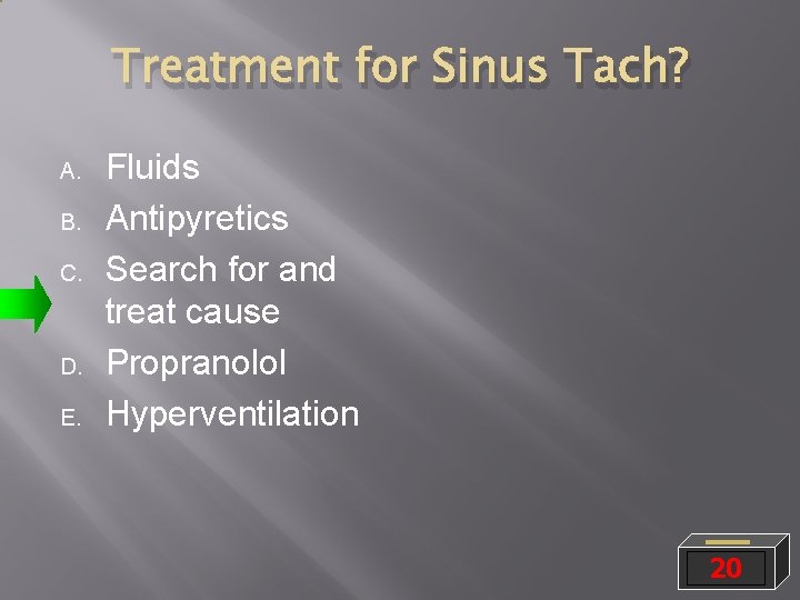
Treatment for Sinus Tach? A. B. C. D. E. Fluids Antipyretics Search for and treat cause Propranolol Hyperventilation 20
Initial treatment for stable SVT? A. B. C. D. E. Adenosine Cardioversion Defibrillation Amiodarone Vagal maneuvers 20
Initial treatment for stable VT? A. B. C. D. E. Defibrillation Vagal manuevers Procainamide Atropine Cardioversion 20
Initial tx for unstable VT? A. B. C. D. E. Defibrillation Epinephrine Amiodarone Cardioversion Adenosine 20
Initial tx for unstable SVT? A. B. C. D. E. Defibrillation Cardioversion Amiodarone Vagal maneuvers Adenosine 20
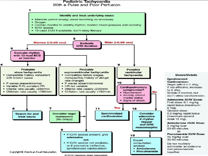
PALS Tachycardia Algorithm. Copyright © American Heart Association
Learning Objectives � � � Differentiate SVT from ST Recognize and manage pediatric tachycardia with a pulse and adequate vs. poor perfusion Describe how and when to use vagal maneuvers, adenosine, and synchronized cardioversion for the treatment of SVT
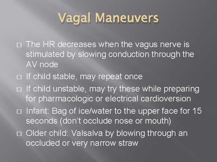
Vagal Maneuvers � � � The HR decreases when the vagus nerve is stimulated by slowing conduction through the AV node If child stable, may repeat once If child unstable, may try these while preparing for pharmacologic or electrical cardioversion Infant: Bag of ice/water to the upper face for 15 seconds (don’t occlude nose or mouth) Older child: Valsalva by blowing through an occluded or very narrow straw
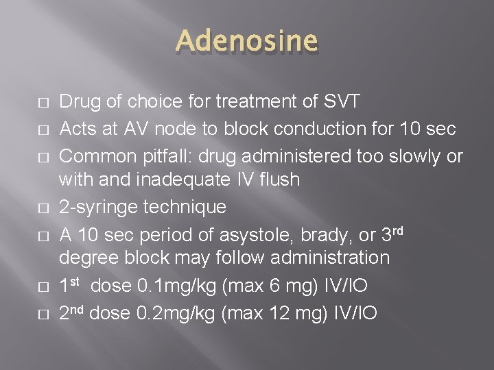
Adenosine � � � � Drug of choice for treatment of SVT Acts at AV node to block conduction for 10 sec Common pitfall: drug administered too slowly or with and inadequate IV flush 2 -syringe technique A 10 sec period of asystole, brady, or 3 rd degree block may follow administration 1 st dose 0. 1 mg/kg (max 6 mg) IV/IO 2 nd dose 0. 2 mg/kg (max 12 mg) IV/IO
Synchronized Cardioversion � � � Defibrillators can deliver unsynchronized and synchronized shocks Unsynchronized: shock delivered any time during the cardiac cycle; used for defibrillation because there is no organized QRS Synchronized: used for cardioversion from SVT and VT with a pulse; shock delivery is timed to coincide with the R wave of the QRS; goal is to prevent VF that results when you shock during the T wave
Cardioversion Pitfalls � � Must select sync mode prior to EACH charge If using paddles, must press both buttons simultaneously When you press shock button, the unit may seem to pause before delivering shock (while waiting for capture)—keep holding down the buttons (if paddles) until shock delivered If the R waves are low amplitude, may need to increase the gain or select a different ECG lead to achieve capture
Learning Objectives � � � Differentiate SVT from ST Recognize and manage pediatric tachycardia with a pulse and adequate vs. poor perfusion Describe how and when to use vagal maneuvers, adenosine, and synchronized cardioversion for the treatment of SVT
Let’s practice some scenarios with the PEM Fellows � � � Station #1: Huma Station #2: Jeremy Station #3: Jen Station #4: Tara Station #5: Meghan Station #6: Danielle