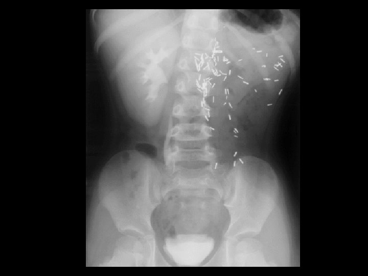
Osteopetrosis Tc99 m sulfur colloid i v intense
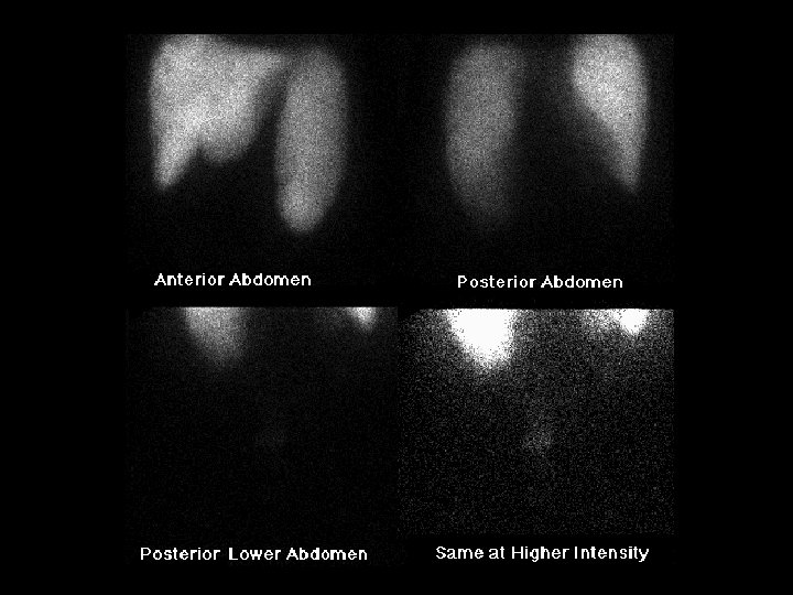


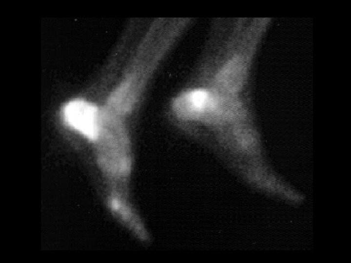


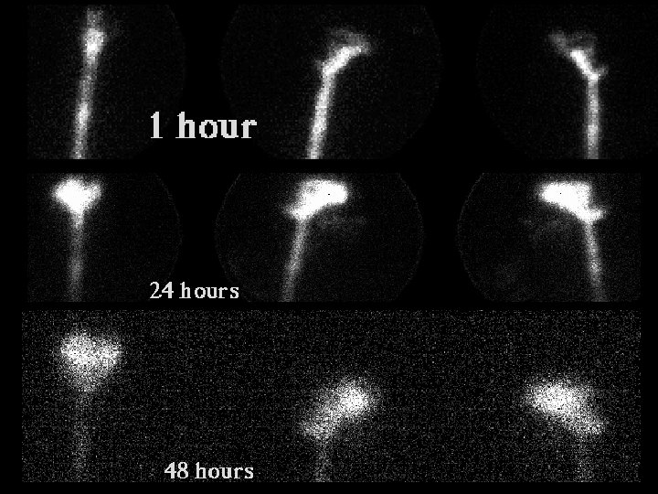


 • Findings: – Intense uptake in the medial")


- Slides: 100
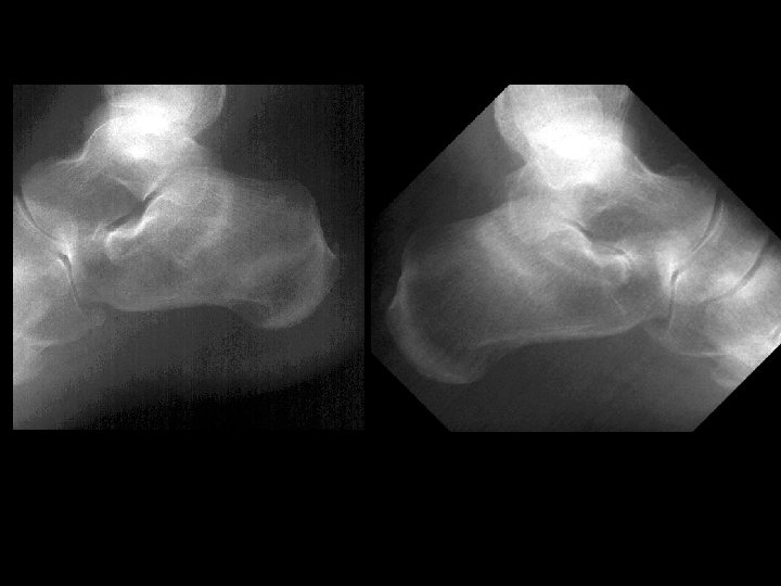
Osteopetrosis • • • Tc-99 m sulfur colloid, i. v. intense liver/spleen uptake, little or none in the bone marrow Trabecular --> compact bone --> Diffuse inc bone density Anemia w/extramedullary hematopoiesis ddx: (adults) – – – myelofibrosis sclerotic metastases renal osteodystrophy multiple myeloma Paget’s
“Brain Death” • 20 m. Ci Tc-99 m DTPA, i. v. • “No evidence of effective cerebral perfusion” (this study does not evaluate the brainstem effectively) • Good perfusion of external carotid arteries, facial structure = “Hot nose sign” • This test used to aid in the determination of brain death • Ddx: – NONE! – This is an Aunt Minnie!
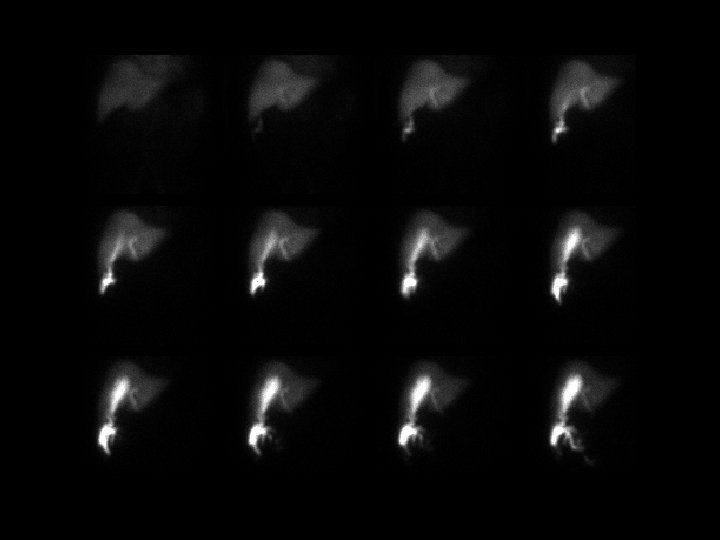
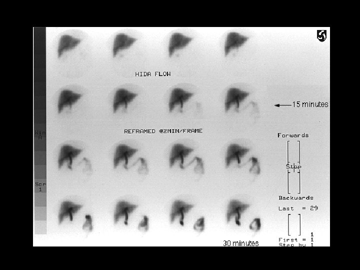
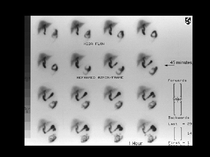
Biliary Leak • • 3. 3 m. Ci Tc 99 m mebrofenin, i. v. leakage of tracer in the gallbladder fossa, subhepatic space, and pericolic gutter treatment: place stent crossing the area of leak ddx: – NONE! – This is an Aunt Minnie!
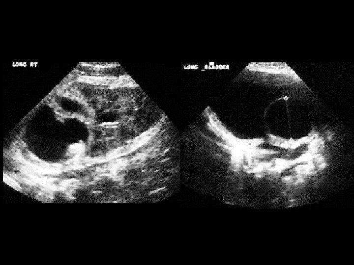
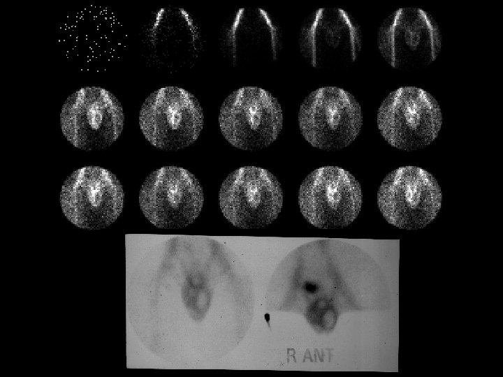
Renal Duplication • • 1. 1 m. Ci Tc-99 m MAG 3, i. v. delayed uptake and no excretion in the right upper pole and filling defect in the bladder dilated upper pole and ectopic ureterocele by US Weigert-Meyer Rule – upper pole obstructs, ureter inserts medially and caudally – lower pole refluxes, inserts normally
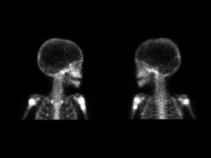
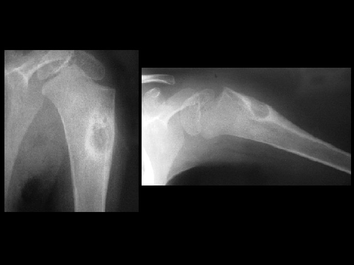
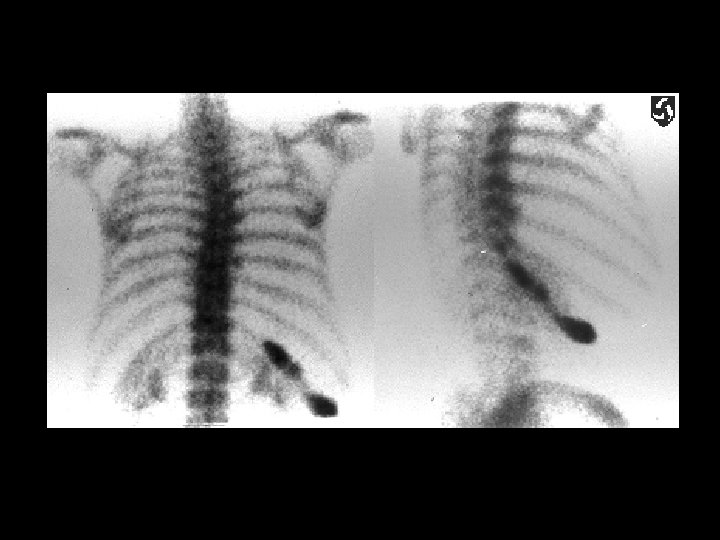
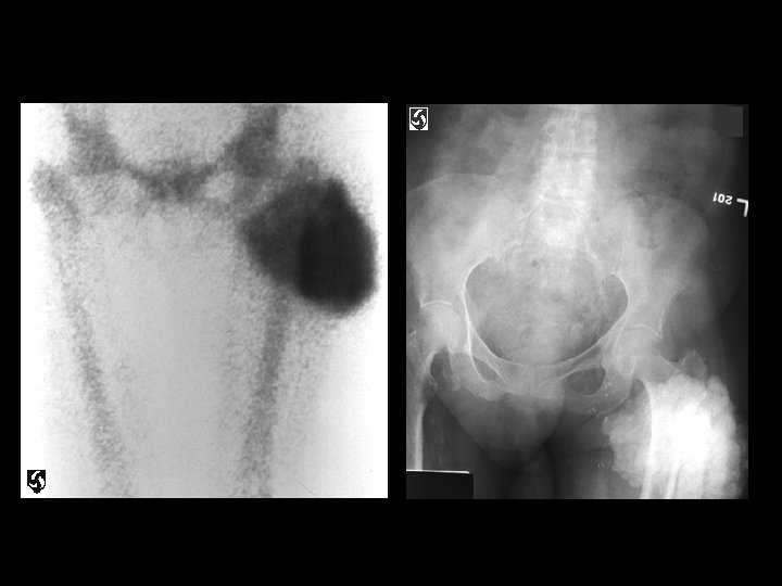
Osteoblastoma • • • 4. 4 m. Ci Tc-99 m MDP, i. v. focal intense activity in the proximal humeral metaphysis eccentric oval lytic lesion with periosteal reaction also occurs in the posterior elements of the spine and long bones histologically similar to osteoid osteoma ddx: – – giant cell tumor ABC non-ossifying fibroma osteosarcoma
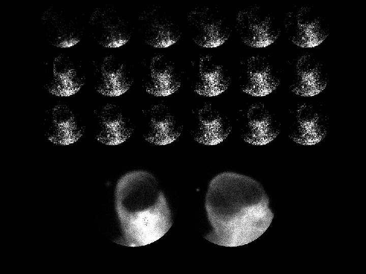
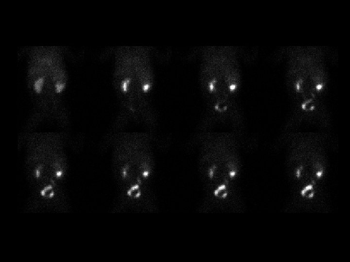
Testicular Torsion • • • 15. 2 m. Ci Tc-99 m pertechnetate, i. v. increased flow surrounding testicle but no flow within bull’s eye appearance valuable in children epididymitis has increased flow and activity ddx: – abscess – hematoma
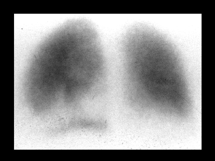
Pulmonary gallium uptake due to Chemo. Rx • • Ga-67 citrate, 5 -10 m. Ci, i. v. Diffuse pulmonary uptake, normal bone, lacrimal glands, liver, and spleen uptake correlate with normal CXR ddx: – pneumonia (PCP in AIDS) – lymphoma – vasculitis
Tuberculosis & AIDS • • • Ga-67 citrate 5 -10 m. Ci, i. v. images obtained at 72 hrs diffuse lymph node activity in the chest & abd intense splenic activity gallium binds transferrin (liver) and lactoferrin (salivary and lacrimal glands, nasopharynx, spleen, and bone marrow) ddx: – lymphoma – sarcoid – infection (abscesses)
Focal Nodular Hyperplasia • • • 5. 2 m. Ci Tc-99 m SC focally increased uptake in the medial segment of the left hepatic lobe activity washes out activity related to hepatic perfusion and distribution of normal Kupffer cells most lesions are “cold” (mets, HCC, hemangioma, adenoma) ddx: – NONE! – This is an Aunt Minnie!
Meckel’s diverticulum • • • 2. 2 m. Ci Tc-99 m pertecnetate, i. v. abnormal focus of activity in the anterior right lower quadrant appears at the same time as normal gastric activity ddx: – active GI bleeding from non-Meckel’s source – ectopic gastric mucosa in jejunal tic (adults) – duplication cyst – inflammation around intussusception (false +)
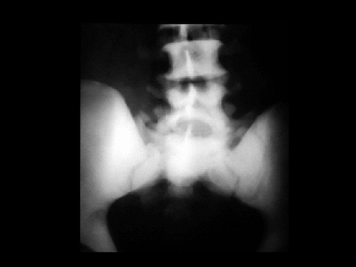
Horeshoe kidney • Findings: – Bone scan demonstrates abnormal location and configuration of kidneys • ddx: – NONE! – This is an Aunt Minnie!
Acute cholecystitis • Findings: – Normal hepatic uptake – Prompt visualization of CBD and small bowel – Non-visualization of GB despite morphine administration – “Rim sign” along inferior right lobe • ddx: – NONE! – This is an Aunt Minnie!
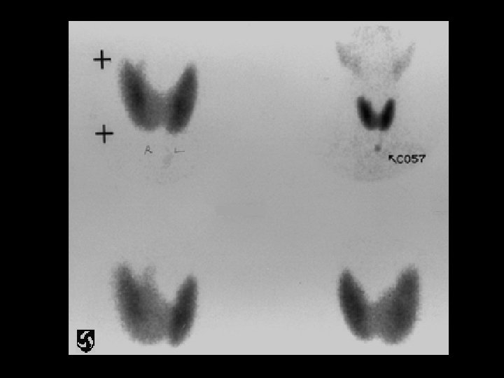
Graves dz • Findings: – Enlarged thyroid – Intense diffuse uptake – sx: hyperthyroid • ddx: – NONE! – This is an Aunt Minnie!
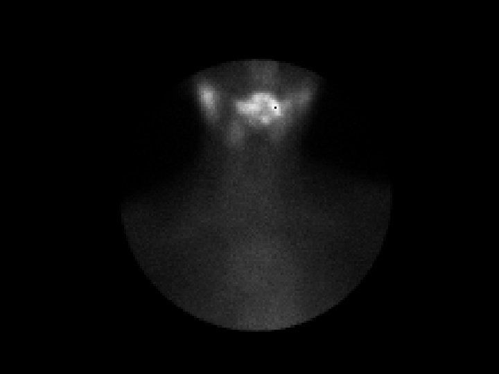
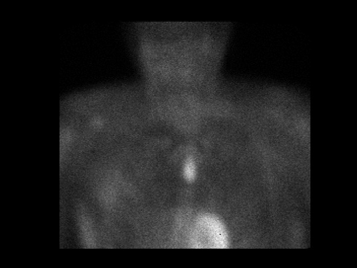
Parathyroid adenoma • Findings: – 99 m. Tc MIBI scan – Intense focal uptake overlying left upper lobe – Persisent after thyroid washout – sx: hypercalemia • ddx: – NONE! – This is an Aunt Minnie!
Low probability for acute PE • Findings: – Perfusion defect in the posterior left lung – Linear activity along the lung periphery beyond perfusion defect = “stripe sign” • ddx: – NONE! – This is an Aunt Minnie!
Fibrous dysplasia • Findings: – Intense activity involving a single enlarged rib • ddx: – Focal trauma – Primary malignancy – Metastasis
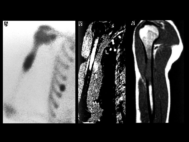
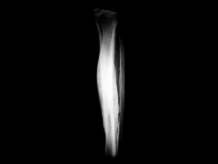
Metastatic Ewing’s sarcoma • Findings: – Intense radiotracer activity in the humeral diaphysis – MR shows an aggressive lesion with cortical disruption and soft tissue edema • ddx: – Lymphoma – Osteosarcoma – Metastasis – osteomyelitis
Hypertrophic osteoarthropathy • Findings: – Diffuse increased linear activity along the long bones • ddx: – Pulmonary disease – Pachydermoperiostosis – Vascular insufficiency – Thyroid acropachy – Fluorosis
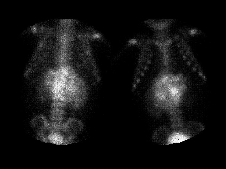
Sickle cell anemia • Findings: – Bone scan shows increased activity in the kidneys, a small spleen, and knees • ddx: – NONE! – This is an Aunt Minnie!
Tumoral calcinosis • Findings: – Intense mass-like uptake about the proximal lateral thigh – Plain film shows soft tissue calcification • ddx: – Myositis ossificans – Heterotopic ossification – Parosteal osteosarcoma
Absent thyroid activity • Findings: – No thyroid activity seen on a 99 m-Tc study • ddx: – Hyperthyroid • painful: – subacute thyroiditis • painless: – exogenous thyroid – Hypothyroid • painless: – Hashimotos's – ablated thyroid – Recent I+ study – Congenital absence (v. rare)
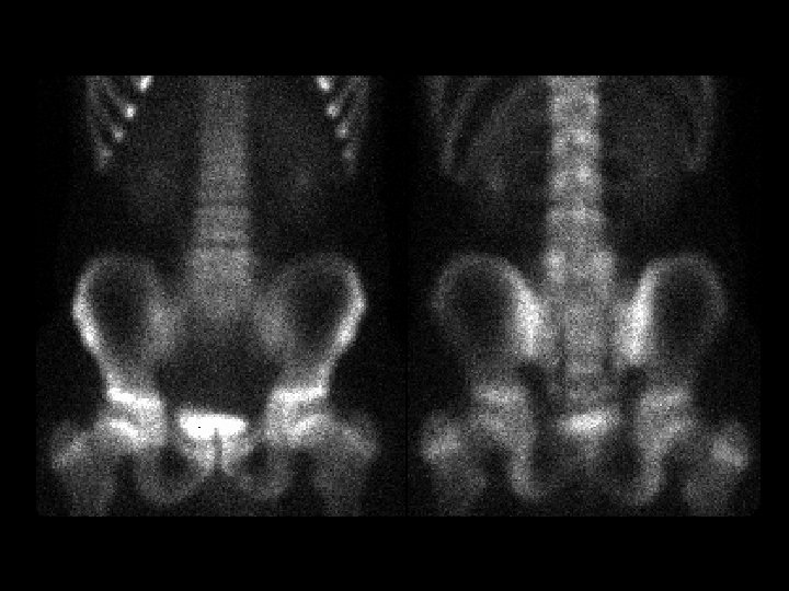
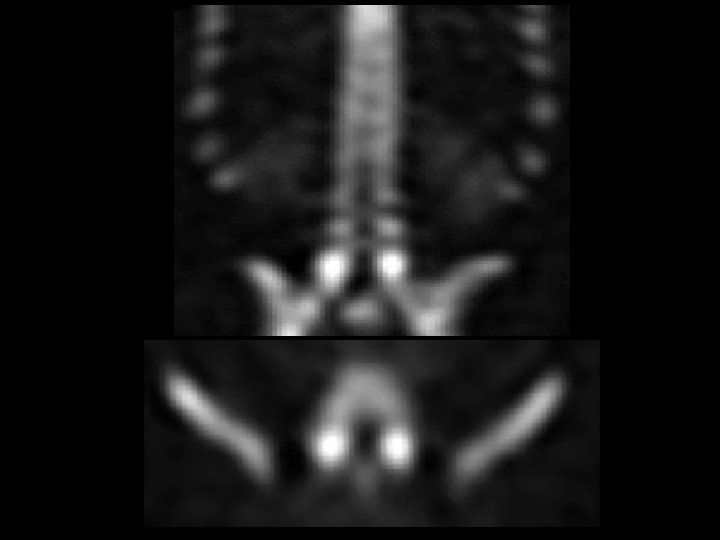
Bilateral pars fractures • Findings: – Planar images of the spine demonstrate subtle increased activity at L 5 – Coronal and axial SPECT images are diagnostic • ddx: – NONE! – This is an Aunt Minnie!
Bilateral calcaneous stress fractures • Findings: – Bone scan shows intense activity in both calcanei = non-specific – Plain films are diagnositic • ddx: – NONE! – This is an Aunt Minnie!
Calcinosis cutis • Findings: – Bone scan shows diffuse activity along the posterior and lateral soft tissues of the buttocks and upper thighs – Normal calcium levels • ddx: – Scleroderma – Dermatamyositis – Polymyositis – SLE – Electrical burns – Idiopathic calcinosis universalis
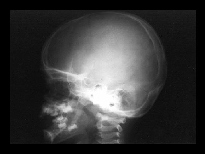
CSF leak • Findings: – Abnormal tracer activity inferior and anterior to skull base – Put pledgets in nose and position pt. to maximize leak – Pledget : plasma > 1. 5 is diagnostic • ddx: – NONE! – This is an Aunt Minnie
Ectopic parathyroid adenoma • Findings: – Delayed MIBI scan shows focal activity in the upper midline chest – Parathyroid scans MUST include a view of the chest for this reason • ddx: – NONE! – This is an Aunt Minnie!
Ectodermal dysplasia • Findings: – Absense of primary teeth – Prominence of secondary teeth – X-linked recessive = males >>> females • ddx: – NONE! – This is an Aunt Minnie!
Engelmann’s disease • Findings: – Increased diaphyseal activity and expansion of the lower extremities – Radiographs show marked of the diaphyseal corticies affecting both periosteal and endosteal surfaces – Progressive diaphyseal dysplasia; autosomal dominant, variable expression • ddx: – Osteopetrosis – Melorhostosis – Hyperphosphatasia – Fibrous dysplasia
Stress fracture • Findings: – Three phase bone scan is positive at the left 4 th MC head • ddx: – Healing fracture – Osteomyelitis – Osteoid osteoma – Charcot joint
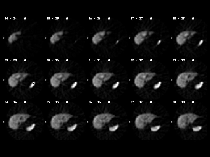
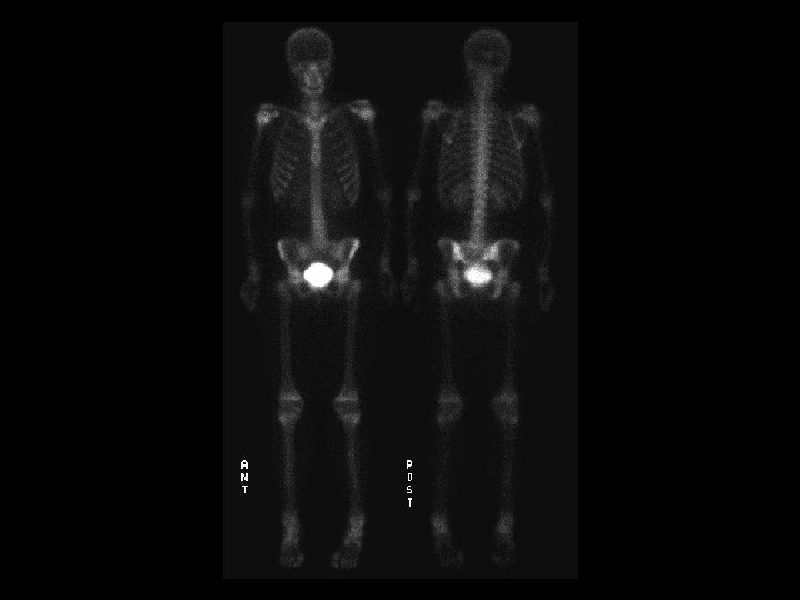
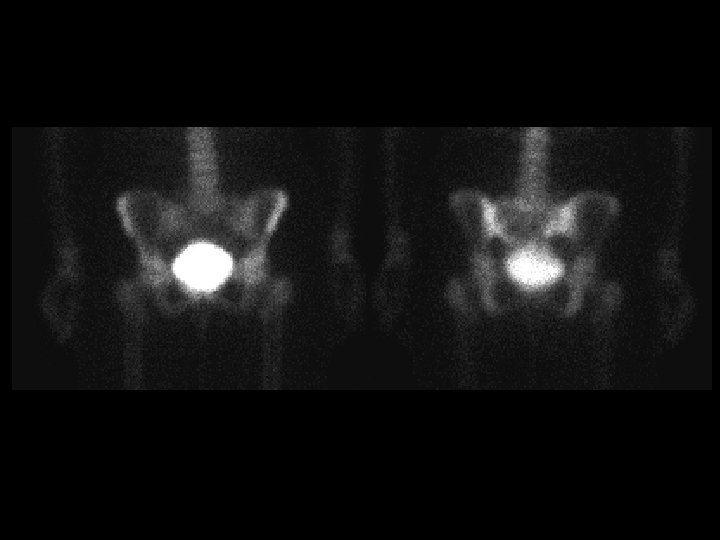
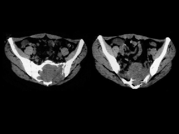
Giant cell tumor of the sacrum • Findings: – Cold lesion of the left sacrum • ddx: – RCC metastasis – Multiple myeloma – Abscess – Hematoma
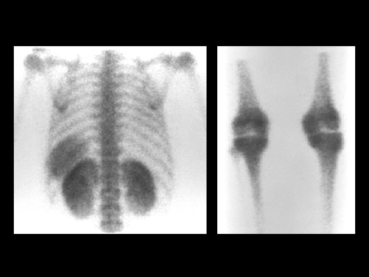
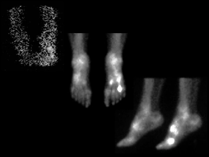
Hypertrophic osteoarthropathy • Finidings: – Linear uptake in the lower extremities, less so in the upper • ddx: – Primary: • Pachydermoperiostitis – Secondary • Pulmonary • Venous insufficiency • Thyroid acropachy • Renal osteodystrophy
Idiopathic pulmonary hemosiderosis • Findings: – Intense lung and renal activity = hemosiderosis & nephrocalcinosis • ddx: – prior lung scintigraphy – alveolar microlithiasis – secondary pulmonary hemosiderosis and ossification • mitral regurgitation
Mucous plug of the left mainstem bronchus • Findings: – DTPA ventilation scan shows no activity in the left lung – Abrupt cut-off of the left mainstem bronchus • ddx: – Intrinsic or extrinsic obstructing lesion – Surgical absence
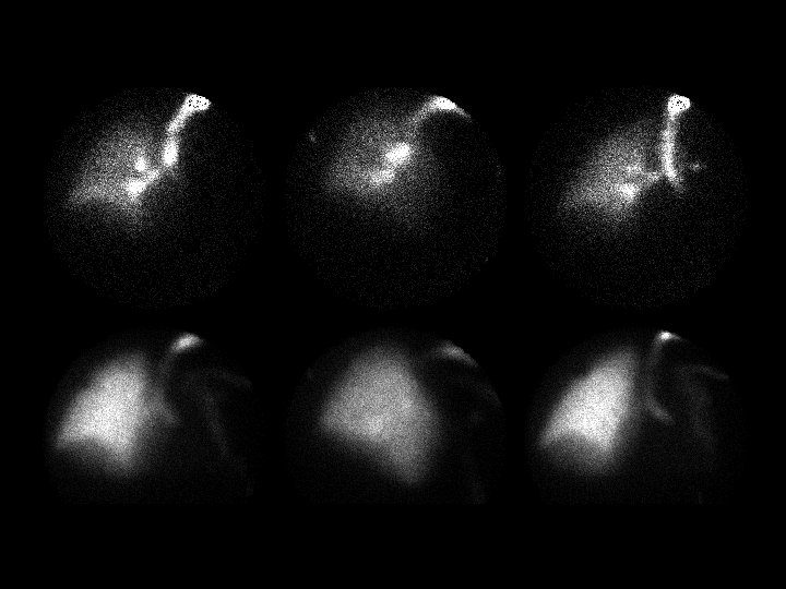
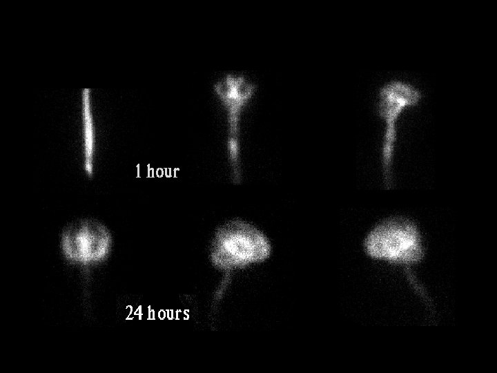
Normal pressure hydrocephalus • • • Findings: – Tracer reflux into ventricles – persistent ventricular activity > 24 hrs Dose / tracer: 0. 5 m. Ci In-111 DTPA Technique: – LP – inject – posterior image over TL spine confirm injection in 15 -30 min – image at 4, 24, 48 hrs (2, 12, 24 hrs kids) Normal study: – basilar cisterns - 4 hrs – convexities - 24 hrs ddx: – obstructive, communicating hyrocephalus (e. g. meningitis)
Off-peak bone scan • Findings: – fuzzy images, lots of soft tissue activity = scatter – camera peak energy set for Co-57 (122 ke. V) instead of Tc-99 m (140 ke. V) • ddx: – NONE! – This is an Aunt Minnie!
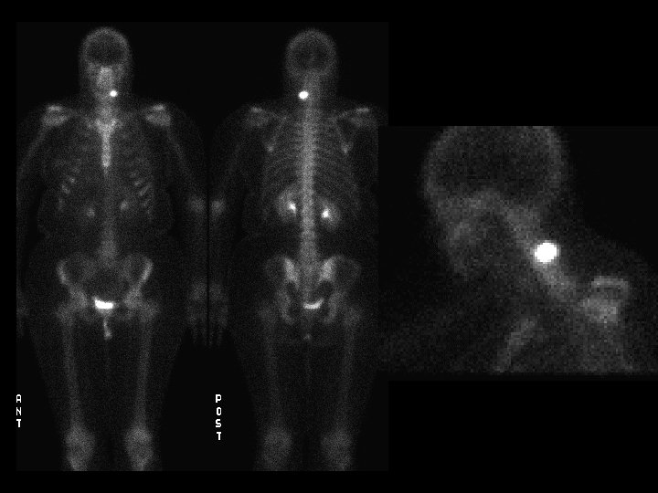
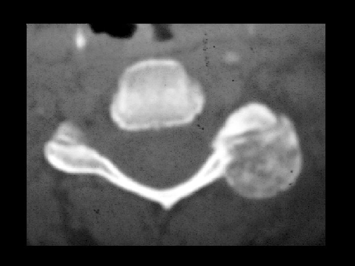
Osteoblastoma • Findings: – Very intense uptake in the posterior left cervical spine – CT scan is diagnositc • ddx: (intense uptake) – Osteiod osteoma – Fracture – Paget's disease – Metastasis – Osteosarcoma
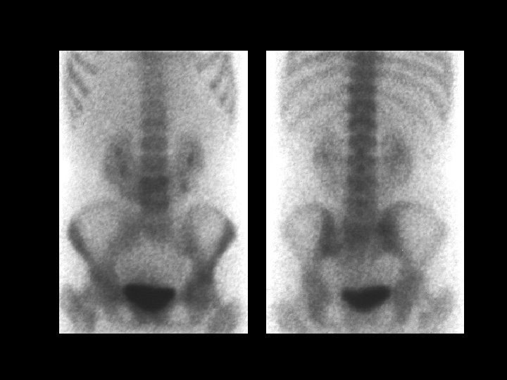
Radiation hypoplasia • Findings: – Bone scan shows a small left ileum, right convex scoliosis, and missing left kidney – Remainder of skeleton is symmetric • ddx: – Hemihypertrophy – Beckwith Wiedemann – Klippel-Trenaunay
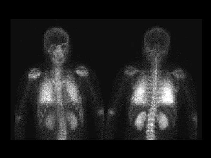
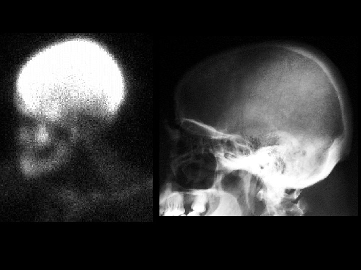
Renal osteodystrophy • Findings: – Diffuse skull uptake – Plain film shows a granular appearance = "salt & pepper" • ddx: – Mets (prostate, breast, lung, lymphoma, bladder) – Metabolic bone disease (hyperpara, osteomalacia, rickets, hyp vit D) – Paget's disease – Myelofibrosis – Mastocytosis – Aplastic anemia
Right to left shunt • Findings: – Pulmonary perfusion scan shows renal and bone marrow activity • Causes: – Intracardiac • Eisenmenger’s • PFO – AVM • ddx: – Free pertechnatate • Positive brain uptake
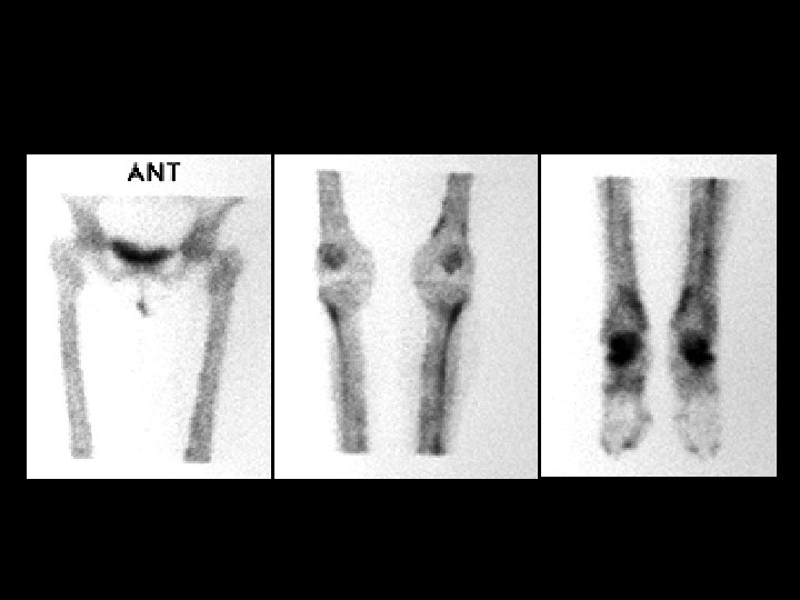
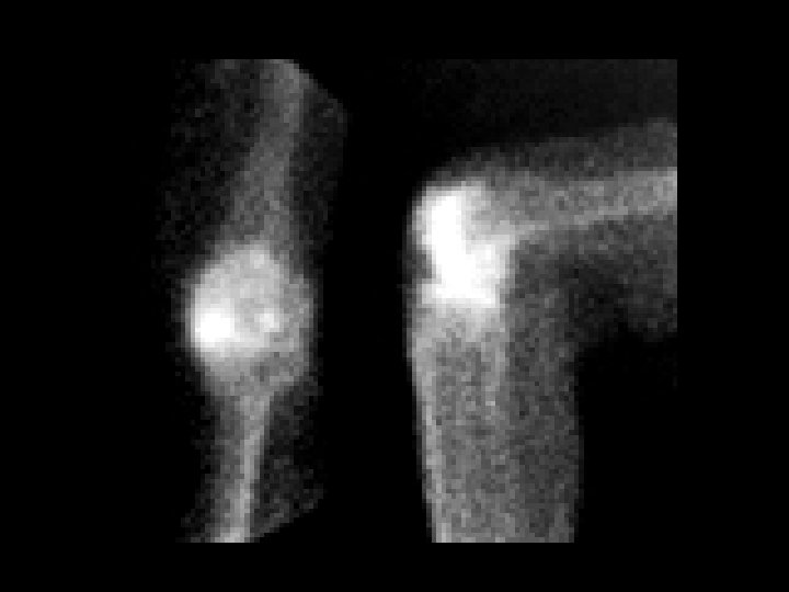
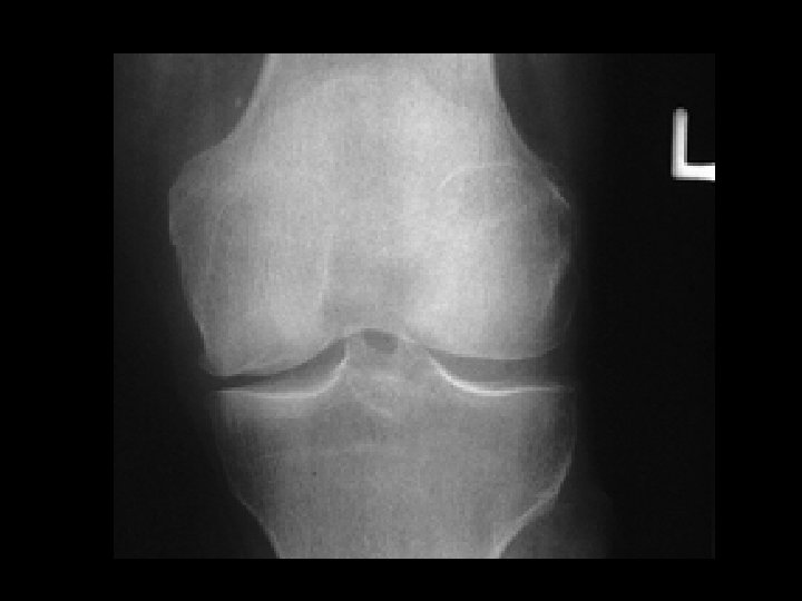
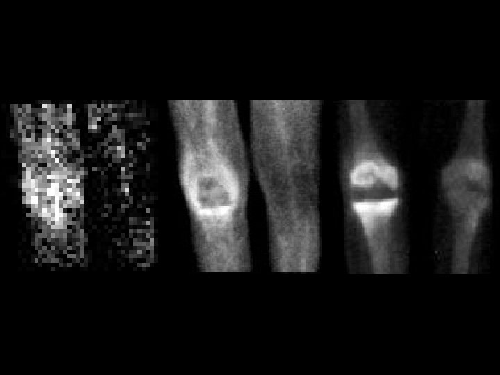
Spontaneous osteonecrosis of the knee (SONK) • Findings: – Intense uptake in the medial femoral condyle – Plain radiograph shows flattening of the medial femoral condyle and subchondral lucency • ddx: – Fracture – Osteoarthritis – Osteomyelitis – Tumor
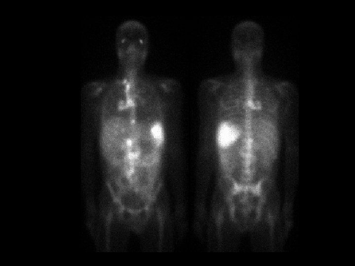
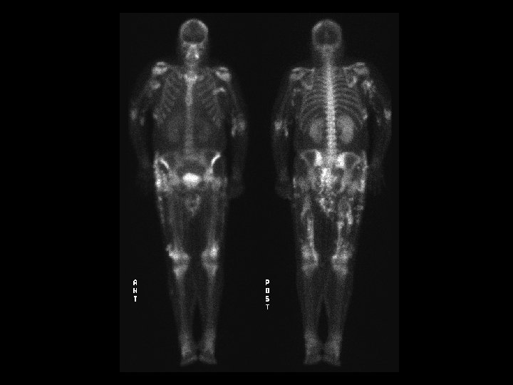
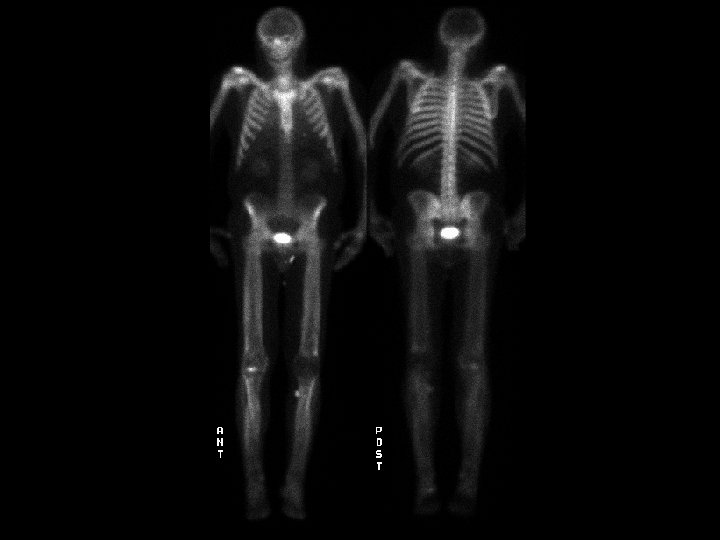
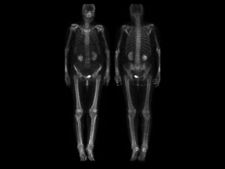
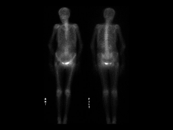
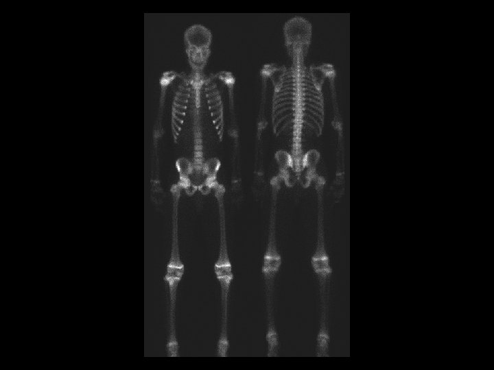
Superscan • • Findings: – Diffusely increased radiotracer activity of the skeleton – No renal or soft tissue uptake ddx: – Diffuse metastates – Metabolic • Renal osteodystrophy
Septic arthritis • Findings: – Positive three phase bone scan of the right knee • ddx: – Charcot joint – Inflammatory arthritis
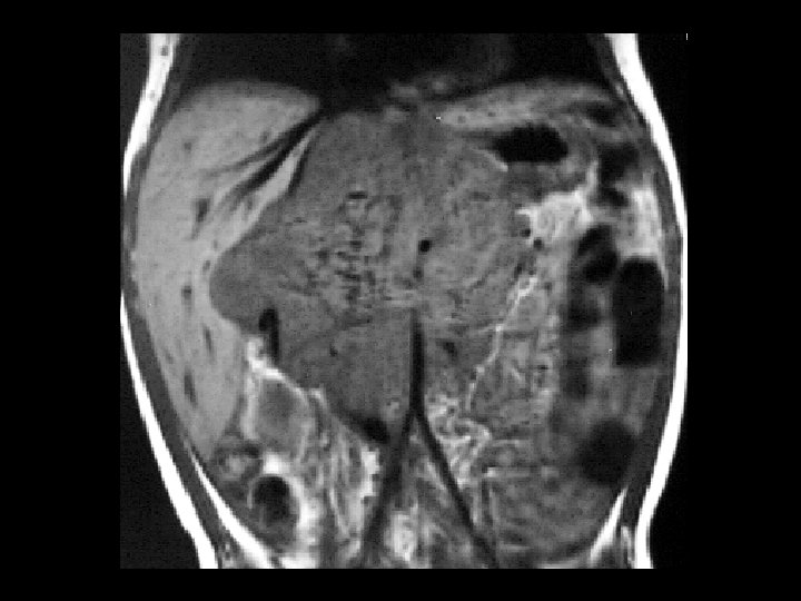
Neuroblastoma • Findings: – Bone scan shows increased uptake in the mid abdomen of a child – MR scan shows a corresponding soft tissue mass with scattered T 1 low sign foci = Ca 2+ • ddx: – NONE! – This is an Aunt Minnie!