Musculoskeletal Pathology Assoc Professor Jan Laco MD Ph
- Slides: 67
Musculoskeletal Pathology Assoc. Professor Jan Laco, MD, Ph. D
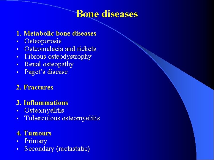
Bone diseases 1. Metabolic bone diseases • Osteoporosis • Osteomalacia and rickets • Fibrous osteodystrophy • Renal osteopathy • Paget’s disease 2. Fractures 3. Inflammations • Osteomyelitis • Tuberculous osteomyelitis 4. Tumours • Primary • Secondary (metastatic)
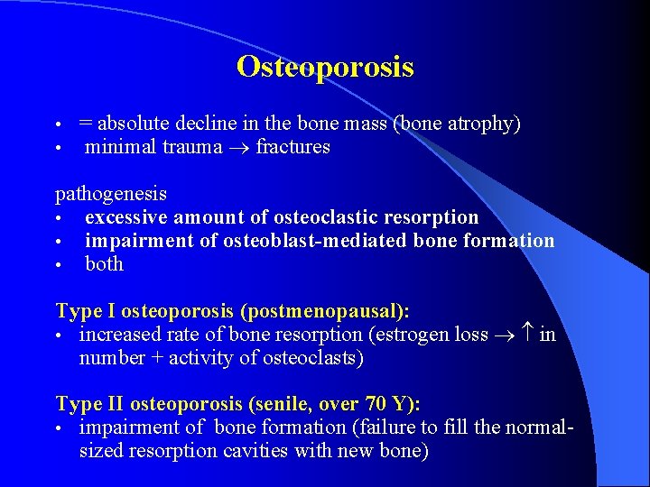
Osteoporosis • • = absolute decline in the bone mass (bone atrophy) minimal trauma fractures pathogenesis • excessive amount of osteoclastic resorption • impairment of osteoblast-mediated bone formation • both Type I osteoporosis (postmenopausal): • increased rate of bone resorption (estrogen loss in number + activity of osteoclasts) Type II osteoporosis (senile, over 70 Y): • impairment of bone formation (failure to fill the normalsized resorption cavities with new bone)
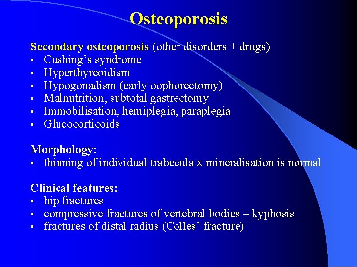
Osteoporosis Secondary osteoporosis (other disorders + drugs) • Cushing’s syndrome • Hyperthyreoidism • Hypogonadism (early oophorectomy) • Malnutrition, subtotal gastrectomy • Immobilisation, hemiplegia, paraplegia • Glucocorticoids Morphology: • thinning of individual trabecula x mineralisation is normal Clinical features: • hip fractures • compressive fractures of vertebral bodies – kyphosis • fractures of distal radius (Colles’ fracture)
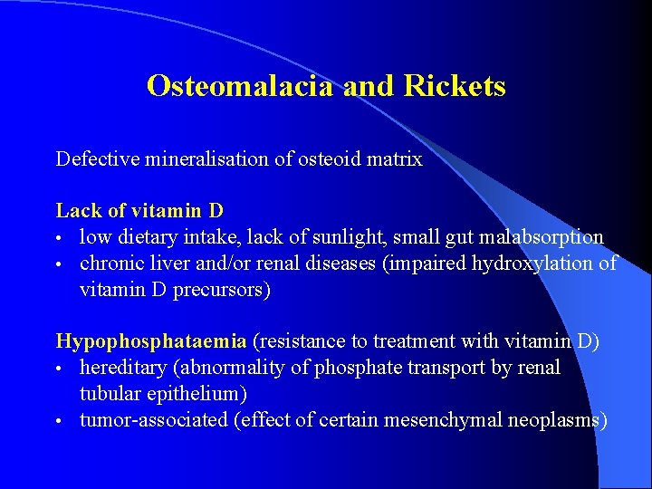
Osteomalacia and Rickets Defective mineralisation of osteoid matrix Lack of vitamin D • low dietary intake, lack of sunlight, small gut malabsorption • chronic liver and/or renal diseases (impaired hydroxylation of vitamin D precursors) Hypophosphataemia (resistance to treatment with vitamin D) • hereditary (abnormality of phosphate transport by renal tubular epithelium) • tumor-associated (effect of certain mesenchymal neoplasms)
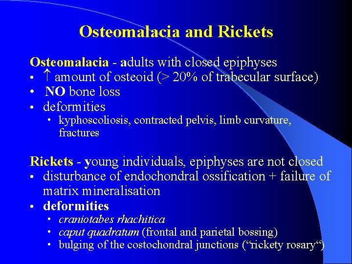
Osteomalacia and Rickets Osteomalacia - adults with closed epiphyses • amount of osteoid (> 20% of trabecular surface) • NO bone loss • deformities • kyphoscoliosis, contracted pelvis, limb curvature, fractures Rickets - young individuals, epiphyses are not closed • disturbance of endochondral ossification + failure of matrix mineralisation • deformities • craniotabes rhachitica • caput quadratum (frontal and parietal bossing) • bulging of the costochondral junctions (“rickety rosary“)
Fibrous osteodystrophy = osteitis fibrosa cystica generalisata von Recklinghausen pathogenesis severe hyperparathyreoidism (esp. primary) PTH osteoclastic resorption of bone Mi: outline of bony trabeculae shows deep indentations filled with numerous osteoclasts and connective tissue • severe cases brown tumors (a misnomer) = areas in which trabecular bone has been completely eroded away and replaced by vascular fibrous tissue with many osteoclasts and hemosiderin depositions (brown color) • • clinical features: bone pain, pathological fractures, hypercalcaemia
Paget’s disease of bone • • = osteitis deformans Sir James Paget (1882) enlargement + deformities of bones > 45 years one bone x many bones sites: skull, pelvis, spine etiology ? ? ? (viral infection)
Paget’s disease of bone • Gross: • bones enlarged and thickened (thick calvarium) and softer (deformities – bowing of lower limbs) • skull deformities (narrowing of foramina of cranial nerves neuropathies, deafness) • Micro: • episodes of bone resorption + subsequent reparation • 1. osteolytic phase – prominent osteoclastic resorption, giant multinucleated osteoclasts • 2. mixed / osteoblastic phase – continuing resorption and osteoblastic new bone formation • 3. osteosclerotic or burned-out phase – large irregular trabeculae with mosaic-like appearance due to numerous cement lines !!! greater risk of developing osteosarcoma !!!
Fractures = breaks in continuity of bone • infraction = incomplete fracture • pathological fracture: • bone disease (osteoporosis, cysts, tumours) minor trauma or spontaneously fracture Fracture healing: hematoma between ends of bone + necrosis of BM organization of hematoma - granulation tissue connection of bone ends by fibrous tissue (fibrous callus) woven bone + cartilage in fibrous tissue provisional callus remodelling to mature lamellar bone definite callus • Impaired fracture healing • • infection, poor immobilization, non-vital bone ends • exuberant callus pseudo-arthrosis
Osteomyelitis = infective inflammation of bone (Staphylococci, Streptococci. . . ) • infection way: • directly (open fractures, operations) • hematogeneously • pathogenesis • acute pyogenic inflammation of BM rise of interstitial pressure blood supply ischaemic necrosis • inflammation subperiosteal space (subperiosteal abscess) periosteal blood vessels shear off ischemic necrosis • perforation of periosteum soft tissues and skin - sinus track, portions of dead bone can be discharged onto the skin • • • adults sites: mandible (periapical infection, fracture) complications • sepsis, amyloidosis, pyogenic arthritis, skin squamous carcinoma
Tuberculous osteomyelitis • children, blood-borne infection • long bones, vertebrae • bone destruction + granulomatous lesion with caseation • Tuberculosis of the spine (Pott’s disease) • sharp anterior angulation of the spine (kyphosis) • caseation may spread to the paravertebral soft tissues and track down along the psoas muscle to form an inguinal subcutaneous mass
Bone metastases • Much more common than primary bone tumours • Usually multiple • • Osteolytic (bone destruction) Osteoplastic (reactive bone formation) – prostatic carcinoma • Tumors • prostatic carcinoma, breast carcinoma, renal cell carcinoma, carcinoma of stomach, thyroid carcinoma
Primary bone tumours – classification less common, usually solitary diagnosis: microscopy + age + site + RTG 1. Bone-forming tumours • Osteoma • Osteoid osteoma • Osteoblastoma • Osteosarcoma 2. Cartilage-forming tumours • Osteochondroma • Chondrosarcoma 3. Other tumours • Giant cell tumor of bone • Ewing’s sarcoma 4. Tumour-like lesions • Fibrous dysplasia • Bone cysts
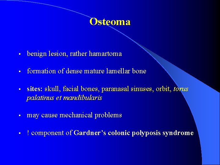
Osteoma • benign lesion, rather hamartoma • formation of dense mature lamellar bone • sites: skull, facial bones, paranasal sinuses, orbit, torus palatinus et mandibularis • may cause mechanical problems • ! component of Gardner’s colonic polyposis syndrome
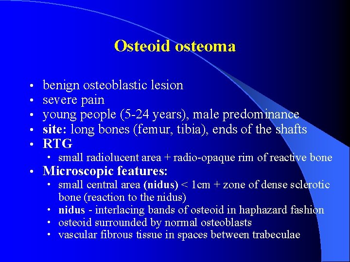
Osteoid osteoma • • • benign osteoblastic lesion severe pain young people (5 -24 years), male predominance site: long bones (femur, tibia), ends of the shafts RTG • small radiolucent area + radio-opaque rim of reactive bone • Microscopic features: • small central area (nidus) < 1 cm + zone of dense sclerotic bone (reaction to the nidus) • nidus - interlacing bands of osteoid in haphazard fashion • osteoid surrounded by normal osteoblasts • vascular fibrous tissue in spaces between trabeculae
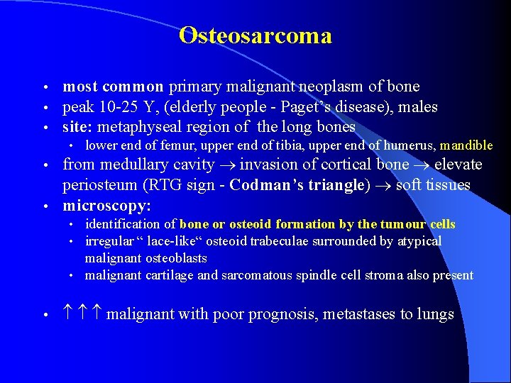
Osteosarcoma • • • most common primary malignant neoplasm of bone peak 10 -25 Y, (elderly people - Paget’s disease), males site: metaphyseal region of the long bones • lower end of femur, upper end of tibia, upper end of humerus, mandible from medullary cavity invasion of cortical bone elevate periosteum (RTG sign - Codman’s triangle) soft tissues • microscopy: • • identification of bone or osteoid formation by the tumour cells • irregular “ lace-like“ osteoid trabeculae surrounded by atypical malignant osteoblasts • malignant cartilage and sarcomatous spindle cell stroma also present • malignant with poor prognosis, metastases to lungs
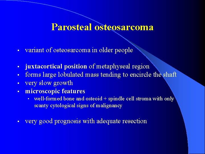
Parosteal osteosarcoma • variant of osteosarcoma in older people • • juxtacortical position of metaphyseal region forms large lobulated mass tending to encircle the shaft very slow growth microscopic features • well-formed bone and osteoid + spindle cell stroma with only scanty cytological signs of malignancy • very good prognosis with adequate resection
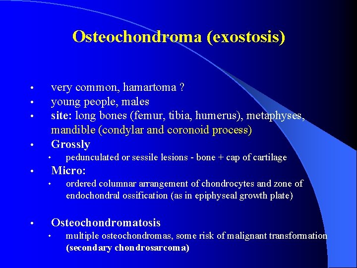
Osteochondroma (exostosis) • • very common, hamartoma ? young people, males site: long bones (femur, tibia, humerus), metaphyses, mandible (condylar and coronoid process) Grossly • • Micro: • • pedunculated or sessile lesions - bone + cap of cartilage ordered columnar arrangement of chondrocytes and zone of endochondral ossification (as in epiphyseal growth plate) Osteochondromatosis • multiple osteochondromas, some risk of malignant transformation (secondary chondrosarcoma)
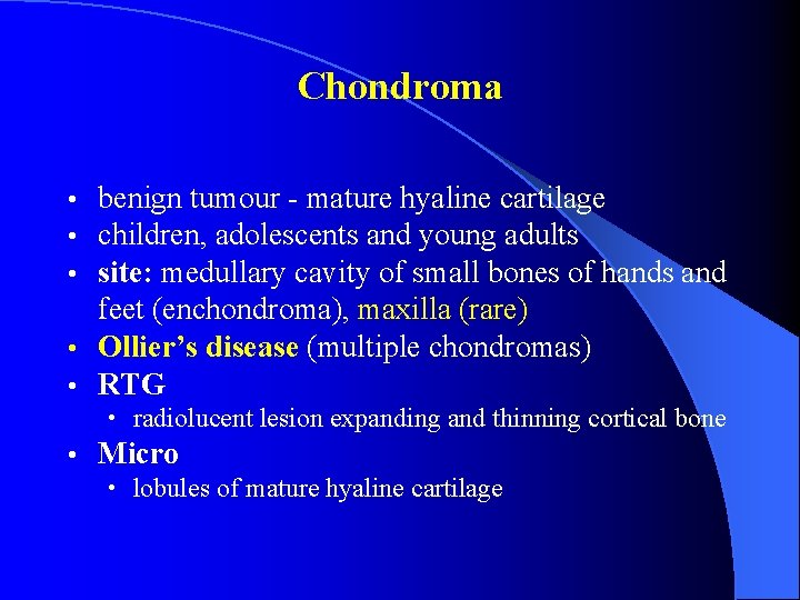
Chondroma benign tumour - mature hyaline cartilage children, adolescents and young adults site: medullary cavity of small bones of hands and feet (enchondroma), maxilla (rare) • Ollier’s disease (multiple chondromas) • RTG • • radiolucent lesion expanding and thinning cortical bone • Micro • lobules of mature hyaline cartilage
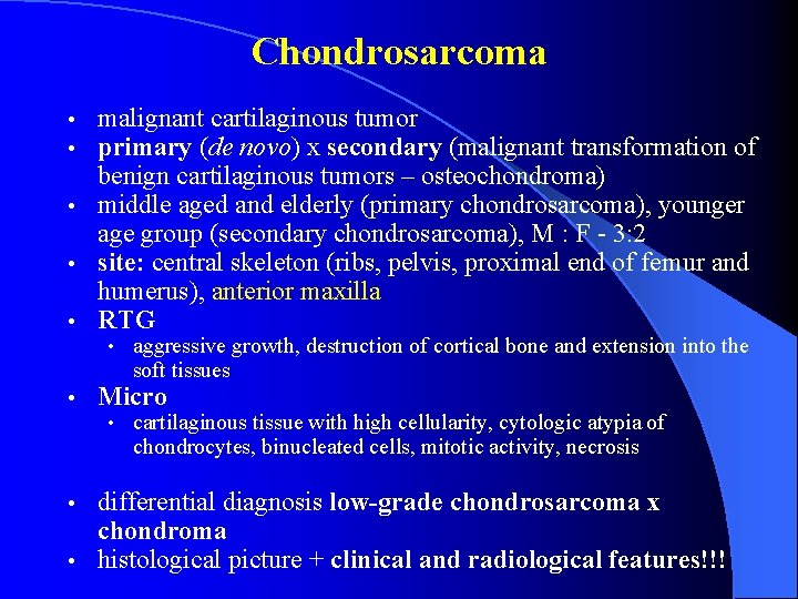
Chondrosarcoma malignant cartilaginous tumor primary (de novo) x secondary (malignant transformation of benign cartilaginous tumors – osteochondroma) • middle aged and elderly (primary chondrosarcoma), younger age group (secondary chondrosarcoma), M : F - 3: 2 • site: central skeleton (ribs, pelvis, proximal end of femur and humerus), anterior maxilla • RTG • • • aggressive growth, destruction of cortical bone and extension into the soft tissues • Micro • cartilaginous tissue with high cellularity, cytologic atypia of chondrocytes, binucleated cells, mitotic activity, necrosis differential diagnosis low-grade chondrosarcoma x chondroma • histological picture + clinical and radiological features!!! •
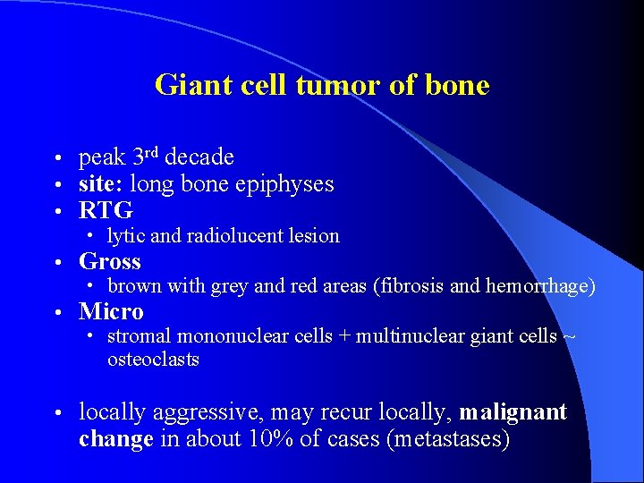
Giant cell tumor of bone • • • peak 3 rd decade site: long bone epiphyses RTG • lytic and radiolucent lesion • Gross • brown with grey and red areas (fibrosis and hemorrhage) • Micro • stromal mononuclear cells + multinuclear giant cells ~ osteoclasts • locally aggressive, may recur locally, malignant change in about 10% of cases (metastases)
Ewing’s sarcoma • • group of primitive neuroectodermal tumours (PNET) peak 5 -20 years, males site: most often long bones and pelvis, mandible (body) RTG • lytic destruction of the bone • Gross • grayish white, focal necrosis and haemorrhage • Micro • small uniform round cells with glycogen (PAS +) • clinical features: pain and swelling • • very aggressive tumor with poor prognosis metastatic spread to lungs and other bones
Fibrous dysplasia • non-neoplastic condition • children and adolescents • site: ribs, femur, tibia, maxilla, mandibula, humerus • monostotic x polyostotic form • micro • well-demarcated localized area of bone replaced by fibrous tissue with spicules of woven bone of fish-hook or Chinese letters shape • Mc. Cune-Albright´s syndrome = polyostotic FD + skin pigmentation + precocious puberty
Bone cysts • Solitary bone cyst • unicameral cavity lined by smooth fibrous membrane • mandible • Aneurysmal bone cyst • multiple spaces filled with red blood cells separated by thin fibrous septa with numerous osteoclasts • rare in jaws
Joint diseases 1. Congenital defects • Dysplasia coxae congenita 2. Trauma 3. Degenerative diseases • Osteoarthritis 4. Inflammation • Infective arthritis • Lyme disease • Tuberculous arthritis • Rheumatoid arthritis • Gout 5. Tumours • Pigmented villonodular synovitis
Dysplasia coxae congenita • autosomal recessive disorder • hypoplasia of hip joint with congenital subluxation or luxation • untreated may lead to early secondary osteoarthritis
Trauma • disruption of synovial membrane – bleeding into joint cavity (hemarthros) • organization of hematoma fibrous adhesions limiting joint movements or ankylosis (join space replaced by metaplastic bone) • hemarthros often complicates disorders of coagulation (hemophilia)
Osteoarthritis • • • most common of joint diseases (14% of adults) breakdown of the articular cartilage weight-bearing joints • hip, knee, intervertebral joints, TMJ (asymptomatic) • clinical features • pain, joint deformity, limitation of movement, crepitus (creaking sound heard on movement of the joint) • RTG • narrowing of joint space • Two main groups: • primary OA: no known associated condition • secondary OA: known associated event or disease causally related to OA (congenital disorders, inflammation, trauma)
Osteoarthritis Morphology: erosive changes of joint cartilage (“fibrillation“) + flaking off of small portions of cartilage • complete loss of cartilage – polishing of the denuded bone (smooth ivory-like surface) • thickening of subchondral bone plate, synovial fluid under pressure may enter into small defects in the bone – forming of subchondral pseudocysts • bony outgrowths at the margins of the articular cartilage (osteophytes) • •
Infective arthritis • • bacteria (St. aureus, N. gonorrhoeae) routes of infection: • blood spread (most common) • direct penetration (trauma) • direct spread (osteomyelitis) • morphology • hyperemia + acute inflammatory infiltrate in synovial membrane, exudate accumulation in the joint cavity (pyarthros) • lysosomal enzymes released from neutrophils may cause severe damage of the articular cartilage – secondary osteoarthritis
Lyme disease spirochaete Borrelia burgdorferi transmitted to humans via tick bites • skin + nervous system + heart + joints • 3 clinical stages: • • stage I: • macular skin lesion (erythema migrans) + low-grade fever, headache, arthralgia, muscle pain, LNpathy • stage II: bloodstream dissemination of borrelia • • nervous system (meningitis, meningoencephalitis, neuritis) heart (atrioventricular block, pericarditis) • stage III: • • chronic disease (months to years after initial infection) arthritis (large joints, similar to rheumatoid arthritis) nervous system (encephalopathy) skin (acrodermatitis chronica atrophicans Herxheimer)
Tuberculous arthritis • • children associated with infection (haematogeneous dissemination) • site: knee, hip, elbow and ankle • clinical features • insidious development of pain, swelling and limitation of movement, other signs of inflammation mild or absent • Micro • synovial hyperplasia, tuberculous granulomas in 90% of cases
Rheumatoid arthritis • common multisystem autoimmune disease • 3 x commonly in premenopausal women as in males • site: small joints of the hands and feet, knees, hips, involvement frequently symmetrical, TMJ - minor symptoms • rheumatoid factors (Ig. M directed against Fc´ portion of Ig. G) positive in 95% pts.
Rheumatoid arthritis • • • inflammatory hyperplasia of synovial membrane with increased vascularity, cellularity and synovial fluid production inflammatory infiltrate (lymphocytes, plasma cells, lymphoid follicles proliferation of inflammed hypervascular granulation tissue termed pannus degradation of underlying articular cartilage fibrous adhesions limit joint movement, formation of metaplastic bone (ankylosis) deformities of joints (ulnar deviation of fingers) periarticular rheumatoid nodules in 30% • central area of fibrinoid necrosis + macrophages and fibroblasts arranged in a palisaded fashion) • complication: secondary amyloidosis
Variants of RA • Juvenile rheumatoid arthritis (JRA): < 16 Y • large joints predominantly involved, RFs often negative • Still’s disease (systemic JRA) • fever, leucocytosis, enlargement of liver, spleen and lymph nodes • Felty’s syndrome: RA + splenomegaly + neutropenia
Gout disorder of purine metabolism leading to hyperuricaemia peak 30 – 60 Y, males deposition of monosodium urate crystals in articular cartilage, synovial membrane and periarticular soft tissues (tophi, tophaceous gout) • clinical features: • • acute gouty arthritis: metatarsophalangeal joint of the big toe (70%), ankle, knee, wrist, elbow joint is red, hot, swollen, very painful and tender • chronic gouty arthritis: follows recurrent episodes of acute gouty arthritis, progressive erosion of cartilage and bone – limited joint function • Micro • deposits of urate crystals + inflammatory infiltrate + foreign body-type giant cells
Pigmented villonodular synovitis • benign tumor rather than inflammatory condition • site: knee joint (80%) • clinical symptoms: mild pain, swelling, tenderness • Gross • brown-coloured thickened synovium thrown into small villous folds • Micro • diffuse proliferation of mononuclear cells with osteoclast-like multinuclear giant cells and depositions of haemosiderin
Diseases of tendons, tendon sheaths and bursae • 1. Degenerative ganglion • 2. Inflammatory tendovaginitis bursitis
Ganglion • area of myxoid degeneration of connective tissue of tendon sheath • on extensor surfaces of hand feet • Morphology: thin walled pseudocyst containing mucoid fluid
Tendovaginitis • = inflammation of tendon sheath • purulent, rheumatoid (serofibrinous), tuberculous • Tendovaginitis stenosans (de. Quervain) = stenosis of tendon sheath by accumulation of fibrocartilaginous tissue – discontinual movement of affected finger (digitus saltans)
Bursitis • bursae around shoulder, elbow and knee • acute bursitis (mechanical overload) • serofibrinous exudate • chronic bursitis (repeated traumatisation) • fibrous thickening of wall, hyperplasia of synovial lining and fibrin deposits
Diseases of skeletal muscle • Muscle atrophy • Muscle dystrophy • Myasthenia gravis • Inflammatory disorders (myositis)
Muscle atrophy • Generalized: malnutrition, hypopituitarism, immobilisation • Localized: immobilisation of one limb, denervation (trauma, neuritis, poliomyelitis) • Microscopic features: decrease in size of muscle fibers
Muscle dystrophy • heterogeneous group of genetically determined disorders • spontaneous progressive degeneration of muscle fibers • Micro • different size of muscle fibers (combination of atrophy and hypertrophy) • degenerative changes (fragmentation of the sarcoplasma, necrosis) • regeneration (cells with basophilic cytoplasm and more nuclei), fibrosis, later lipomatosis
Muscle dystrophy Duchenne-type dystrophy: • X-linked recessive disorder – males affected only • lack of dystrophin • early childhood (5 years) • pelvifemoral groups of muscles affected earliest (frequent falls, gait disturbances, difficulty in rising) • progress to other muscle groups (wheelchair bound at 10 to 12 Y) • death at 20 Y (respiratory difficulties, pneumonia) Becker-type dystrophy: • much milder clinical picture • dystrophin is abnormal
Myasthenia gravis • • acquired autoimmune disorder Ab bind to acetylcholine receptors on motor endplate – defect of neuromuscular transmission • association with hyperplasia of thymus in 60% and with thymoma in 20% • • • Clinical features abnormal muscle fatiguability, muscle weakness (eyelids) respiratory muscles may be severely affected • Microscopic features: no light-microscopic abnormalities
Myositis = inflammation of muscles • Myositis ossificans • tumor-like lesion preceded by trauma • central area of plump fibroblasts surrounded by zone of immature woven and mature lamellar bone • must be distinguished from extraskeletal osteosarcoma
Soft tissue tumors • in non-skeletal mesodermal tissues such as adipose tissue, fibrous tissue, muscle, blood vessels and peripheral nerves • • • benign intermediate: locally aggressive x rarely metastasizing malignant • from primitive mesenchymal stem cells • classification according to their differentiation lines (e. g. liposarcoma is not a tumor arising from lipoblast but exhibiting lipoblastic differentiation)
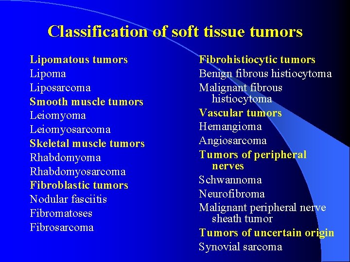
Classification of soft tissue tumors Lipomatous tumors Lipoma Liposarcoma Smooth muscle tumors Leiomyoma Leiomyosarcoma Skeletal muscle tumors Rhabdomyoma Rhabdomyosarcoma Fibroblastic tumors Nodular fasciitis Fibromatoses Fibrosarcoma Fibrohistiocytic tumors Benign fibrous histiocytoma Malignant fibrous histiocytoma Vascular tumors Hemangioma Angiosarcoma Tumors of peripheral nerves Schwannoma Neurofibroma Malignant peripheral nerve sheath tumor Tumors of uncertain origin Synovial sarcoma
Lipoma • • common benign tumor subcutaneous tissue of the trunk and limbs, oral cavity middle-aged and elderly soft, slowly growing mass • Micro: well-defined lobules of mature adipose tissue • • • Lipoma variants: Angiolipoma Spindle cell lipoma
Liposarcoma • • • most common malignant soft tissue tumor adults (peak 40 -60 Y) site: lower limb and retroperitoneal space • • • Key diagnostic feature: multivacuolated lipoblast subtypes: Well-differentiated liposarcoma (atypical lipomatous tumor): closely resembles lipoma Myxoid liposarcoma Round cell liposarcoma Pleomorphic liposarcoma Dedifferentiated liposarcoma Mixed liposarcoma • • •
Smooth muscle tumors Leiomyoma skin, subcutaneous tissue, uterus, gastrointestinal tract Micro bundles of well-differentiated smooth muscle cells with bright eosinophilic cytoplasm and blunt-ended nuclei Leiomyosarcoma mesentery, retroperitoneal space, wall of large veins, skin, subcutaneous tissue, deep soft tissues of limbs signs of malignancy large size, high mitotic rate, necrosis, cellular pleomorphism
Rhabdomyoma • adult type: head and neck of middle aged individuals • round or polygonal cells with eosinophilic cytoplasm and abundant cytoplasmatic glycogen • genital type: polypoid growth of the cervix, vagina or vulva in middle age • large rhabdomyoblasts with cross-striations • fetal type: boys < 3 Y, head and neck • mixture of immature skeletal muscle and myxoid stroma • cardiac rhabdomyoma: in infancy • fatal by the age of 5 years, association with tuberous sclerosis in about 50% • multiple nodules in heart ventricles • “spider cells“
Rhabdomyosarcoma most common malignant soft tissue tumour in infants and young children • Dg. : demonstration of rhabdomyoblasts • • Embryonal rhabdomyosarcoma: most common • early childhood, head and neck, genitourinary system • Bothryoid rhabdomyosarcoma (grape-like) • ER with polypoid configuration and myxoid consistency • Alveolar rhabdomyosarcoma: 10 to 20 Y • muscles of limbs and trunk • Pleomorphic rhabdomyosarcoma: adults • limbs of adults
Nodular fasciitis • benign fibroblastic proliferation • adolescents and young adults • rapidly growing nodule within subcutaneous tissue, forearm • Micro • immature fibroblasts arranged in short bundles, numerous mitoses, cellular pleomorphism not present
Superficial fibromatoses Palmar fibromatosis (Dupuytren’s contracture) • middle-aged men • nodular thickening of palmar aponeurosis - flexion deformities of fingers Plantar fibromatosis (Ledderhose’s disease) • nodular thickening of plantar aponeurosis Penile fibromatosis (Peyronie’s disease) • abnormal curvature of penis Micro: • nodules of well-differentiated fibroblasts arranged in long sweeping bundles
Deep fibromatoses (desmoid tumors) Abdominal • abdominal wall, young adults, women who have borne children, detected in peripartum or postpartum period, sometimes in surgical scars Intra-abdominal • young adults, mesentery, + Gardner’s syndrome Extra-abdominal • the most aggressive, adults in third and fourth decades, pectoral and pelvic girdles General features • deep intramuscular location, large size (up to 10 -15 cm), infiltrative growth pattern, high risk of recurrence after excision
Fibrosarcoma • • • uncommon malignant neoplasm middle aged adults deep soft tissues of lower limbs and trunk Micro bundles of spindle shaped cells arranged at angles to one another (“herring-bone pattern“), frequent mitoses Infantile fibrosarcoma • within first two years of life, better prognosis
Benign fibrous histiocytoma (dermatofibroma) • common lesion, most frequently on skin of lower leg • papule or nodule, often deeply pigmented Micro • within the mid-dermis, spindle cells arranged in curious whorled pattern (storiform pattern)
Malignant fibrous histiocytoma (MFH) • deep soft tissues of limbs, retroperitoneum • irregularly arranged plump, eosinophilic, spindle-shaped cells with darkly staining and often bizarre nuclei, numerous mitoses, interspersed macrophage-like cells, storiform pattern in some areas MFH represents a morphological pattern shared by wide variety of poorly differentiated malignant neoplasms • = heterogeneous group of unrelated lesions (pleomorphic sarcomas, dedifferentiated carcinomas, highgrade lymphomas etc. ) •
Haemangioma • hamartomas rather than true neoplasms Capillary haemangioma • skin, mass of proliferated capillaries Pyogenic granuloma • rapidly developing polypoid nodule, many small capillaries similar to those seen in granulation tissue, myxoid stroma, surface often eroded, common in oral cavity Cavernous haemangioma • skin, liver, aggregates of large dilated thin-walled vessels lined by flattened endothelium
Angiosarcoma Irregular anastomosing vascular spaces lined by endothelial cells with large hyperchromatic nuclei, highly aggressive tumor with poor prognosis Kaposi’s sarcoma HHV 8, often patients with AIDS, multiple bluish plaques and nodules on the skin of lower extremities, spindleshaped cells with the presence of red cells containing slits and haemosiderin deposition
Schwannoma (neurilemmoma) • smooth lobulated lesion attached to a nerve Microscopic features: two patterns recognized: • Antoni A – compact areas formed by regular interlacing bundles of uniform spindle-shaped cells, often foci of nuclear palisading, Verocay bodies • Antoni B – looser open areas, small cells with rounded nuclei
Neurofibroma • not infrequently multiple, part of neurofibromatosis • infiltrates and expands the affected nerve Microscopic features • spindle-shaped cells with elongated wavy nuclei set in myxoid stroma
Malignant peripheral nerve sheath tumor (MPNST) • adults • most common locations: neck, forearm, lower leg, buttock • large mass producing enlargement of a major nerve Micro • relatively uniform spindle-shaped cells with hyperchromatic nuclei and high mitotic activity
Synovial sarcoma term “synovial sarcoma“ is a misnomer young adults (15 -40 Y) from extraarticular soft tissues, often near joint capsules and tendons • Microscopic features: tumour consists of two cell types: • • Epithelial (forming nests and gland-like spaces) • Spindle (arranged in sheets) • classification • Biphasic (both cell types) • Monophasic (spindle or epithelial cells only) • frequent recurrences, metastases in lymph nodes and lungs