
Musculoskeletal Integrity Program Research Opportunities Adele L Boskey
Musculoskeletal Integrity Program Research Opportunities Adele L Boskey, Ph. D Director, Musculoskeletal Integrity Program (HSS) Starr Chair in Mineralized Tissue Research (HSS) Professor, Program in Physiology, Biophysics, and Systems Biology (Weill) Professor, Biochemistry (Weill) Professor, Bioengineering Field (Cornell, Ithaca)
Laboratories in the Program l l Mineralized tissue laboratory Adele L Boskey Spectroscopy and imaging laboratory Nancy Pleshko Camacho Molecular Imaging laboratory Phillip Mayer-Kuckuk Biomechanics and biomaterials laboratory Timothy M. Wright
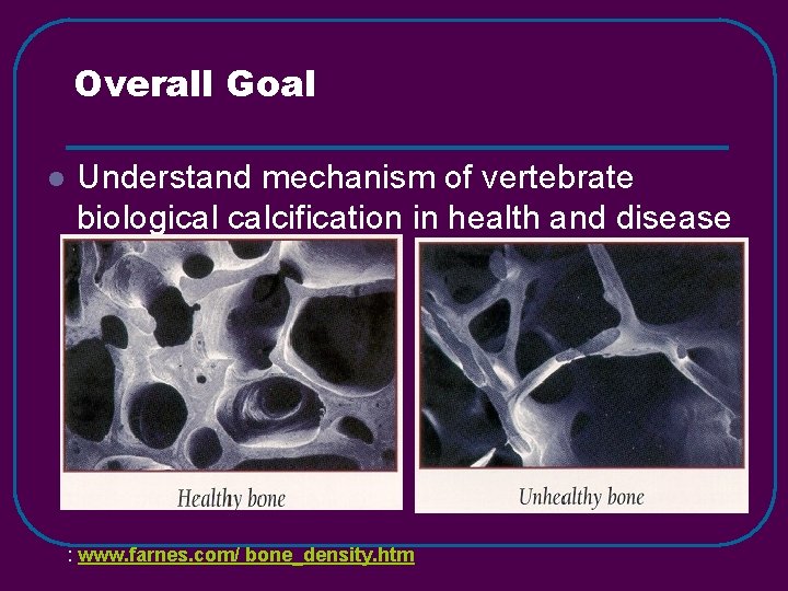
Overall Goal l Understand mechanism of vertebrate biological calcification in health and disease : www. farnes. com/ bone_density. htm
Diseases of Interest l l l Osteoporosis Osteogenesis Imperfecta Osteoarthritis Juvenile dermatomyositis Skeletal dysplasias
Research Questions l l l What causes mineral to form at specific sites in mineralizing tissues? How do extracellular matrix proteins affect the formation and proliferation of mineral crystals? Which genes regulate initial mineral deposition? Mineral crystal growth? How do mineral and matrix properties change in osteoporosis (with and without treatment)? What molecular changes occur in bone and cartilage during aging and in disease? How can we apply the concepts learned from in vitro analyses to tissue engineering of bones, teeth and cartilage?
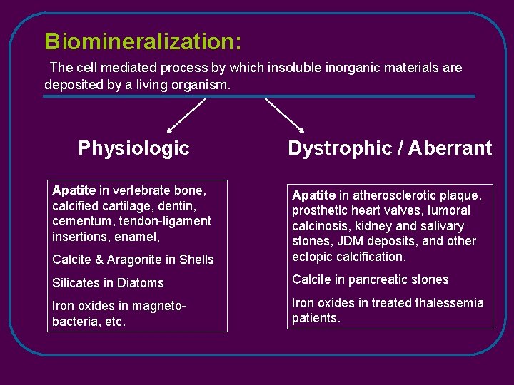
Biomineralization: The cell mediated process by which insoluble inorganic materials are deposited by a living organism. Physiologic Dystrophic / Aberrant Apatite in vertebrate bone, calcified cartilage, dentin, cementum, tendon-ligament insertions, enamel, Calcite & Aragonite in Shells Apatite in atherosclerotic plaque, prosthetic heart valves, tumoral calcinosis, kidney and salivary stones, JDM deposits, and other ectopic calcification. Silicates in Diatoms Calcite in pancreatic stones Iron oxides in magnetobacteria, etc. Iron oxides in treated thalessemia patients.
Common Features of Biomineralization l l The mineral may be amorphous or crystalline Mineral deposits in an oriented fashion on an oriented matrix (template) Cells and matrix regulate the mineralization process Matrix proteins associated with biomineralization are anionic
Bone and Dentin: Mineralization Facts l l l Debate. ACP? The mineral is a poorly crystalline analog of OCP? other hydroxyapatite Mineral crystals deposit in an oriented fashion on an oriented collagen matrix Mineralization is regulated by osteoblasts Debate do and Noncollagenous matrix – proteins. phosphoproteins • The functions of the matrix proteins are redundant. simply regulate Pi • Physicochemicallevels effects (Ca*P P/PP) have or do, they important other function?
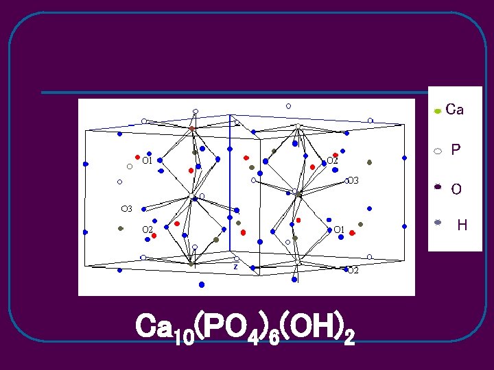
Ca P O H Ca 10(PO 4)6(OH)2
Why some collagenous tissues mineralize while others do not? l Exposure and/or modification of nucleators l Removal or modification of inhibitors
Bone and Dentin ECM protein Families associated with Mineralization l l l SIBLINGs SLRPS Gla-Proteins Phospholipids (Enzymes)
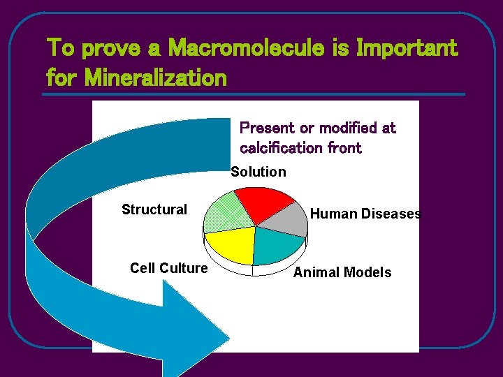
To prove a Macromolecule is Important for Mineralization Present or modified at calcification front Solution Structural Cell Culture Human Diseases Animal Models
Hypothesis l Post-translational modifications of extracellular matrix proteins enables them to regulate the mineralization process in bones, teeth, and other tissues. ØFragmentation ØPhosphorylation ØDephosphorylation Ø(Interaction with collagen)
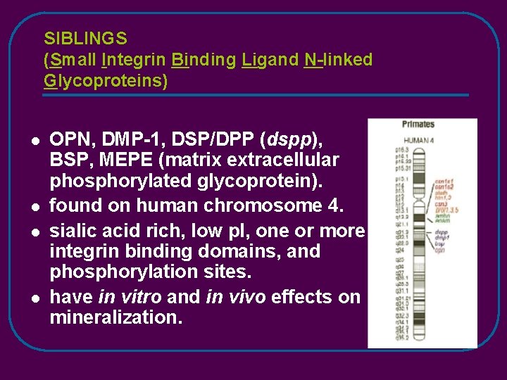
SIBLINGS (Small Integrin Binding Ligand N-linked Glycoproteins) l l OPN, DMP-1, DSP/DPP (dspp), BSP, MEPE (matrix extracellular phosphorylated glycoprotein). found on human chromosome 4. sialic acid rich, low p. I, one or more integrin binding domains, and phosphorylation sites. have in vitro and in vivo effects on mineralization.
ASARM peptides l The SIBLINGs all contain domains with an acidic serine-aspartate-rich motif (ASARM). These domains are thought to interact with HA crystals in regulation of the mineralization process.
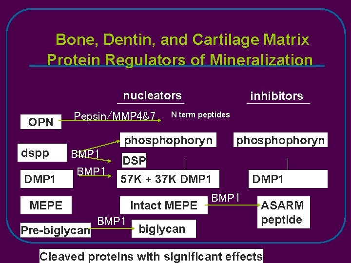
Bone, Dentin, and Cartilage Matrix Protein Regulators of Mineralization nucleators OPN dspp DMP 1 Pepsin/MMP 4&7 inhibitors N term peptides phosphophoryn BMP 1 DSP 57 K + 37 K DMP 1 MEPE Pre-biglycan phosphophoryn Intact MEPE BMP 1 biglycan BMP 1 DMP 1 ASARM peptide Cleaved proteins with significant effects
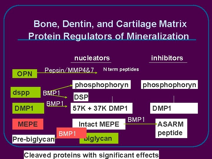
Bone, Dentin, and Cartilage Matrix Protein Regulators of Mineralization nucleators OPN dspp DMP 1 Pepsin/MMP 4&7 inhibitors N term peptides phosphophoryn BMP 1 DSP 57 K + 37 K DMP 1 MEPE Pre-biglycan phosphophoryn Intact MEPE BMP 1 biglycan BMP 1 DMP 1 ASARM peptide Cleaved proteins with significant effects
N 2 gas Ca. Cl 2 P SD Double Diffusion P SD Ca SD P SD Ca SD (NH 4)2 HPO 4 N 2 gas
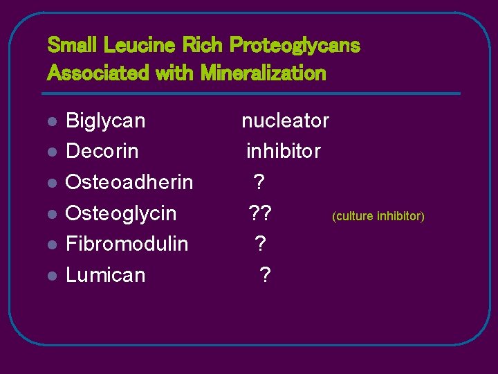
Small Leucine Rich Proteoglycans Associated with Mineralization l l l Biglycan Decorin Osteoadherin Osteoglycin Fibromodulin Lumican nucleator inhibitor ? ? ? (culture inhibitor) ? ?
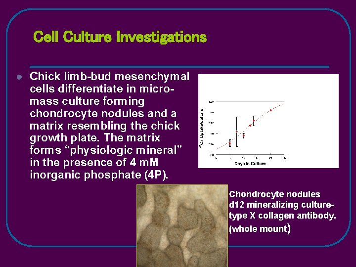
Cell Culture Investigations l Chick limb-bud mesenchymal cells differentiate in micromass culture forming chondrocyte nodules and a matrix resembling the chick growth plate. The matrix forms “physiologic mineral” in the presence of 4 m. M inorganic phosphate (4 P). Chondrocyte nodules d 12 mineralizing culturetype X collagen antibody. (whole mount)
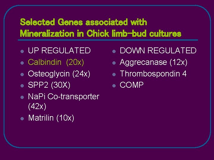
Selected Genes associated with Mineralization in Chick limb-bud cultures l l l UP REGULATED Calbindin (20 x) Osteoglycin (24 x) SPP 2 (30 X) Na. Pi Co-transporter (42 x) Matrilin (10 x) l l DOWN REGULATED Aggrecanase (12 x) Thrombospondin 4 COMP
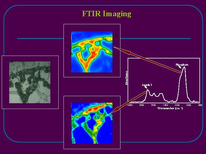
FTIR Imaging Absorbance Phosphate 2000 Amide I 1800 1600 1400 1200 Wavenumber (cm -1) 1000 800
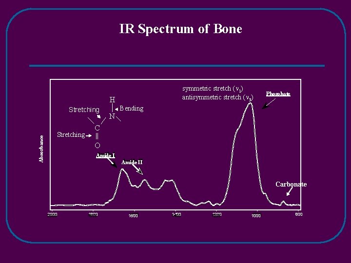
IR Spectrum of Bone symmetric stretch ( 1) antisymmetric stretch ( 3) H Absorbance Stretching N Phosphate Bending C O Amide II II Amide Carbonate 2000 1800 1600 1400 1200 1000 800
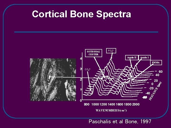
Cortical Bone Spectra OSTEONAL CENTER PO 43 Amide II 4 Amide I PMMA CO 32 - 60 40 2 1 DI ST AN -40 -60 20 CE 0 -20 0 800 1000 1200 1400 1600 1800 2000 WAVENUMBERS (cm-1) Paschalis et al Bone, 1997 ( m ) 3
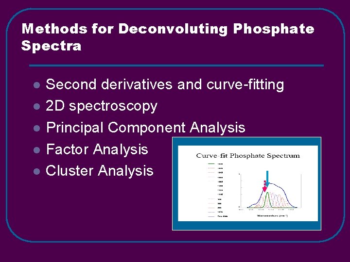
Methods for Deconvoluting Phosphate Spectra l l l Second derivatives and curve-fitting 2 D spectroscopy Principal Component Analysis Factor Analysis Cluster Analysis
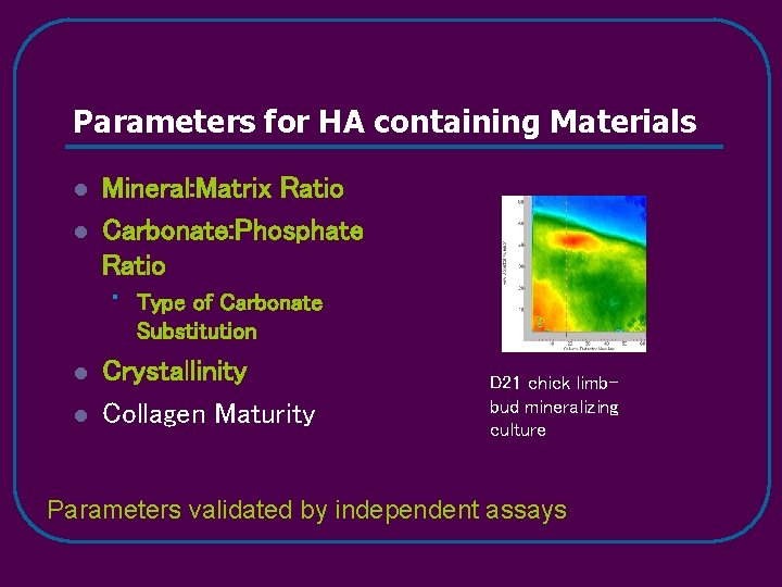
Parameters for HA containing Materials l l Mineral: Matrix Ratio Carbonate: Phosphate Ratio • l l Type of Carbonate Substitution Crystallinity Collagen Maturity D 21 chick limbbud mineralizing culture Parameters validated by independent assays
Osteoporosis Studies l Material properties (determined by FTIRI) predict why individuals with the same BMD do or do not suffer fragility fractures. • Human studies – • Baboon studies – age changes • Sheep studies - therapies
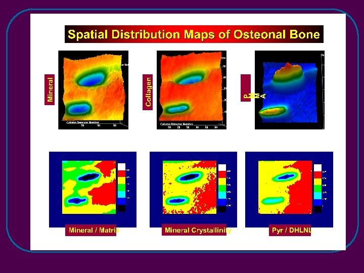
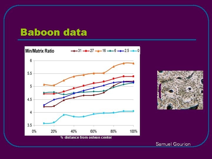
Baboon data Age (yrs) % distance from osteon center Samuel Gourion
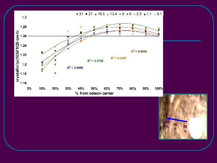
100 % 50 % 10 %
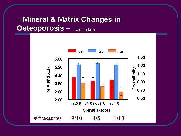
– Mineral & Matrix Changes in Osteoporosis – Dan Faibish Cryst XLR 6. 00 1. 50 5. 20 1. 30 Crystallinity M: M and XLR M: M 4. 40 3. 60 2. 80 2. 00 0. 90 0. 70 0. 50 <-2. 5 to -1. 5 >-1. 5 Spinal T-score # fractures 1. 10 9/10 4/5 1/10
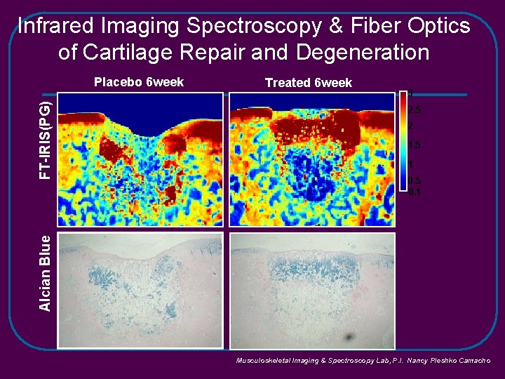
Infrared Imaging Spectroscopy & Fiber Optics of Cartilage Repair and Degeneration Treated 6 week 3 2. 5 2 1. 5 1 0. 5 0. 1 Alcian Blue FT-IRIS(PG) Placebo 6 week Musculoskeletal Imaging & Spectroscopy Lab, P. I. Nancy Pleshko Camacho
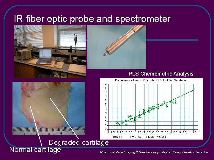
IR fiber optic probe and spectrometer PLS Chemometric Analysis Mankin Grade Degraded cartilage Normal cartilage Musculoskeletal Imaging & Spectroscopy Lab, P. I. Nancy Pleshko Camacho
Techniques Routinely Used l l l l Analytical chemistry X-ray diffraction Mechanical testing Micro-computed tomography Cell culture – including si. RNA Infrared Imaging, Raman Imaging, Fiber-optic Probe In vivo molecular imaging
NIH Support l l DE 04141 AR 037661 AR 043125 AR 046121
- Slides: 35