Microscopic Examination of Urine DrMohamed Mahmoud Nour Eldein


 (400 x) – – >")
 Leukocytes -")


- Slides: 53
Microscopic Examination of Urine Dr/Mohamed Mahmoud Nour Eldein Ph. D Biochemistry n Assistant Professor of Biochemistry Faculty of Medicine Umm AL-Qura University
Microscopic examination n Microscopic urinalysis is done simply pouring the urine sample into a test tube and centrifuging it (spinning it down in a machine) for a few minutes. The top liquid part (the supernatant) is discarded. The solid part left in the bottom of the test tube (the urine sediment) is mixed with the remaining drop of urine in the test tube and one drop is analyzed under a microscope
Microscopic Examination Abnormal Findings Per High Power Field (HPF) (400 x) – – > 3 erythrocytes > 5 leukocytes > 2 renal tubular cells > 10 bacteria Per Low Power Field (LPF) (200 x) > 3 hyaline casts or > 1 granular cast – > 10 squamous cells (indicative of contaminated specimen) – Any other cast (RBCs, WBCs) Presence of: – Fungal hyphae or yeast, parasite, viral inclusions – Pathological crystals (cystine, leucine, tyrosine) – Large number of uric acid or calcium oxalate crystals –
Microscopic Examination Cells Erythrocytes - “Dysmorphic” vs. “normal” (> 10 per HPF) Leukocytes - Neutrophils (glitter cells) - Eosinophils More than 1 per 3 HPF Hansel test (special stain) Epithelial Cells - Squamous cells - Renal tubular epithelial cells - Transitional epithelial cells Indicate level of contamination Few are normal - Oval fat bodies Abnormal, indicate Nephrosis
Microscopic Examination RBCs
Microscopic Examination RBCs
Microscopic Examination WBCs
Microscopic Examination Squamous Cells
Microscopic Examination Tubular Epithelial Cells
Microscopic Examination Transitional Cells
Microscopic Examination Transitional Cells
Microscopic Examination Oval Fat Body
Microscopic Examination LE Cell
Microscopic Examination Bacteria & Yeasts Bacteria - Bacteriuria More than 10 per HPF Yeasts - Candidiasis Most likely a contaminant but should correlate with clinical picture. Viruses - CMV inclusions Probable viral cystitis.
Microscopic Examination Bacteria
Microscopic Examination Yeasts
Microscopic Examination Yeasts
Microscopic Examination Cytomegalovirus
casts n Urinary casts are cylindrical aggregations of particles that form in the distal nephron, dislodge, and pass into the urine. In urinalysis they indicate kidney disease. They form via precipitation of Tamm-Horsfall mucoprotein which is secreted by renal tubule cells.
Microscopic Examination Casts
Types of casts n Acellular casts n Cellular casts Hyaline casts Granular casts Waxy casts Fatty casts Pigment casts Crystal casts Red cell casts White cell casts Epithelial cell cast
Microscopic Examination Casts Erythrocyte Casts: Glomerular diseases Leukocyte Casts: Pyuria, glomerular disease Degenerating Casts: - Granular casts - Hyaline casts - Waxy casts - Fatty casts (oval fat body casts) Nonspecific (Tamm-Horsfall protein) Nonspecific Nephrotic syndrome
n
Red cell casts n The presence of red blood cells within the cast is always pathologic, and is strongly indicative of glomerular damage. n They are usually associated with nephritic syndromes.
Microscopic Examination RBCs Cast - Histology
Microscopic Examination RBCs Cast
Microscopic Examination RBCs Cast - Histology
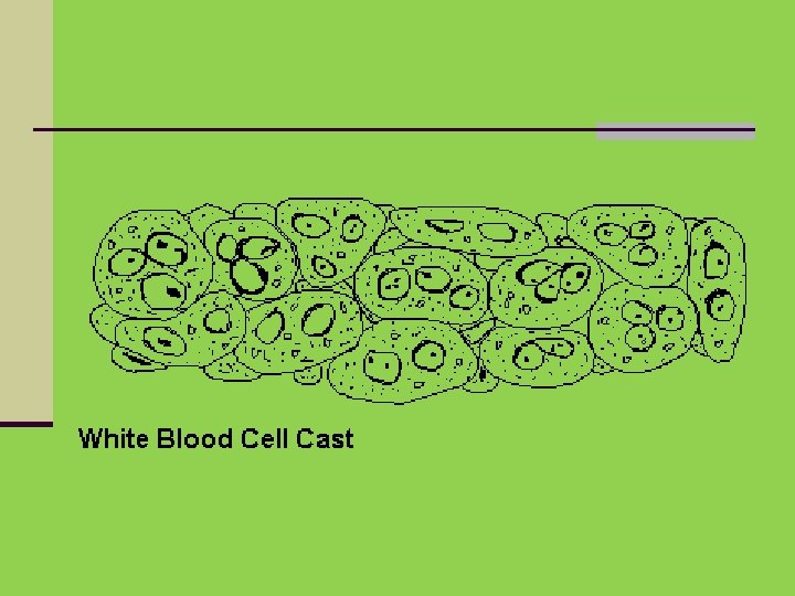
White blood cell casts n Indicative of inflammation or infection, n pyelonephritis n acute allergic interstitial nephritis, n nephrotic syndrome, or n post-streptococcal acute glomerulonephritis
Microscopic Examination WBCs Cast
Epithelial casts n This cast is formed by inclusion or adhesion of desquamated epithelial cells of the tubule lining. These can be seen in n acute tubular necrosis and n toxic ingestion, such as from mercury, diethylene glycol, or salicylate.
Microscopic Examination Tubular Epith. Cast
Microscopic Examination Tubular Epith. Cast
Granular casts n Granular casts can result either from the breakdown of cellular casts or the inclusion of aggregates of plasma proteins (e. g. , albumin) or immunoglobulin light chains n indicative of chronic renal disease
Microscopic Examination Granular Cast
Hyaline casts n The most common type of cast, hyaline casts are solidified Tamm-Horsfall mucoprotein secreted from the tubular epithelial cells n Seen in fever, strenuous exercise, damage to the glomerular capillary
Microscopic Examination Hyaline Cast
Waxy casts n waxy casts suggest severe, longstanding kidney disease such as renal failure(end stage renal disease).
Waxy casts n
Microscopic Examination Waxy Cast
Fatty casts n Formed by the breakdown of lipid-rich epithelial cells, these are hyaline casts with fat globule inclusions They can be present in various disorders, including n nephrotic syndrome, n diabetic or lupus nephropathy, n Acute tubular necrosis
Fatty casts n
Microscopic Examination Fatty Cast
Crystal casts n Though crystallized urinary solutes, such as oxalates, urates, or sulfonamides, may become enmeshed within a hyaline cast during its formation. n The clinical significance of this occurrence is not felt to be great.
Contents of normal urine m/s n Contains few epithelial cells, occasional RBC’s, few crystals.
Crystals in urine Crystals in acidic urine Ø Uric acid Ø Calcium oxalate Ø Amorphous urate Ø Cystine Ø Leucine Crystals in alkaline urine Ø Ammonium Ø Magnesium Ø phosphates (Triple phosphate crystals) Ø Amorphous phosphate Ø Calcium carbonate
Microscopic Examination Uric acid crystals n
Microscopic Examination Calcium Oxalate Crystals
Microscopic Examination Calcium Oxalate Crystals Dumbbell Shape
Microscopic Examination Amorphous Urates Crystals
Microscopic Examination Triple Phosphate Crystals
Microscopic Examination Amorphous phosphate Crystals
Microscopic Examination Cystine Crystals