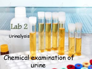
Chemical examination of urine Routine chemical examination of


- Slides: 35

Chemical examination of urine

Routine chemical examination of urine has changed dramatically since the early days of urine testing, due to the development of reagent strip method for chemical analysis. Reagent strips currently provide a simple, rapid means for performing medically significant chemical analysis of urine, including p. H, protein, glucose, ketones, blood, bilirubin, urobilinogen, nitrite, leukocytes, and specific gravity

A small amount of protein (50 – 150 mg / 24 hrs) appears daily in the normal urine, or 10 mg/dl in any single specimen which not appear in routine urinalysis procedure. More than 150 mg/day is defined as Proteinuria. This amount of protein is form of: 1. 40% consist of albumin, which may escape from the glomerulus membrane & not reabsorbed. 2. 40% of (tamm–Horsfall) muccoprotein which is secreted from the renal tubule and other secretions from genitalia. 3. 20% other traces of non-plasma proteins.

Proteinuria Is defined as the presence of detectable amount of proteins in urine. Ø Causes of proteinuria 1. Renal (Glomerular membrane damage), which may be: – Primary: due to primary glomerular defect as glomerulnephritis. – Secondary: - due to external disease that affects the glomerular function as: 1 - SLE 2 - Drug 3 - Septicemia 2. Prerenal Proteinuria Over flow / over load, increase of LMW protein such as multiple myeloma. Ex. Bence Jones protein.

Proteinuria 3. Tubular proteinuria: Caused by renal tubular dysfunction. Proteins of low molecular weight are normally filtered at the glomerulus of the kidney and are then normally reabsorbed by the tubular cells. Low-molecular-weight proteins' appearing in larger quantities than this is tubular proteinuria, which points to failure of reabsorption by damaged tubular cells.

Fever Emotional Cold Later months of pregnancy Postural (as long standing & exercises

This test is based on the protein-error-of-indicators principle. The dipstick is impregnated with citrate buffer (PH = 3. 0) containing Bromphenol blue which is yellow at p. H 3. 0 and blue at p. H 4. 2, at p. H (3) the indicator is mostly unionized.

If protein is present in the urine into which it is dipped, the ionized fraction binds to the protein, thereby causing more dye to ionize until equilibrium is reached; hence, the impregnated strip has less yellow and bluer color as the protein concentration increases. This reaction is seen visually as a change from yellow to green (a mixture of yellow dye plus blue dye appears green). The color is compared with that of the protein content from (30 – 1000 mg /dl). At a constant p. H, the development of any green color is due to the presence of protein. Colors range from yellow for "Negative" through yellow-green and green to green-blue for "Positive" reactions.

Remark : Trace positive results (which represent a slightly hazy appearance in urine) are equivalent to 10 mg/100 ml or about 150 mg/24 hours (the upper limit of normal) 1+ corresponds to about 200 -500 mg/24 hours 2+ to 0. 5 -1. 5 gm/24 hours 3+ to 2 -5 gm/24 hours 4+ represents 7 gm/24 hours or greater. False (+ve) may be due to increased urine p. H

a - Heat denaturation for protein precipitation. b - Sulfosalicylic acid Neg. No turbidity (1+) Turbidly, No granules. (2+) Turbidity, granulation, flocculation. (3+) Turbidity, granulation (4+) Clumps of proteins. Precipitation by heat is a better semi-quantitative method but, it’s not a highly sensitive. Sulfosalicylic acid test is a more sensitive precipitation test as it can detect albumin, globulins, and Bence-Jones protein at low

Bence Jones protein appears in urine of multiple myeloma patients. 1. Heat the urine between 40 – 6 C, so precipitation will occur 2. Then when heating is continued till 10 C, the precipitation will disappear (clear). 3. If you cool the urine till 40 – 6 C the precipitation will occur again.

If both are +ve then proteinuria is present If dipstick 1+ and sulfosalicylic negative then there is probably no pathologic concentration of protein. If dipstick negative and sulfosalicylic positive then the protein may be Bence Jones protein or one of the heavy chain proteins and should confirmed by immunologic method.

Under normal conditions, almost all of glucose filtered by glomerulus is reabsorbed in the proximal convoluted tubule, by an active process to maintain the plasma concentration of glucose. Less than 0. 1% of glucose normally filtered by the glomerulus appears in urine (< 130 mg/24 hr). If the blood glucose concentration is increased, reabsorption of glucose ceases & glucose appears in urine. Glycosuria (excess sugar in urine) generally means diabetes mellitus . • Threshold substances Substances that are completely absorbed by the tubules when their plasma concentration is normal and not completely absorbed by the tubules if their plasma concentration exceeds normal levels.

Glycosuria may be due to 1. Reabsorption defect 2. Increase Blood glucose, in the following cases: Diabetes mellitus Alimentary glycosuria (transitory), after meal. Stress in which elevation of epinephrine leads to increase glycogenolysis, and cortisol increase gluconeogenesis. Pancreatic disease affect insulin-secreting gland. Decrease reabsorption ability.

Tests for sugar : 1 - Benedicts test ” 2 - reagent strip

Normally, there is no blood or Hb in normal urine. Presence of them in urine is referred to hematuria, hemoglobinuria or myoglobinuria. Kidney problems Lower Urinary tract • Renal disease problem • Renal calculi • Infection • Renal tumor • Trauma • Calculi • toxins that damage • Trauma the glomeruli • Bleeding disorders & blood disease • Leukemia • Hemophilia • Thrombocytopeni

Note If hematuria, cast and proteinuria are present then the origin of problem is kidney. • • As a result of intravascular hemolysis Hemolytic anemia Sever burns Transfusion reaction Poisoning Sever physical exercises Infections with hemolytic bacteria

The presence of myoglobin, which is heme protein of muscles, facilitate the movement of oxygen within muscles. Hence it will appear in urine in case of: Muscular trauma Convulsions Prolonged coma

RBCs, Hb, and myoglobin will give +ve reaction. RBCs will give a spotted reaction pattern & will appear in microscopic test. Hb & myoglobin will give diffused reaction pattern; ammonium sulfate will differentiate between them, which precipitate Hb but not myoglobin. In urine sample, both give normal RBCs microscopically (0– 2)

A positive nitrite test indicates that bacteria may be present in significant numbers in urine. Gram negative rods such as E. coli are more likely to give a positive test. Negative test can not exclude the presence of bacteria. Cystitis Pyelonephritis

Bilirubin derived from Hb, is conjugated in the liver and excreted in the bile. Conversion to stereobilinogen (faecal urobilinogen) takes place in the intestinal lumen. Some reabsorbed urobilinogen is excreted in the urine.

10% 90%

Normal urine has a small amount of Urobilinogen 0 – 4 mg / day Urobilin 10 – 130 mg / day. While no bilirubin is present Conjugated bilirubin will appear if the normal degradation cycle is obstructed by the bile duct or when the integrity of liver is damaged allowing, leakage of conjugated bilirubin into the circulation such as cholestasis & hepatitis.

1. Reagent strip reaction Diazonium salt + bilirubin Azodye( Diazonium Compound color) 2. Tablet contain diazonium salt 3. Examine the color produced from the conversion of bilirubin to biliverdin. a- Oxidation test (Harrison Spot test) = Fouchet test Filter paper is soaked in saturated Ba. Cl 2, dried, cut in strip. When performing the test, the lower half of the strip is embedded in urine sample & then removed, apply one drop of (Fe. Cl 3 + TCA) (Fouchet reagent) in the line separated the wet & dried half. +ve result found as greenish color of the cut off line.

b- Smith iodine test 5 ml urine + 2 ml of 0. 7 iodine prepared in 95% ethyl alcohol. +ve green ring at the junction between the two fluids. c- Shake test: This test neither specific nor sensitive. +ve yellow foam p-dimethyl aminobenzaldehyde (Ehrlich’s reagent) +ve result with urobilinogen (red color).

Clinical significance Ketones are 3 intermediate product of fat metabolism which are Acetone (78%), Aceotacetic acid (20%), Beta-hydroxybutyric acid (2%). Sodium nitroprusside react with aceotacetic acid Ketonurea occurs in Diabetes acidosis Starvation Excessive Carbohydrate loss.

A positive leukocyte esterase test results from the presence of WBCs either as whole cells or as lysed cells. Pyuria can be detected even if the urine sample contains damaged or lysed WBC's. A -ve leukocyte esterase test means that an infection is unlikely Microscopic exam and/or urine culture need not be done to rule out significant bacteriuria.

Reagent strips consist of chemical-impregnated absorbent pads attached to a plastic strip. A color-producing chemical reaction takes place when the absorbent pad comes in contact with urine. The reactions are interpreted by comparing the color produced on the pad with a chart supplied by the manufacturer. By careful comparison of the colors on the chart and the strip, a semiquantitative value of trace, 1_, 2_, 3_, or 4_ can be reported. • Several colors or intensities of a color for each substance being tested appear on the chart. • An estimate of the milligrams per deciliter present is available for appropriate testing areas.

1. Dipping the reagent strip completely, but briefly, into a well-mixed specimen 2. Removing excess urine from the strip by running the edge of the strip on the container 3. Waiting the specified length of time for reactions to take place, and comparing the colored reactions against the manufacturer’s chart using a good light source.

Formed elements such as red and white blood cells sink to the bottom of the specimen and will be undetected in an unmixed specimen. Allowing the strip to remain in the urine for an extended period may cause leaching of reagents from the pads. Excess urine remaining on the strip after its removal from the specimen can produce a run over between chemicals on adjacent pads, producing distortion of the colors. To ensure against run over, blotting the edge of the strip on absorbent paper and holding the strip horizontally while comparing it with the color chart is recommended.

The amount of time needed for reactions to take place varies between tests and manufacturers, and ranges from an immediate reaction for p. H to 120 seconds for leukocytes. For the best semiquantitative results, the manufacturer’s stated time should be followed; however, when precise timing cannot be achieved, manufacturers recommend that reactions be read between 60 and 120 seconds, with the leukocyte reaction read at 120 seconds. A good light source is essential for accurate interpretation of color reactions. The strip must be held close to the color chart without actually being placed on the chart.

Reagent strips and color charts from different manufacturers are not interchangeable. Specimens that have been refrigerated must be allowed to return to room temperature prior to reagent strip testing, as the enzymatic reactions on the strips are temperature dependent Be alert for the presence of interfering substances. Understand the principles and significance of the test, read package inserts. Relate chemical findings to each other and to the physical and microscopic urinalysis results.

Store with desiccant in an opaque, tightly closed container. Store below 30_C; do not freeze. Do not expose to volatile fumes. Do not use past the expiration date. Do not use if chemical pads become discolored. Remove strips immediately prior to use.

Test open bottles of reagent strips with known positive and negative controls every 24 hr. Resolve control results that are out of range by further testing. Test reagents used in backup tests with positive and negative controls. Perform positive and negative controls on new reagents and newly opened bottles of reagent strips. Record all control results and reagent lot numbers.

 Normal bilirubin
 Dari hasil tes ternyata urin pak yudha mengandung glukosa
 Entamoeba histolytica cyst labeled
 Centrifuged
 Urine manjal niram
 General urine examination
 Pasty stool
 Centrifuged
 Urine chemical formula
 Triple phosphate crystals in urine
 Routine activities theory
 Slidetodoc.com
 Urine analyzers
 Peer massage
 Reading daily routine
 Composition of urine
 Vasa recta
 You should not open a persuasive request with a question.
 Ma routine quotidienne in english
 Hypertonic urine
 Introduction of urinary system
 Kaprekar's routine
 Amorfe zouten in urine
 Urine chain of custody form
 Urine characteristics
 Pemeriksaan bilirubin urine metode harrison
 Urine anion gap calculator
 Daily life lunch
 Idexx upc test instructions
 Proteinoria
 Ketones in urine moderate
 Urine diversion
 What does cloudy urine look like
 Routine data sources
 Pushups
 Bile salts in urine
