Major Themes of Anatomy Physiology Anatomical Terminology I


 Plane")


 Cranial Cavity Ventral Cavity (Belly – Side) Spinal")


 membrane, cell connections • Review cell")


- Slides: 50
Major Themes of Anatomy & Physiology Anatomical Terminology
I. A & P Overview A. Anatomy – Latin “to cut up” – the study of the structures of the body and their relationship to one another. – Gross or macroscopic anatomy – the study of large structures without the aid of a microscope (can be seen with the naked eye). • Systemic anatomy – the study of the body by systems. • Regional anatomy – the study of structures in a specific given area. • Surface anatomy – study of internal body structures as related to the overlying skin.
– Microscopic anatomy – study of structures too small to see without a microscope. • Cytology – the study of cells • Histology – the study of tissues – Developmental anatomy – the study that traces structural changes in an individual from conception to old age. • Embryology- study of the developmental changes in an organism from conception to birth
B. Physiology – the study of the functioning of the body and its structures – “how the body structures work” – emphasizing the maintenance of constant conditions (homeostasis). • Consider the operations of particular systems: – Renal physiology – kidney – Neurophysiology – brain – Cardiovascular physiology – heart
C. Principle of Complementarity – the structure is built for a specific function – “Structure is for function”. – Anatomy and physiology should be mentioned together because the structure reflects the function and vice versa Examples • Teeth – humans vs. a cat – canine teeth are different because they have different needs functions
Histology example SIMPLE CILIATED COLUMNAR EPITHELIAL cilia HISTOLOGY- PRINCIPLE OF COMPLEMENTARITY GOBLET CELLS IN TISSUE SECRETE MUCUS CILIATED TO MOVE MUCUS AND FILTER PARTICLES
II. Structure – Levels of Organization • • Atoms compose molecules Molecules compose macromolecules Macromolecules compose organelles Organelles compose cells Cells compose tissues Tissues compose organs Organs compose organ systems Organ systems compose the organism
• Levels of Organization • Atom molecules macromolecules organelles cell tissue organ system organism • Organ systems – 11 in the human body (see overview in textbook) • You need to be familiar with all of these definitions from Biology.
SUPPORT AND MOVEMEMENT • Integumentary system • Skeletal system • Muscular system
COMMUNICATION AND CONTROL • Nervous system • Endocrine system
REGULATION & MAINTENANCE • Cardiovascular system • Digestive system • Respiratory system • Urinary system • Lymphatic system
CONTINUITY OF LIFE • Reproductive system – male female
III. Important Scientists A. Aristotle – he was the first to record attempts at studying anatomy by dissecting plants and animals – no humans. B. Hippocrates – considered the father of medicine, interested in medical ethics. “Hippocratic Oath” he did not dissect humans. C. Claudius Galen – wrote an anatomical text used for 1400 years. His work is based on different animals: oxen, swine, dogs, apes, doctor to Roman gladiators D. Andreas Vesalius – known as “The Father of Modern Anatomy”. He wrote the first human anatomy text book, “On the Structure of the Human Body”. E. William Harvey- focused on the role of the path of blood through the heart and circulatory pathways
ANDREAS VESALIUS “FATHER OF MODERN ANATOMY” Gross anatomy. This famous woodcut of a gross dissection appeared in the world's fist anatomy textbook, De Corporis Humani Fabrica (On the Structure of the Human Body) in 1543. This woodcut features the book's author, Andreas Vesalius, who is considered to be the founder of modern anatomy. The body being dissected is called a cadaver
IV. Language of Anatomy A. Anatomical position – this is our set reference point for anatomy. • Standing erect, feet on floor • Arms at side, palms forward, thumb (pollex) is lateral • Head and feet pointed forward.
B. Planes of the Body
B. Planes of the body • Frontal or coronal – divides into a front (anterior) and a back (posterior) • Sagittal – right and left halves. – Mid-sagittal – equal right and left halves. – Parasagittal – unequal right and left halves. • Transverse – horizontal (across), top (superior) and bottom (inferior) halves. “Cross section” = transverse.
Sagittal Plane
Frontal (coronal) Plane
Transverse Plane
C. Directional Terms • Superior – up towards head / Inferior – down towards feet • Anterior - towards front (ventral) / Posterior – towards back (dorsal) • Superficial – close to surface / Deep – further from surface • Proximal – closer to the point of attachment / Distal – further away from the point of attachment (when referring to limbs) • Medial – closer to the midline / lateral – away from the midline
This is another example of a body regions picture, make sure that you know all the structures on page 13 in your textbook.
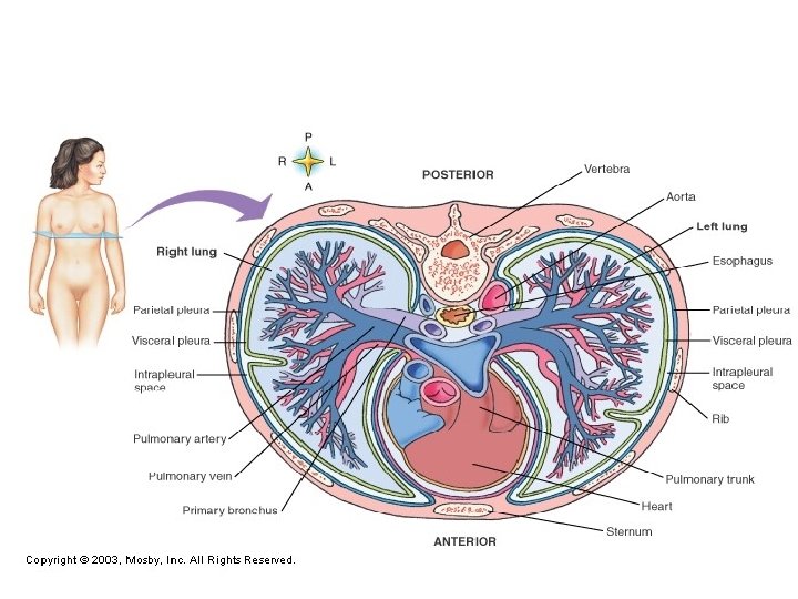
D. BODY CAVITIES Dorsal Cavity (Back) Cranial Cavity Ventral Cavity (Belly – Side) Spinal Cavity Thoracic Cavity Pleural Cavity Left Right Mediastinum Superior Abdominopelvic Cavity Abdominal Pericardial Pelvic
Body Cavities
2 13 10 5 1 11 12 3 6 8 9 This handout is will reviewed in the anatomical terms video 4 7
E. Body Membranes • Body Membranes – thin filmy membrane that lines body cavities and walls or cover an organ. – Parietal membrane – covers or lines the body wall – Visceral membrane – covers or lines each organ Examples- parietal pleural lines the pleural wall of the thoracic cavity visceral pleural lines the lobes of the lungs
V. Homeostasis A. Overview 1. Claude Bernard – French physiologist noticed that body cells survived in a constant environment. (Internal conditions should remain constant. ) 2. Walter Cannon – American physiologist in 1932 came up with the term “ homeostasis”. • Homeostasis – Latin for “the process of standing the same” –the ability for an organism to maintain internal conditions despite external changes. (everything regulatory in the body exists to maintain internal stability)
3. Homeostatic control mechanisms – Feedback control loops – an integrated control network to accomplish self regulation (involuntarily) • Examples: Body temperature, blood pressure, heart rate, oxygen and carbon dioxide levels, thirst, pulse rate, glucose/insulin levels
B. Basic components of feed back loop; pg. 9 • Sensory mechanism – afferent – caries forward • Control center – brain • Effector – causes an effect or efferent – carry away from • Feedback – our check (set point), it will continue until a set point is met
Mechanical example of feedback loop
Physiological example of negative feedback loop
Negative feedback Blood pressure
Glucose regulation
C. Feedback loops – Two types of feed back loops • Negative feed back mechanism Inhibitory – it opposes a change by creating an opposite response. • Example: If cold, heat kicks on (see mechanical ex) – It produces a change that is opposite to the disturbance – Stabilizes physiological imbalance – Responsible for internal stability – Most common- bp, body temp. , insulin/glucose
Negative Feedback, Set Point • Room temperature does not stay at set point of 68 degrees -- it only averages 68 degrees
Physiological example of negative feedback loop
Body Temperature homeostasis Body temperature increases Hypothalamus detects change and causes: 1. increases sweating 2. dilation of skin and blood vessels 3. sweat gland activation Sweating and blood flow cause heat loss Body temperature returns toward normal range
Body temperature decreases Hypothalamus detects change and causes: 1. decreased sweating 2. constriction of skin and blood vessels 3. shivering Decreased sweating and skin blood flow help retain heat Shivering produces heat Body temperature returns toward normal
• Positive feedback mechanism – Stimulatory response – Does not help the body to maintain stability – Accelerates a disturbance – “snowball effect” – Very rare under normal conditions – Necessary mechanism; example: sneezing, labor contractions, blood clot (wound healing) • Rare; example: prolonged high fever, certain cancers
Positive Feedback Loops Oxytocin regulation 2. Nerve impulses from cervix transmit information to brain
Positive Feedback-Fever • If temperature rises above 108 F – metabolic rate increases causing body to produce heat faster still • Temperature increases & cycle repeats again • Fatal at 113 F
• Feed forward mechanism – in digestive system – triggers other mechanisms to start working for digestion.
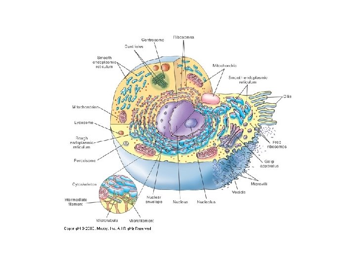
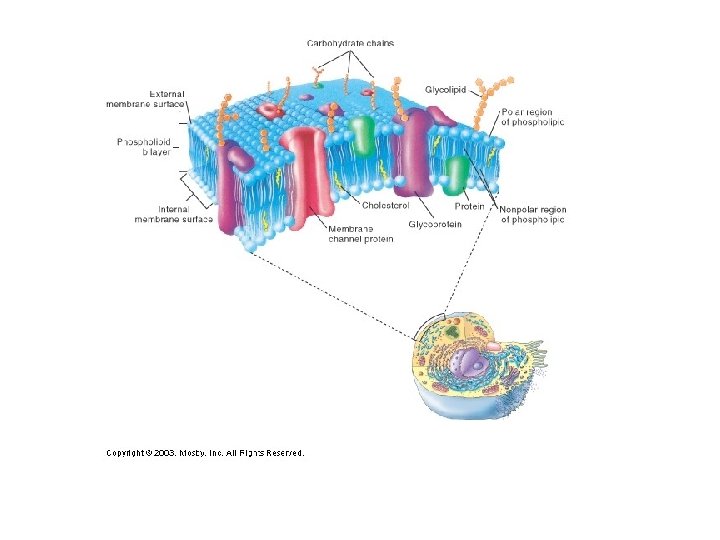
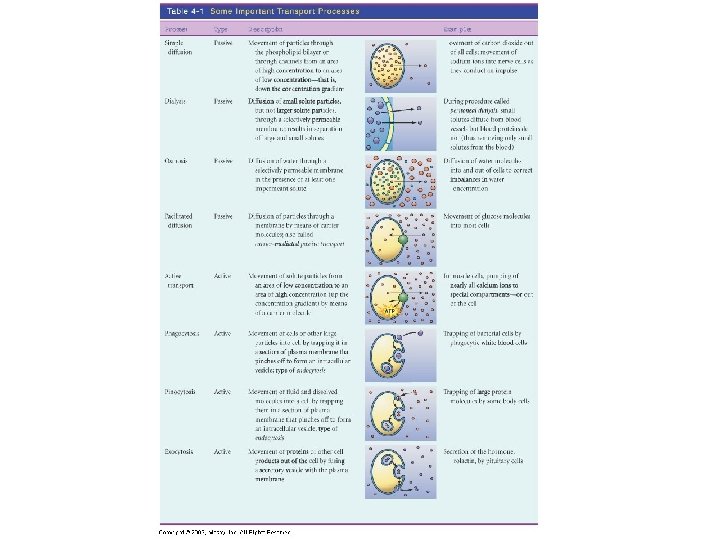
CYTOLOGY/HISTOLOGY • Review animal cell structures, cell (plasma) membrane, cell connections • Review cell physiology (passive and active transport) • Cell shapes- see handout
1 2 6 7 3 8 4 9 5 1. 2. 3. 4. 5. 6. 7. 8. 9. SQUAMOUS CUBOIDAL COLUMNAR STELLATE SPHEROID DISCOID FUSIFORM FIBROUS
VI. GERM LAYERS
VI. Embryonic Germ Layers A. Formation of primary germ layers – Early part of first trimester of development. Three different layers of specialized cells that form primitive germ layers (germ as is germination or to grow) • Ectoderm – outer germ layer that forms peripheral structures. Ex. Epidermis, tooth enamel, cornea, lens, most of the nervous system (brain and spinal cord). • Mesoderm – middle germ layer forms what is in middle. Ex. Dermis, skeletal bones, muscle, cardio • Endoderm – inner germ layer. Ex. lining of tracts, glands, “gut” – digestive organs, respiratory organs, urinary organs.