Hypersensitivity Reactions Allergies Refif alshawk Ph D Lec
 Lec 2&3")


 are proteins or chemicals bound to proteins.")


 Biogenic Amines and proteolytic enzymes Histamine")
 Lipid Mediators • prostaglandin D 2 (PGD 2). Acts as a vasodilator and")
 Cytokines • Mast cells produce many different cytokines that contribute to allergic inflammation")

The Immediate Reaction • The early vascular changes")

 Systemic Anaphylaxis • Anaphylaxis is a systemic immediate hypersensitivity reaction characterized by edema")


 is a consequence of immediate hypersensitivity reactions to")


 the")
, joint pain, lymph node enlargement,")

 and typically elicited in the skin when a low")


- Slides: 46
Hypersensitivity Reactions, Allergies Refif alshawk (Ph. D) Lec 2&3
• Hypersensitivity – responding inappropriately to an antigen • Inflammatory response can have deleterious effects • Tissue injury • Disease • death
• May develop in course of humoral OR cellmediated response • Immediate hypersensitivity – Anaphylactic – Antibody-antigen complexes – Manifests in minutes • Delayed-type hypersensitivity – May occur in days
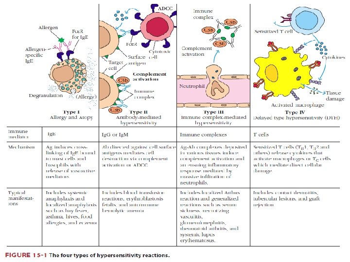
• • Types I : OVERVIEW OF Ig. E-DEPENDENT ALLERGIC REACTIONS All allergic reactions share common feature: the production of Ig. E antibody, which is dependent on the activation of IL-4– producing helper T cells. allergic reactions differ greatly in the types of antigens that elicit these reactions and their clinical and pathologic manifestations. Antigens that elicit immediate hypersensitivity reactions called allergens.
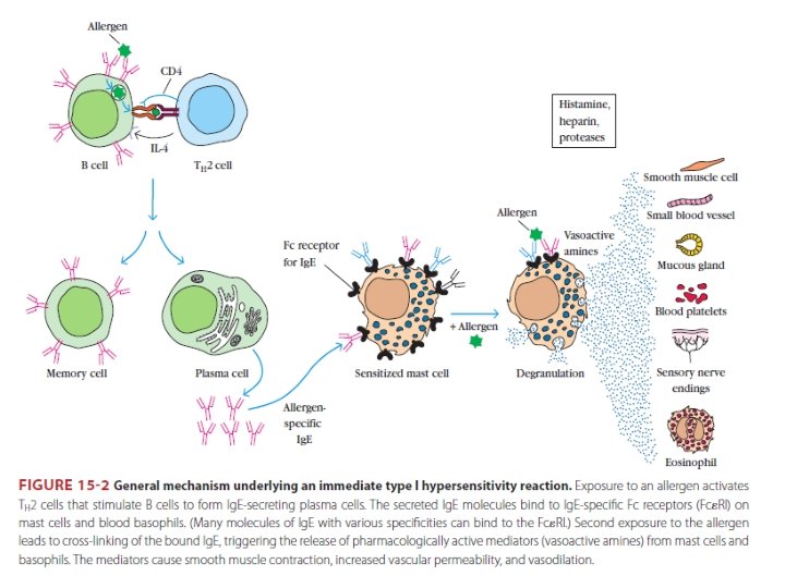
The typical sequence of events in immediate hypersensitivity consists • exposure to an antigen • activation of lymphocytes (TH 2 cells, and B cells) specific for the antigen • production of Ig. E antibody • binding of the antibody to Fc receptors of mast cells • and triggering of the mast cells by re-exposure to the antigen • resulting in the release of mediators from the mast cells • and the subsequent pathologic reaction.
There is a genetic predisposition for the development of allergies. • Atopic individuals produce high levels of Ig. E in response to environmental allergens, whereas normal individuals generally produce other Ig isotypes, such as Ig. M and Ig. G, and only small amounts of Ig. E.
Antigens that elicit immediate hypersensitivity reactions (allergens) are proteins or chemicals bound to proteins. • The important characteristics of allergens that, unlike microbes, they do not generally stimulate the innate immune responses that are associated with macrophage and dendritic cell secretion of TH 1 and TH 17 -inducing cytokines. WHY? ? ? Chronic or repeated cell activation in the absence of strong innate immunity may drive CD 4+ T cells preferentially toward the TH 2 pathway. The chemical nature. include low to medium molecular weight, stability, glycosylation, and high solubility in body fluids. These structural features probably protect the antigens from denaturation and degradation in the gastrointestinal tract and allow them to be absorbed intact. Some nonprotein substances, such as penicillin, can elicit strong Ig. E responses, when react chemically with amino acid residues in self proteins to form hapten carrier conjugates, which induce IL-4– producing helper T cell responses and Ig. E production.
1 - Activation of IL-4–Producing Helper T Cells • In allergic diseases, TH 2 cells are required for differentiation of Ig. E-producing B cells, and play a central role in the inflammatory reaction in tissues. 2 - Activation of B Cells and Switching to Ig. E • B cells specific for allergens are activated by TH 2 cells in lymphoid organs, as in other T cell– dependent B cell responses. In response to cytokines, mainly IL-4, the B cells undergo heavy chain isotype switching and produce Ig. E.
3 - ROLE OF TH 2 CELLS, MAST CELLS, BASOPHILS, AND EOSINOPHILS IN ALLERGIC REACTIONS • TH 2 cells, mast cells, basophils, and eosinophils are the major effector cells of immediate hypersensitivity reactions and allergic disease. + Mast cells, basophils, and eosinophils, in distinction from TH 2 cells, have cytoplasmic granules that contain preformed amines and enzymes, and all three cell types produce lipid mediators and cytokines that induce inflammation. + TH 2 cells contribute to inflammation by secreting cytokines.
4 - Role of TH 2 Cells and Innate Lymphoid Cells in Allergic Disease • TH 2 cells secrete cytokines, including IL-4, IL-5 IL-13 that work in combination with mast cells and eosinophils to promote inflammatory responses to allergens within tissues. • IL-4 secreted by TH 2 cells induces expression of endothelial adhesion molecules VCAM-1 • that promotes the recruitment of eosinophils and additional TH 2 cells into tissues • IL-5 secreted by TH 2 cells activates eosinophils • IL-13 stimulates epithelial cells (e. g. , in the airways) to secrete increased amounts of mucus, and excessive mucus production is also a common feature of these reactions • TH 2 cells also contribute to the inflammation of the latephase reaction.
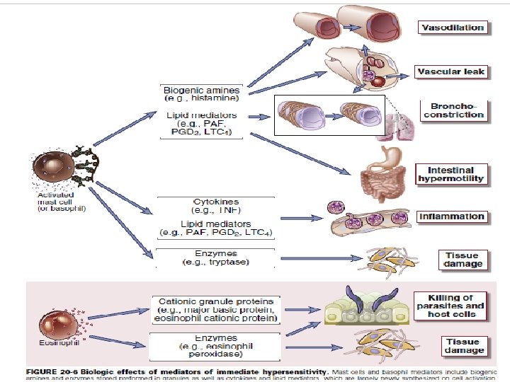
5 - Binding of Ig. E to Mast Cells and Basophils: The Fcε Receptor • Mast cells and basophils express a highaffinity Fc receptor specific for ε heavy chains, called FcεRI, which binds Ig. E. 6 - Activation of Mast Cells • In an individual allergic to a particular antigen, a large proportion of the Ig. E bound to FcεRI on the surface of mast cells is specific for that antigen • Exposure to the antigen will cross-link sufficient Ig. E molecules to trigger mast cell activation • Activation of mast cells results in three types of biologic response: degranulation synthesis and secretion of lipid mediators and synthesis and secretion of cytokines.
7 - Mediators Derived from Mast Cells: 1) Biogenic Amines and proteolytic enzymes Histamine acts by binding to target cell receptors (e. g. , H 1, H 2, H 3) The actions of histamine are contraction of the endothelial cells, leading to increased inter endothelial spaces, increased vascular permeability, and leakage of plasma into the tissues and other effect cause vasodilation also causes contraction of intestinal and bronchial smooth muscle.
2) Lipid Mediators • prostaglandin D 2 (PGD 2). Acts as a vasodilator and a bronchoconstrictor. PGD 2 also promotes neutrophil chemotaxis and accumulation at inflammatory sites. • leukotrienes its degradation products LTD 4 and LTE 4 which cause prolonged bronchoconstriction. (slow-reacting substance of anaphylaxis and are thought to be important mediators of asthmatic bronchoconstriction. • platelet-activating factor (PAF) has direct bronchoconstricting actions.
3) Cytokines • Mast cells produce many different cytokines that contribute to allergic inflammation (the latephase reaction). These cytokines like TNF, IL-1, IL-4, IL-6, CCL 3…. . . and granulocyte-monocyte colony-stimulating factor (GM-CSF). The cytokines that are released from activated mast cells and TH 2 cells are mainly responsible for the inflammation associated with the late-phase reaction. TNF activates endothelial expression of adhesion molecules and together with chemokines accounts for neutrophil and monocyte infiltrates.
8 - Properties of Eosinophils • Cytokines produced by TH 2 cells promote the activation of eosinophils and their recruitment to late-phase reaction inflammatory sites. Eosinophils release granule proteins that are toxic to helminthic parasites and may injure normal tissue
Ig. E- AND MAST CELL–DEPENDENT REACTIONS 1)The Immediate Reaction • The early vascular changes that occur during immediate hypersensitivity reactions are demonstrated by the wheal-andflare reaction). • 2) The Late-Phase Reaction • The immediate wheal-and-flare reaction is followed 2 to 4 hours later by a late-phase reaction consisting of the accumulation of inflammatory leukocytes, including neutrophils, eosinophils, basophils, and helper T cells.
ALLERGIC DISEASES IN HUMANS • The clinical and pathologic manifestations of the diseases depend on the: tissues in which the mast cell mediators have effects as well as the chronicity of the resulting inflammatory process. • The most common forms of these diseases are allergic rhinitis (hay fever), bronchial asthma, atopic dermatitis (eczema), and food allergies.
1) Systemic Anaphylaxis • Anaphylaxis is a systemic immediate hypersensitivity reaction characterized by edema in many tissues and a decrease in blood pressure, secondary to vasodilation. • These effects usually result from the systemic presence of antigen introduced by injection, an insect sting, or absorption across an epithelial surface such as gut mucosa. • The allergen activates mast cells in many tissues, resulting in the release of mediators that gain access to vascular beds throughout the body. • The decrease in vascular tone and leakage of plasma caused by mast cell mediators can lead to a significant decrease in blood pressure or shock, called anaphylactic shock, which is often fatal. • The cardiovascular effects are accompanied by constriction of the upper and lower airways, laryngeal edema • treatment is systemic epinephrine, which can be lifesaving by reversing the bronchoconstrictive and vasodilatory effects of mast cell mediators.
• Bronchial Asthma • Asthma is an inflammatory disease caused by repeated immediate-type hypersensitivity and late-phase allergic reactions in the lung leading to the clinic-pathologic triad of intermittent and reversible airway obstruction, chronic bronchial inflammation with eosinophils, and bronchial smooth muscle cell hypertrophy and hyperreactivity to bronchoconstrictors • (Fig. 20 -9
Asthma • Inflammatory disease • Induce expression of adhesion molecules on endothelial cells for eosinophils and neutrophils ○ Cause significant injury because of toxic enzymes, cytokines ○ Notice sloughing of the pseudostratified ciliated columnar epithelial cells lining the bronchiole
• The pathophysiologic sequence in atopic asthma is probably initiated by mast cell activation in response to allergen binding to Ig. E as well as by TH 2 cells reacting to allergens. • The lipid mediators and cytokines produced by the mast cells and T cells lead to the recruitment of eosinophils, basophils, and more TH 2 cells. • The chronic inflammation in this disease may continue without mast cell activation. • Smooth muscle cell hypertrophy and hyperreactivity are thought to result from leukocyte-derived mediators and cytokines. • Mast cells, basophils, and eosinophils all produce mediators that constrict airway smooth muscle. The most important of the bronchoconstricting mediators are LTC 4, LTD 4, and LTE 4. • Increased mucus secretion results from the action of cytokines, on bronchial epithelial cells. • Corticosteroids may also be given systemically, especially once an attack is under way, to reduce inflammation and bronchial smooth muscle cell relaxants
• Allergic rhinitis, (hay fever) is a consequence of immediate hypersensitivity reactions to common allergens such as plant pollen or house dust mites localized to the upper respiratory tract by inhalation. mucosal edema, leukocyte infiltration with abundant eosinophils, mucus secretion, coughing, sneezing, and difficulty in breathing. • Food allergies are immediate hypersensitivity reactions to ingested foods that lead to the release of mediators from intestinal mucosal and submucosal mast cells of the GI tract, including the oropharynx. pruritis, tissue edema, enhanced peristalsis, increased epithelial fluid secretion, and associated symptoms of oropharyngeal swelling, vomiting and diarrhea. Allergic reactions to many different types of food most common are peanuts and shellfish. • Common allergic reactions in the skin include urticaria and atopic dermatitis. Urticaria, or hives, is an acute wheal-and-flare reaction induced by mast cell mediators and occurs in response to direct local contact with an allergen or after an allergen enters the circulation. • eczema. It is a common skin disorder that may be caused by a late-phase reaction to an allergen in the skin. In the cutaneous late-phase reaction, TNF, IL -4, and other cytokines, probably derived from TH 2 cells and mast cells, act on endothelial cells to promote inflammation.
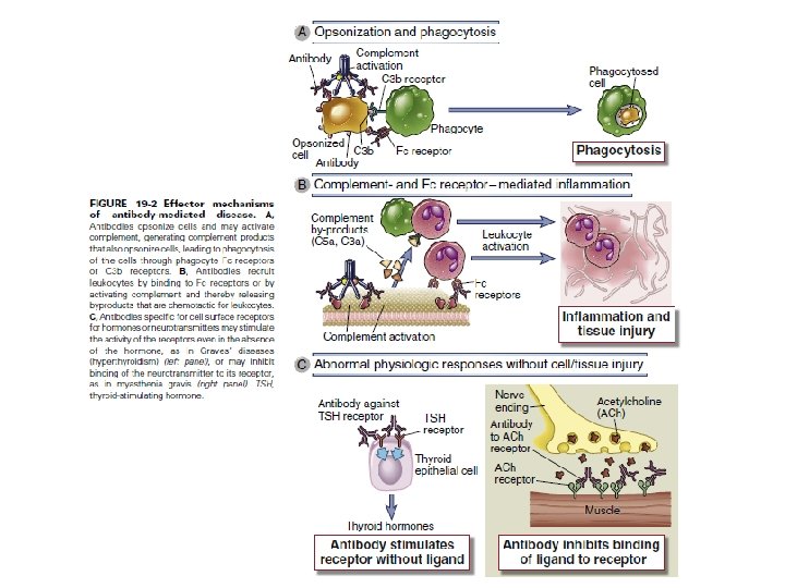
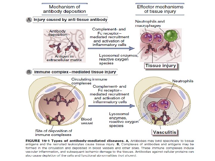
Type II – Antibody-Mediated Cytotoxic Hypersensitivity • involves the binding of Ig. G antibodies to: [cell surface antigens or extracellular matrix molecules]. Antibody bound to a cell-surface antigen can induce death of the antibodybound cell by three distinct mechanisms.
Type II – Antibody-Mediated Cytotoxic Hypersensitivity • First, certain immunoglobulin subclasses can activate the complement system, creating pores in the membrane of a foreign cell. • Secondly, antibodies can mediate cell destruction by antibody dependent cell-mediated cytotoxicity (ADCC), in which cytotoxic cells bearing Fc receptors bind to the Fc region of antibodies on target cells and promote killing of the cells. • Finally, antibody bound to a foreign cell also can serve as an opsonin, enabling phagocytic cells with Fc or C 3 b receptors to bind and phagocytose the antibody-coated cell
Type II – Antibody-Mediated Cytotoxic Hypersensitivity • Transfusion Reactions – antigens that are associated with the blood types are identified as A, B, and H, – Individuals have antibodies to blood types not their own and adults possess Ig. M antibodies to those Ag. – Antibodies directed toward ABH antigens are termed isohemagglutinins – Antibody attaches to RBC and initiates complement system to lyse RBC – After lysis: ○ Hemoglobin detected in plasma, starts to filter through kidneys and found in urine (hemoglobinuria) ○ Hemoglobin converted to bilirubin – toxic at high levels ○ Fever, chills, blood clotting
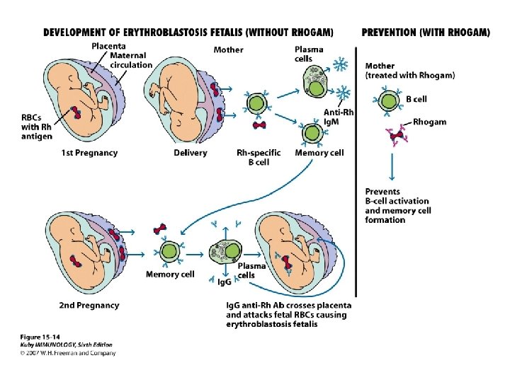
Type II – Antibody-Mediated Cytotoxic Hypersensitivity • Hemolytic disease of newborn – Rh+ fetus, Rh- mother – Ig. G antibodies cross placenta(memory) – Some of these antibodies may be anti-Rh antibodies - Can have severe consequences – Antibodies against ABO blood groups produce less consequences, can be easily treated – Rhogam shot ○ Given to mother ○ Anti-Rh antibodies bind to fetal cells that might have entered mother’s system during birthing process, facilitates clearing before there is a B cell response
Type II – Antibody-Mediated Cytotoxic Hypersensitivity • Hemolytic Anemia Can Be Drug Induced • Certain antibiotics (e. g. , penicillin, cephalosporins, and streptomycin), as well as other well-known drugs • adsorb nonspecifically to proteins on red blood cell membranes, forming a drug protein complex • induce formation of antibodies • inducing complement-mediated lysis • progressive anemia
Type III – Immune complex-mediated hypersensitivity • Complexing of antigen plus antibody facilitates phagocytosis and clearing of antigen • Large amounts of these complexes can lead to tissue damage & Type III hypersensitivity
Conditions associated with the initiation of a type III response include : (1) the presence of antigens capable of generating particularly extensive antigen-antibody lattices, (2) a high intrinsic affinity of antigens for particular tissues (3) the presence of highly charged antigens (which can affect immune complex engulfment) and (4) a compromised phagocytic system.
Symptoms induced such as • fever, urticaria (rashes), joint pain, lymph node enlargement, and protein in the urine. • The resulting inflammatory lesion is referred to as vasculitis if it occurs in a blood vessel, glomerulonephritis if it occurs in the kidney, or arthritis if it occurs in the joints.
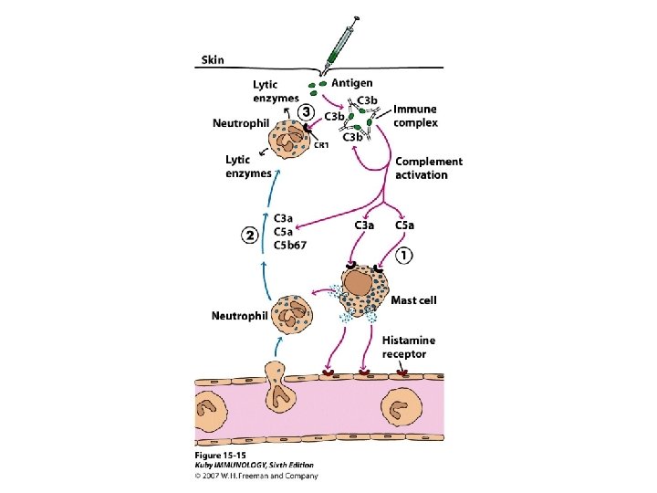
Type III can be localized • Injection of antigen intradermally or subcu into animal that has high level of antibody for that antigen • Arthus reaction • Bug bites
• Local (Arthus reaction) and typically elicited in the skin when a low dose of antigen is injected and immune complexes form locally. Ig. G antibodies are involved, and the resulting activation of complement leads to activation of mast cells and neutrophils, mediator release, and enhanced vascular permeability. This typically occurs in about 12 hours.
Type III can be generalized • Serum sickness ○ After receiving antiserum (serum from another animal that may contain antitoxins for treatment) • Use of monoclonal antibodies for use of cancer treatment ○ Patient developed antibody against mouse monoclonal antibody • Autoimmune diseases – Lupus, Rheumatoid arthritis • Drug reactions – Penicillin, sulfonamides • Infectious disease
• Systemic immune complex acute poststreptococcal glomerulonephritis is a well known immune complex disease. Its onset occurs several weeks after a group A βhemolytic streptococcal infection, particularly of the skin, and often occurs with infection due to nephritogenic types of streptococci. • It is likely that streptococcal antigen– antibody complexes are filtered out by glomeruli, fix complement, and attract neutrophils. This series of events results in an inflammatory process that damages the kidney.
Type IV – Delayed-type Hypersensitivity • Some subpopulations of TH cells encounter antigen, secrete cytokines and induce localized inflammatory response • Most cases are not detrimental(harmful)
Type IV Sensitization phase and Effector phase of DTH
Prolonged DTH can lead to formation of granuloma Tuberculosis test is done this way
Type IV – contact dermatitis
• Refferences : v. Immunology , Kuby, seventh edition v. Medical microbiology, Jawetz, 26 th edition v. Cellular and Molecular Immunology, Abul K. Abbas, 8 th edition.