
Immunodeficiency Refif S Alshawk Autoimmunity system attacks host


- Slides: 19
Immunodeficiency Refif S. Alshawk
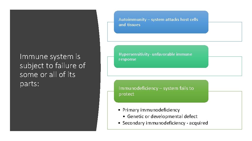
Autoimmunity – system attacks host cells and tissues Immune system is subject to failure of some or all of its parts: Hypersensitivity- unfavorable immune response Immunodeficiency – system fails to protect • Primary immunodeficiency • Genetic or developmental defect • Secondary immunodeficiency - acquired
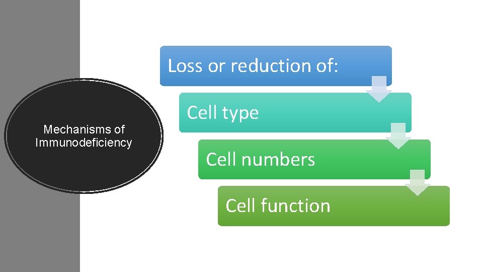
Loss or reduction of: Mechanisms of Immunodeficiency Cell type Cell numbers Cell function
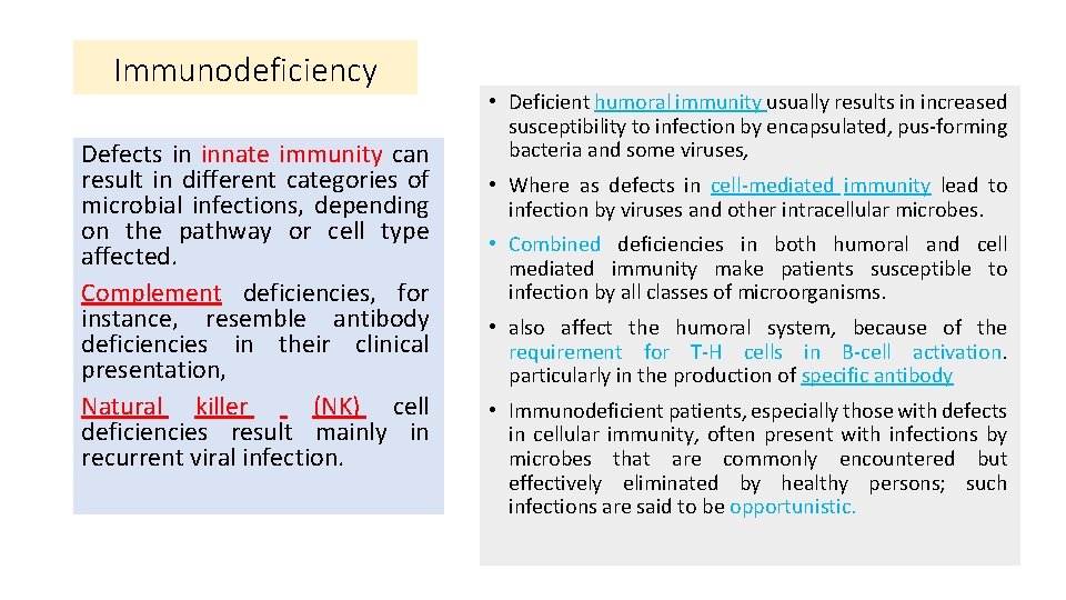
Immunodeficiency Defects in innate immunity can result in different categories of microbial infections, depending on the pathway or cell type affected. Complement deficiencies, for instance, resemble antibody deficiencies in their clinical presentation, Natural killer (NK) cell deficiencies result mainly in recurrent viral infection. • Deficient humoral immunity usually results in increased susceptibility to infection by encapsulated, pus-forming bacteria and some viruses, • Where as defects in cell-mediated immunity lead to infection by viruses and other intracellular microbes. • Combined deficiencies in both humoral and cell mediated immunity make patients susceptible to infection by all classes of microorganisms. • also affect the humoral system, because of the requirement for T-H cells in B-cell activation. particularly in the production of specific antibody • Immunodeficient patients, especially those with defects in cellular immunity, often present with infections by microbes that are commonly encountered but effectively eliminated by healthy persons; such infections are said to be opportunistic.
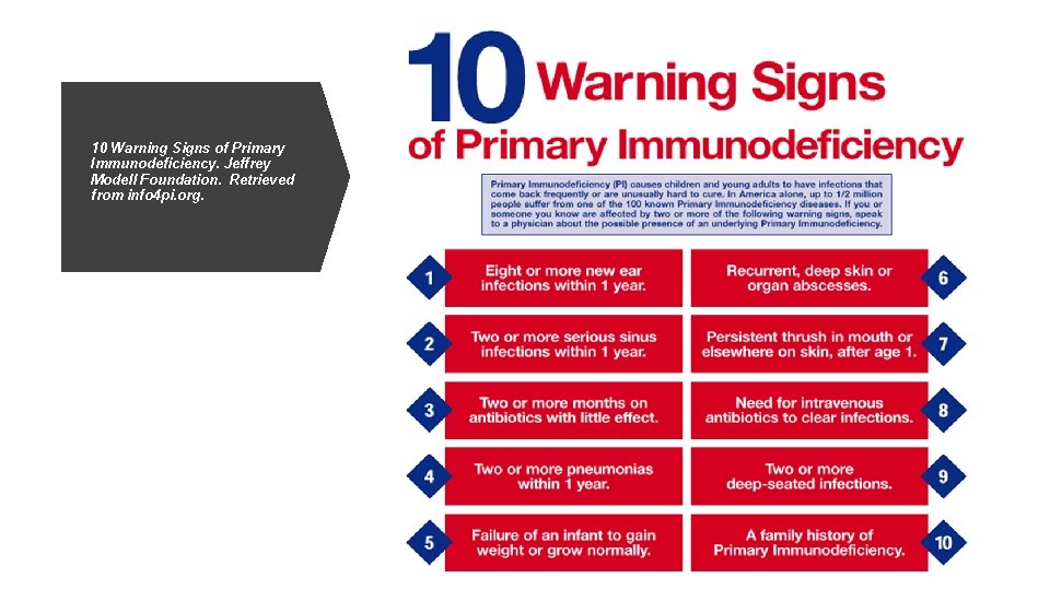
10 Warning Signs of Primary Immunodeficiency. Jeffrey Modell Foundation. Retrieved from info 4 pi. org.
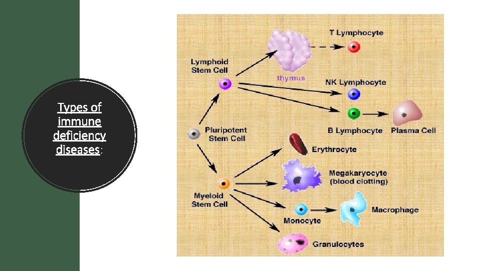
Types of immune deficiency diseases:
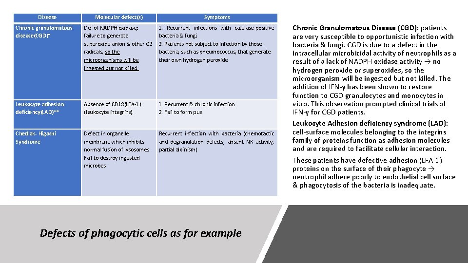
Disease Molecular defect(s) Symptoms Chronic granulomatous disease(CGD)* Def of NADPH oxidase; failure to generate superoxide anion & other O 2 radicals, so the microorganisms will be ingested but not killed. 1. Recurrent infections with catalase-positive bacteria & fungi. 2. Patients not subject to infection by those bacteria, such as pneumococcus, that generate their own hydrogen peroxide. Leukocyte adhesion deficiency(LAD)** Absence of CD 18(LFA-1) (leukocyte integrins). 1. Recurrent & chronic infection. 2. Fail to form pus. Chediak- Higashi Syndrome Defect in organelle membrane which inhibits normal fusion of lysosomes Fail to destroy ingested microbes Recurrent infection with bacteria (chemotactic and degranulation defects, absent NK activity, partial albinism) Defects of phagocytic cells as for example Chronic Granulomatous Disease (CGD): patients are very susceptible to opportunistic infection with bacteria & fungi. CGD is due to a defect in the intracellular microbicidal activity of neutrophils as a result of a lack of NADPH oxidase activity → no hydrogen peroxide or superoxides, so the microorganism will be ingested but not killed. The addition of IFN-γ has been shown to restore function to CGD granulocytes and monocytes in vitro. This observation prompted clinical trials of IFN-γ for CGD patients. Leukocyte Adhesion deficiency syndrome (LAD): cell-surface molecules belonging to the integrins family of proteins function as adhesion molecules and are required to facilitate cellular interaction. These patients have defective adhesion (LFA-1) proteins on the surface of their phagocyte → neutrophil adhere poorly to endothelial cell surface & phagocytosis of the bacteria is inadequate.
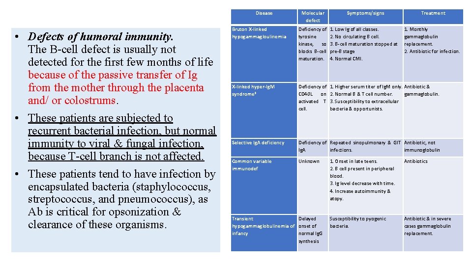
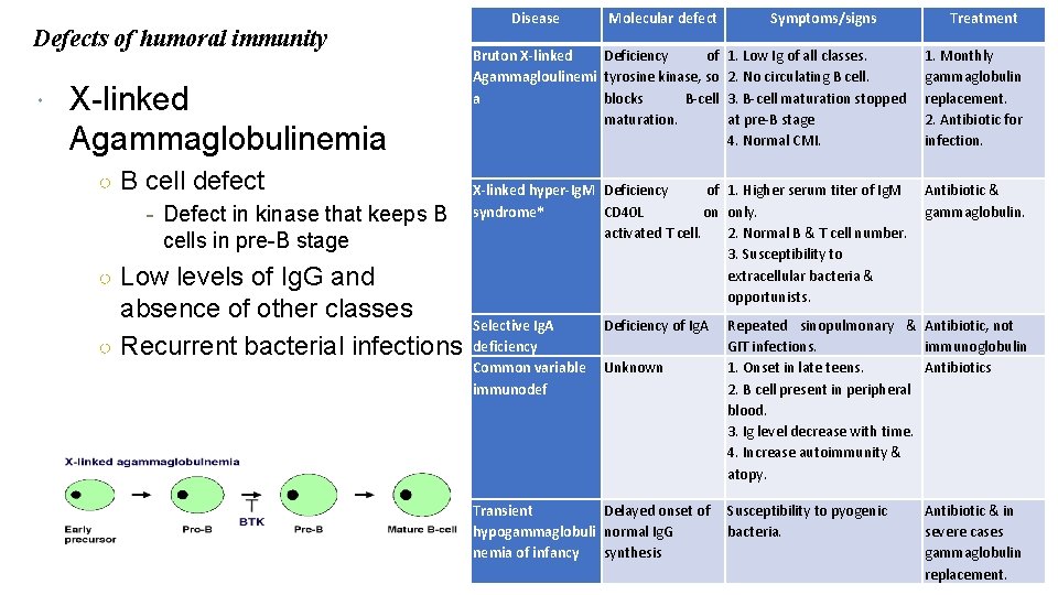
Disease • Defects of humoral immunity. The B-cell defect is usually not detected for the first few months of life because of the passive transfer of Ig from the mother through the placenta and/ or colostrums. • These patients are subjected to recurrent bacterial infection, but normal immunity to viral & fungal infection, because T-cell branch is not affected. • These patients tend to have infection by encapsulated bacteria (staphylococcus, streptococcus, and pneumococcus), as Ab is critical for opsonization & clearance of these organisms. Molecular defect Symptoms/signs Treatment Bruton X-linked hypogammagloulinemia Deficiency of tyrosine kinase, so blocks B-cell maturation. 1. Low Ig of all classes. 2. No circulating B cell. 3. B-cell maturation stopped at pre-B stage 4. Normal CMI. 1. Monthly gammaglobulin replacement. 2. Antibiotic for infection. X-linked hyper-Ig. M syndrome* Deficiency of CD 40 L on activated T cell. 1. Higher serum titer of Ig. M only. Antibiotic & 2. Normal B & T cell number. gammaglobulin. 3. Susceptibility to extracellular bacteria & opportunists. Selective Ig. A deficiency Deficiency of Repeated sinopulmonary & GIT Antibiotic, not Ig. A infections. immunoglobulin Common variable immunodef Unknown Transient Delayed hypogammaglobulinemia of onset of infancy normal Ig. G synthesis 1. Onset in late teens. 2. B cell present in peripheral blood. 3. Ig level decrease with time. 4. Increase autoimmunity & atopy. Antibiotics Susceptibility to pyogenic bacteria. Antibiotic & in severe cases gammaglobulin replacement.
Defects of humoral immunity X-linked Agammaglobulinemia ○ B cell defect - Defect in kinase that keeps B cells in pre-B stage ○ Low levels of Ig. G and absence of other classes ○ Recurrent bacterial infections Disease Molecular defect Bruton X-linked Deficiency of Agammagloulinemi tyrosine kinase, so a blocks B-cell maturation. Symptoms/signs Treatment 1. Low Ig of all classes. 2. No circulating B cell. 3. B-cell maturation stopped at pre-B stage 4. Normal CMI. 1. Monthly gammaglobulin replacement. 2. Antibiotic for infection. X-linked hyper-Ig. M Deficiency of 1. Higher serum titer of Ig. M syndrome* CD 40 L on only. activated T cell. 2. Normal B & T cell number. 3. Susceptibility to extracellular bacteria & opportunists. Antibiotic & gammaglobulin. Selective Ig. A deficiency Common variable immunodef Deficiency of Ig. A Unknown Repeated sinopulmonary & Antibiotic, not GIT infections. immunoglobulin 1. Onset in late teens. Antibiotics 2. B cell present in peripheral blood. 3. Ig level decrease with time. 4. Increase autoimmunity & atopy. Transient Delayed onset of Susceptibility to pyogenic hypogammaglobuli normal Ig. G bacteria. nemia of infancy synthesis Antibiotic & in severe cases gammaglobulin replacement.
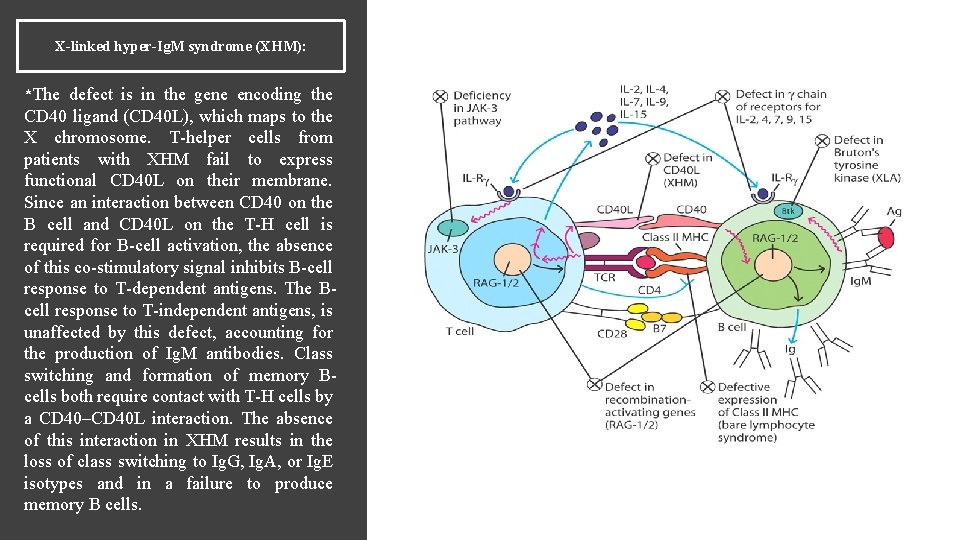
X-linked hyper-Ig. M syndrome (XHM): *The defect is in the gene encoding the CD 40 ligand (CD 40 L), which maps to the X chromosome. T-helper cells from patients with XHM fail to express functional CD 40 L on their membrane. Since an interaction between CD 40 on the B cell and CD 40 L on the T-H cell is required for B-cell activation, the absence of this co-stimulatory signal inhibits B-cell response to T-dependent antigens. The Bcell response to T-independent antigens, is unaffected by this defect, accounting for the production of Ig. M antibodies. Class switching and formation of memory Bcells both require contact with T-H cells by a CD 40–CD 40 L interaction. The absence of this interaction in XHM results in the loss of class switching to Ig. G, Ig. A, or Ig. E isotypes and in a failure to produce memory B cells.
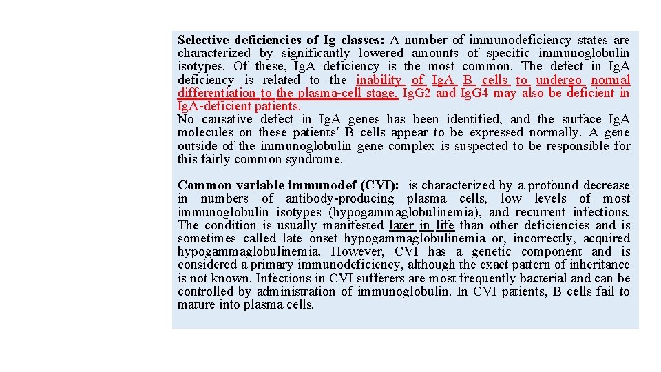
Selective deficiencies of Ig classes: A number of immunodeficiency states are characterized by significantly lowered amounts of specific immunoglobulin isotypes. Of these, Ig. A deficiency is the most common. The defect in Ig. A deficiency is related to the inability of Ig. A B cells to undergo normal differentiation to the plasma-cell stage. Ig. G 2 and Ig. G 4 may also be deficient in Ig. A-deficient patients. No causative defect in Ig. A genes has been identified, and the surface Ig. A molecules on these patients’ B cells appear to be expressed normally. A gene outside of the immunoglobulin gene complex is suspected to be responsible for this fairly common syndrome. Common variable immunodef (CVI): is characterized by a profound decrease in numbers of antibody-producing plasma cells, low levels of most immunoglobulin isotypes (hypogammaglobulinemia), and recurrent infections. The condition is usually manifested later in life than other deficiencies and is sometimes called late onset hypogammaglobulinemia or, incorrectly, acquired hypogammaglobulinemia. However, CVI has a genetic component and is considered a primary immunodeficiency, although the exact pattern of inheritance is not known. Infections in CVI sufferers are most frequently bacterial and can be controlled by administration of immunoglobulin. In CVI patients, B cells fail to mature into plasma cells.
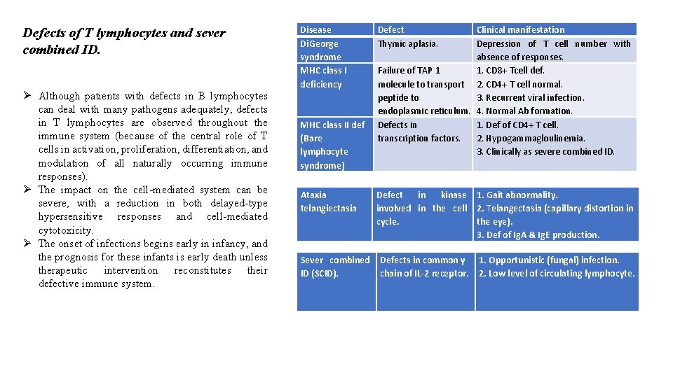
Defects of T lymphocytes and sever combined ID. Ø Although patients with defects in B lymphocytes can deal with many pathogens adequately, defects in T lymphocytes are observed throughout the immune system (because of the central role of T cells in activation, proliferation, differentiation, and modulation of all naturally occurring immune responses). Ø The impact on the cell-mediated system can be severe, with a reduction in both delayed-type hypersensitive responses and cell-mediated cytotoxicity. Ø The onset of infections begins early in infancy, and the prognosis for these infants is early death unless therapeutic intervention reconstitutes their defective immune system. Disease Di. George syndrome MHC class I deficiency MHC class II def (Bare lymphocyte syndrome) Ataxia telangiectasia Sever combined ID (SCID). Defect Thymic aplasia. Clinical manifestation Depression of T cell number with absence of responses. Failure of TAP 1 1. CD 8+ Tcell def. molecule to transport 2. CD 4+ T cell normal. peptide to 3. Recurrent viral infection. endoplasmic reticulum. 4. Normal Ab formation. Defects in 1. Def of CD 4+ T cell. transcription factors. 2. Hypogammagloulinemia. 3. Clinically as severe combined ID. Defect in kinase 1. Gait abnormality. involved in the cell 2. Telangectasia (capillary distortion in cycle. the eye). 3. Def of Ig. A & Ig. E production. Defects in common γ chain of IL-2 receptor. 1. Opportunistic (fungal) infection. 2. Low level of circulating lymphocyte.
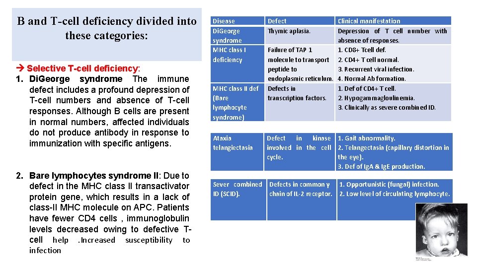
B and T-cell deficiency divided into these categories: Selective T-cell deficiency: 1. Di. George syndrome The immune defect includes a profound depression of T-cell numbers and absence of T-cell responses. Although B cells are present in normal numbers, affected individuals do not produce antibody in response to immunization with specific antigens. 2. Bare lymphocytes syndrome II: Due to defect in the MHC class II transactivator protein gene, which results in a lack of class-II MHC molecule on APC. Patients have fewer CD 4 cells , immunoglobulin levels decreased owing to defective Tcell help . Increased susceptibility to infection Disease Di. George syndrome MHC class I deficiency MHC class II def (Bare lymphocyte syndrome) Ataxia telangiectasia Sever combined ID (SCID). Defect Thymic aplasia. Clinical manifestation Depression of T cell number with absence of responses. Failure of TAP 1 1. CD 8+ Tcell def. molecule to transport 2. CD 4+ T cell normal. peptide to 3. Recurrent viral infection. endoplasmic reticulum. 4. Normal Ab formation. Defects in 1. Def of CD 4+ T cell. transcription factors. 2. Hypogammagloulinemia. 3. Clinically as severe combined ID. Defect in kinase 1. Gait abnormality. involved in the cell 2. Telangectasia (capillary distortion in cycle. the eye). 3. Def of Ig. A & Ig. E production. Defects in common γ chain of IL-2 receptor. 1. Opportunistic (fungal) infection. 2. Low level of circulating lymphocyte.
B and T-cell deficiency divided into these categories: Combined partial B and T-cell deficiency: Ataxia telangiectasia Defect in kinase involved in cell cycle Associated with a lack of coordination of movement (ataxis) and dilation of small blood vessels of the facial area (telangiectasis). T-cells and their functions are reduced to various degrees. B cell numbers and Ig. M concentrations are normal to low. Disease Di. George syndrome MHC class I deficiency Defect Thymic aplasia. MHC class II def (Bare lymphocyte syndrome) Defects in transcription factors. Ataxia telangiectasia Defect in kinase 1. Gait abnormality. involved in the cell 2. Telangectasia (capillary distortion in cycle. the eye). 3. Def of Ig. A & Ig. E production. Sever combined ID (SCID). Clinical manifestation Depression of T cell number with absence of responses. Failure of TAP 1 1. CD 8+ Tcell def. molecule to transport 2. CD 4+ T cell normal. peptide to endoplasmc 3. Recurrent viral infection. reticulum. 4. Normal Ab formation. Defects in common γ chain of IL-2 receptor. 1. Def of CD 4+ T cell. 2. Hypogammagloulinemia. 3. Clinically as severe combined ID. 1. Opportunistic (fungal) infection. 2. Low level of circulating lymphocyte.
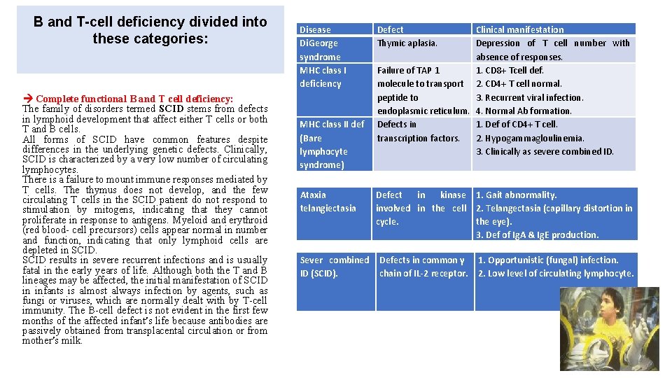
B and T-cell deficiency divided into these categories: Complete functional B and T cell deficiency: The family of disorders termed SCID stems from defects in lymphoid development that affect either T cells or both T and B cells. All forms of SCID have common features despite differences in the underlying genetic defects. Clinically, SCID is characterized by a very low number of circulating lymphocytes. There is a failure to mount immune responses mediated by T cells. The thymus does not develop, and the few circulating T cells in the SCID patient do not respond to stimulation by mitogens, indicating that they cannot proliferate in response to antigens. Myeloid and erythroid (red blood- cell precursors) cells appear normal in number and function, indicating that only lymphoid cells are depleted in SCID results in severe recurrent infections and is usually fatal in the early years of life. Although both the T and B lineages may be affected, the initial manifestation of SCID in infants is almost always infection by agents, such as fungi or viruses, which are normally dealt with by T-cell immunity. The B-cell defect is not evident in the first few months of the affected infant’s life because antibodies are passively obtained from transplacental circulation or from mother’s milk. Disease Di. George syndrome MHC class I deficiency MHC class II def (Bare lymphocyte syndrome) Ataxia telangiectasia Sever combined ID (SCID). Defect Thymic aplasia. Clinical manifestation Depression of T cell number with absence of responses. Failure of TAP 1 1. CD 8+ Tcell def. molecule to transport 2. CD 4+ T cell normal. peptide to 3. Recurrent viral infection. endoplasmic reticulum. 4. Normal Ab formation. Defects in 1. Def of CD 4+ T cell. transcription factors. 2. Hypogammagloulinemia. 3. Clinically as severe combined ID. Defect in kinase 1. Gait abnormality. involved in the cell 2. Telangectasia (capillary distortion in cycle. the eye). 3. Def of Ig. A & Ig. E production. Defects in common γ chain of IL-2 receptor. 1. Opportunistic (fungal) infection. 2. Low level of circulating lymphocyte.
Defects of complement. Deficiencies of complement or its regulation as in these cases: Deficiencies in components or functions, grouped as (early component, late component or alternative cascade deficiencies). These may result in: 1. Hereditary angioedema. Absence of C 1 -inhibitor C 1 act on C 4 ↑C 4 a ↑ vasoactive (C 3 a & C 5 a) capillary permeability and edema in several organs. 2. Recurrent infections Patient with C 3 deficiency are particularly susceptible sepsis with pyogenic bacteria such as S. aurous. Those with reduced C 6, C 7 or C 8 are prone to bacteremia with Neisseria meningitides or Neisseria gonorrhoeae. 3. Autoimmune diseases: Patient with C 2 & C 4 deficiencies have autoimmune diseases. C 3 deficiency are associated with SLE Components Deficiency Signs/diagnosis Classic pathway C 1 q, C 1 r, C 1 s, C 4, C 2 1. Marked increase in immune complex disease 2. Increased infection with pyogenic bacteria. Both pathways C 3 Def of regulatory proteins. 1. Recurrent bacterial infection. 2. Immune complex disease. C 5, C 6, C 7, C 8 Recurrent meningococcal & gonococcal infections. C 1 -INH (hereditary angioedema). 1. Overuse of C 1, C 4 or C 2. 2. Edema at mucosal surfaces.
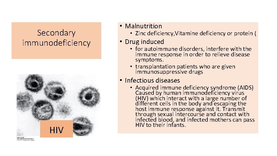
Secondary immunodeficiency • Malnutrition • Zinc deficiency, Vitamine deficiency or protein ( • Drug induced • for autoimmune disorders, interfere with the immune response in order to relieve disease symptoms. • transplantation patients who are given immunosuppressive drugs • Infectious diseases HIV • Acquired immune deficiency syndrome (AIDS) Caused by human immunodeficiency virus (HIV) which interact with a large number of different cells in the body and escaping the host immune response against it. Transmit through sexual intercourse and contact with infected blood, and infected mothers can pass HIV to their infants.
Refferences : v. Immunology , Kuby, seventh edition 2013 v Immunology , Kuby, eighth edition 2019 v. Medical microbiology, Jawetz, 26 th edition v. Cellular and Molecular Immunology, Abul K. Abbas, 8 th edition.