General Thoracic Surgery CardioThoracic Department Papworth Hospital University


 N 1 MTS peribronchial")


 (most common 75%) Hemoptysis (33%) Pain (dolore) (50%")

 1) syndromes similar to myasthenia gravis")


 It is better to evaluate the vascular invasion")

 Higher specificity has the PET con FDG (F 18 fluoro")


 Surgical treatment Not responder chemo")


")


 • The main indication is in patients with")
. Aberrant signalling from the")


- Slides: 71
General Thoracic Surgery Cardio-Thoracic Department Papworth Hospital University of Cambridge Hospital Trust Cambridge, UK Phase III Lecture Programme Oncology lung cancer Marcello Migliore
History In 1910 Alton Oschner, esteemed surgeon, recalled that as a student at Washington University he was asked to witness an autopsy of a patient with lung cancer, having been told lung cancer was so rare that he may never see another case. He saw the next case 17 years later. Within the next 6 months 8 more cases were seen at that hospital and this began what he called an epidemic.
IA IB IIIA
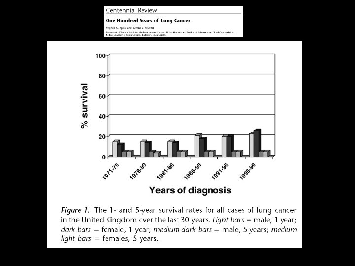
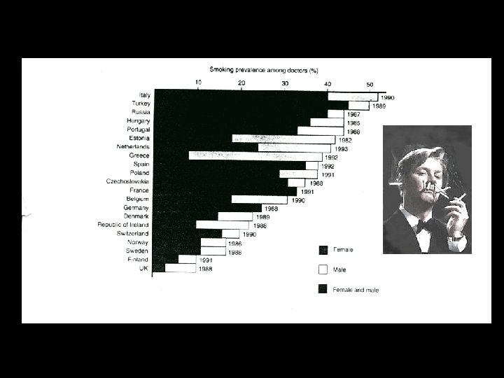
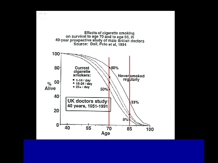
Epidemiology • 1950 Doll and Hill in the British Medical Journal confirmed suspicions that lung cancer was associated with cigarette smoking
Epidemiology • In 1930 in the USA, lung cancer rates were less than 5 per 100, 000 • In 1998 the death rate per 100, 000 population for men reached 77. 2 in Belgium and 75. 5 in Scotland.
Epidemiology What is going on in the developing countries • In 1994 the rate of lung cancer was similar to that of the USA in 1930 • In 1999 the rate of lung cancer was 14. 1 per 100, 000 in developing countries and 71. 4 in developed countries
Lung cancer Only 25% of patients will undergo surgery with the hope of curing the disease Most 75% will not have surgery
Lung cancer c p TNM
TNM T factor T 1 < 3 cm T 2 > 3 cm, invading visceral pleura, > 2 cm from carina T 3 spread to the chest wall, < 2 cm from carina T 4 spread to the heart, two nodules in the same lobe, + pleural fluid for malignant cell
T factor T 2 : > 3 cm, or invading visceral pleura, > 2 cm from carina
T factor T 3 spread to the chest wall, < 2 cm from carina T 4 spread to the heart, two nodules in the same lobe, + pleural fluid for malignant cell
TNM N factor N 0 Absence of nodal metastasis (MTS) N 1 MTS peribronchial and ilar nodes - same side N 2 MTS mediastinal (same side) and sub carinal nodes N 3 MTS mediastinal and ilar contro lateral, supraclavear and scalene
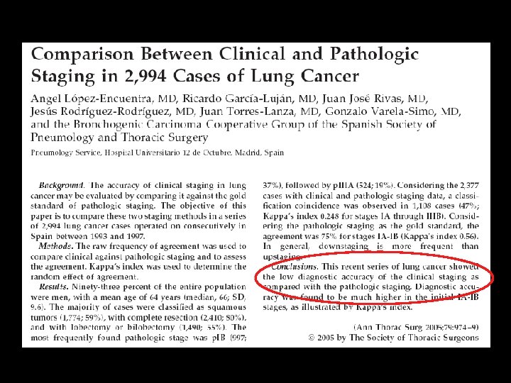
Lung cancer Why it is important to know the status of nodal diseases Ann Thorac Surg 2002; 73: 1545 -51
c. TNM N T 2 N 3 N 2
VIDEO - MEDIASTINOSCOPY Washington University School of Medicine review of 2137 mediastinoscopies Morbidity and mortality rates was 0. 6% and 0. 2% respectively Sensitivity of 85. 2% in the accurate staging of N 2 and N 3 disease when used preoperatively in patients with lung cancer.
TNM M factor Distant MTS Or two nodules in different lobes
Lung cancer - STAGE and T N M STAGE Tumor Nodes Metastasis IA IB T 1 T 2 N 0 M 0 IIA IIB T 1 T 2 T 3 N 1 N 0 M 0 M 0 III A T 3 T 1 -3 N 1 M 0 N 2 M 0 IIIB T 1 -4 T 4 N 3 N 0 -3 M 0 IV T 1 -4 N 0 -3 M 1 Mountain 1997, AJCC, UICC
Lung cancer Pathological and clinical characteristics 1. Squamous a. Obctructive pneumonitis b. Lung or lobar collapse d. 1/3 are in the periphery of the lung e. 20% are escavated 2. Adenocarcinoma a. 2/3 are in the periphery b. < 3 cm c. Bronchioloalveolar type have diffuse pattern 25 -40% 30 -50% 3. A great cell (undifferenciated) 1. 60% are in the periphery 2. 2/3 > 4 cm 4. Small cell 1. 80% abnormal lung ilum 2. 2/5 parecnchimal changes 10 -20 % 15 -25 %
Lung cancer Clinical presentation 1. Broncho-pulmonary 2. Extrapulmonary but intrathoracic 3. Extrapulmonary metastatic 4. Extra pulmonary non metastatic ( i. e. paraneoplastic)
Lung cancer Bronchopulmonary Symptoms Cough (tosse) (most common 75%) Hemoptysis (33%) Pain (dolore) (50% poor prognostic sign) Anorexia and weight loss (poor prognostic sign) Shortness of breath Hoarseness (1 -8%)
Lung cancer Clinical Signs • “clubbing” is the most common • Pleural effusion • Pulmonary hypertrophic osteopathy (2 -12% of all patients with lung cancer)
Extra pulmonary non metastatic ( i. e. paraneoplastic) 1) syndromes similar to myasthenia gravis 2) polimyositis b. Cushing Syndrome - small cell c. SIADH – small cell d. Hypercalcemia – squamous cell e. Gynecomastia – small cell f. Gonadotropin – indifferenciated great cell
Lung cancer Diagnosis Invasive Not invasive
. . . In every new lung nodule it is necessary to achieve a final pathologic diagnosis to exclude the presence of cancer EVIDENCE BASED MEDICINE
Lung cancer – diagnosis Chest x ray Generally radiographic alterations are present 7 months before symptoms reveal the tumor It shows nodules > 1 cm The most common finding is the coin lesion It failed to demonstrate a reducing in lung cancer mortality
CHEST X RAY
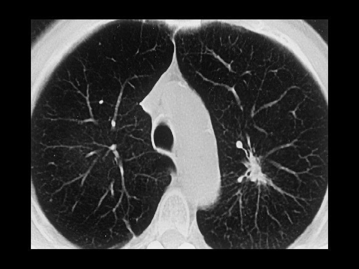
Lung cancer - diagnosis CT of the chest a. The best method to evaluate mediastinal adenopathy and renal glands b. Invasion of thoracic wall is not always correctly diagnosed c. Paraesophageal nodes and inferior pulmonary vein are not visible d. Nodes < 1 cm have 7% possibility to be malignant e. Nodes > 1 cm have 55 -65% possibility to be malignant
Lung cancer Diagnosis CT of the chest Three studies revealed that using Spiral CT as a screening method it was possible to discover 84 -93 % of lung cancer in the initial stage (IA) with a low percentage of false positive (20%).
Lung cancer Diagnosis Magnetic Resonance (MR) It is better to evaluate the vascular invasion
MR – Brain metastasis
PET (Positron Emission Tomography) Higher specificity has the PET con FDG (F 18 fluoro deossiglucosio) (sensibility 95% - specificity 85%) but it is useful in nodules greater than 6 mm. It reduces the need to send the patient for an invasive method such as mediastinoscopy.
CT thorax PET scan N 3
Lung cancer Diagnosis Sputum cytology Sensitive in 20 - 70%, but it is correlated with the position of the tumour Squamous carcinoma is more common to be positive than small cell or adenocarcinoma. When cytology is positive a final diagnosis in 85% is possible
Lung cancer Diagnosis Invasive diagnosis
RIGID FLEXIBLE
BRONCOSCOPY Haemoptsis Intraluminal mass Biopsy
Lung cancer and pleural effusion Thoracocentesis
Lung cancer - diagnosis CT GUIDED BIOPSY Percutaneous o transbronchial In peripheric lesion is accurate in 85 -95%
CT- FNA: 85 -95% sensitive Complications ?
Lung cancer – invasive diagnosis VATS Thoracotomy
Lung cancer TREATMENT SURGERY
Lung cancer – preoperative tests Lung function is mandatory if the patient needs an operation High surgical risk 1. FEV 1 < 40% 2. Predicted postoperative FEV 1 < 30% 4. DLCO < 40% 5. PCO 2 > 45 mm. Hg
The goal of surgical treatment in oncology is twofold • to achieve long term survival with a good quality of life • to avoid recurrence
Surgical treatment of lung cancer Indications • c. TNM • Lung Function Tests • Intra-operative
Lung cancer - Surgical treatment • • Wedge Segmentectomy Lobectomy Pneumonectomy Sleeve Resection Limphadenectomy Lung transplantation (Bronchioloalveolar ca. ) VATS - ?
Surgical Treatment Stage I o II Lobectomy + Nodal dissection
VATS Lobectomy
Age and lung resection
Surgical Treatment Stage IIIA Neoadjuvant Therapy: Responder (down staging) Surgical treatment Not responder chemo therapy
23. 9 %!!! mortality
Bronchiolo – alveolar carcinoma Stadio IV T 4 N 0 M 1 Bilateral Lung TX
Lung cancer TREATMENT CHEMO RADIO OTHER (stent, laser, photodynamic therapy)
Chemotherapy Advanced NSCLC • Over the last 10 years new agents have provided a better median survival of 7 -10 months with 35 -40% alive at 1 year.
Chemotherapy Small Cell Lung Cancer • • The most frustrating cancer to treat 1970 mean survival was 5 months Nowadays 18 -20 months (limited disease) Nowdays 6 -9 months (extensive disease)
Radiotherapy • As a curative modality, radiotherapy has been disappointing. • The combination of chemo and radiotherapy has provided better median and long term survival than radiotherapy alone • Radiotherapy has proven most successful in palliating lung cancer symptoms in up to 80% of instances.
Other (stent, laser, photodymanic therapy, cryosurgey) • The main indication is in patients with the occlusion of the main bronchus or lobar bronchi • Also indicated in patients in early-stage disease who are not candidates for surgical procedures. It may also be used to reduce symptoms in late-stage disease.
NEW APPROACHES • Mutation in Epidermal Growth Factor Receptor (EGFR). Aberrant signalling from the EGFR is known in lung cancer. • Two oral inhibitors of EGF, geftinib and erlotinib, inhibit the EGFR and both have demonstrated antitumor activity. • Responses to these agent are few. Mainly nonsmokers, women, BAC.
NEW APPROACHES Immunotherapy uses drugs that boost the patient's immune system to fight cancer. Gene Therapy. Research is underway to determine if methods can be developed to attack genetic mutations that are causing cancer. Angiogenesis Inhibitors. Under investigation are agents that inhibit the formation of new blood vessels (called angiogenesis). The spread of new blood vessels is controlled by compounds called growth factors, such as vascular endothelial cell growth factor (VEGF). Angiogenesis inhibitors are agents that literally turn off the growth factors themselves or their receptors.
SUMMARY – in general • One hundred year ago lung cancer was rare • Only 25 % of patients with lung cancer will have the chance to have the cancer removed, and therefore the hope of a cure. The majority will undergo oncologic treatment. • Chest x ray modification is present 7 months before the first symptoms. Chest x ray reveals only nodules > 1 cm. • Modern diagnostic tools are “latecomers” and their main role is to evaluate the possibility of surgical treatment. • All the non invasive diagnostic tests do not determine the histology. (evidence based medicine).
SUMMARY – surgical treatment • Patients in stage I and II can undergo surgery after the evaluation of the respiratory condition • Patients in stage IIIA are potentially resectable, but they first need neoadjuvant therapy
SUMMARY – surgical treatment • VATS is probably the best treatment for patients in stage I • To avoid or discuss in detail with the patient if R pneumonectomy is indicated following neoadjuvant treatment for the high mortality risks • In case of advanced BAC Lung Transplantation is an option.
SUMMARY – non surgical treatment • Chemotherapy is the only option for SCLC • Radiotherapy is successful in palliating lung cancer symptoms • The combination of chemo and radiotherapy has provided better median and long term survival.
To achieve the best long term survival it is necessary to reduce the time interval between The first clinical symptoms and the final diagnosis and treatment
2008 Anatomical lung resection is the most appropriate treatment to achieve better long term survival adding the hope of curing the cancer in patients at the initial stage with good lung function
The future
The future in the treatment of lung cancer • early diagnosis • Surgery associated with otherapies