WALL OF THORAX PLEURA LUNGS Week 7 Review


. Rib")

 Axillary artery and brachial plexus pass between anterior/middle & posterior")


 Run inferioranteriorly from rib above to")


. Hemiazygos (left inferior side). Accessory")
 Located")


![Pleura [of the Lung] A serous (watery) membrane – a “sack” – think of](https://slidetodoc.com/presentation_image/2eb92b406816df723005b2a6d4b7d8dd/image-25.jpg "Pleura [of the Lung] A serous (watery) membrane – a “sack” – think of")
![Pleura [of the Lung] (cont. ) Parietal pleura blue Visceral pleura pink Areas of](https://slidetodoc.com/presentation_image/2eb92b406816df723005b2a6d4b7d8dd/image-26.jpg "Pleura [of the Lung] (cont. ) Parietal pleura blue Visceral pleura pink Areas of")
![Pleura [of the Lung] (cont. ) A “recess” is different than a point of](https://slidetodoc.com/presentation_image/2eb92b406816df723005b2a6d4b7d8dd/image-27.jpg "Pleura [of the Lung] (cont. ) A “recess” is different than a point of")
![Pleura [of the Lung] (cont. ) Know the names of the recesses. Know that](https://slidetodoc.com/presentation_image/2eb92b406816df723005b2a6d4b7d8dd/image-28.jpg "Pleura [of the Lung] (cont. ) Know the names of the recesses. Know that")


 APICAL (2)ANTERIOR (3)POSTERIOR")
APICO-POSTERIOR (2)ANTERIOR (3)SUPERIOR (4)INFERIOR LOBE")


 A patient presents with having difficulty gripping and holding objects. Physical")
 If a person aspirates something into their trachea, where will it")


 Heart sounds Skeleton of the heart Conduction system of")
- Slides: 57
WALL OF THORAX PLEURA & LUNGS Week 7 Review
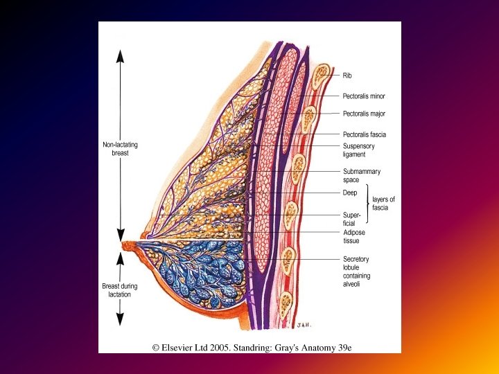
Mammary Glands Lab: Responsible for learning male & female anatomy. Modified sweat gland of the skin superficial structure. Each gland terminates as a Lactiferous Ducts at the nipple Suspensory ligament [of the breast]. Connected to superficial fascia – not muscle
Vessels of the Breast Internal thoracic artery – medial portion. Lateral thoracic artery – lateral portion.
Lymph Drainage of Breast Key points: Drainage of Axillary Tail Humeral & Pectoral Lateral portion of breast Pectoral Axillary Nodes Medial portion of the breast Parasternal lymph nodes which can cross the midline. The breast actually continues into the axillary region…
Sternum – Joint Facts Clavicle & Sternum articulate in a complex joint with a meniscus. Rib #1 articulates with the manubrium via a synchondrosis joint. Ribs 2 -7 articulate using synovial joints. Ribs 8 -10 articulate using synchondrosis with 7 th Costal Cartilage
Vertebrae & Ribs Review on your own: Thoracic vertebrae (including their unique characteristics). Rib bones (including parts and features). 7 -True, 5 -False, and 2 -Floating Ribs Clavicle
Scalene Muscles Study more next term. Three muscles: Anterior, middle, and posterior. Origin: Transverse processes of the cervical vertebrae. Insertion: Anterior & middle: 1 st rib. Posterior: 2 nd rib.
Scalene Muscles (cont. ) Axillary artery and brachial plexus pass between anterior/middle & posterior scalene muscles.
Sternum Manubrium. Body – 4 fused sternebrae. Xiphoid process. Sternal Angle T 4 Sternalis muscle – rarely present – its significance in imaging studies is it causes a false positve for breast cancer.
Review on Your Own Thoracic inlet & outlet and significance. Outlet sealed off by diaphragm but inlet not sealed; lungs can stick up above 1 st rib. Structures of the upper limb including the subclavius muscle.
Arterial Divisions Subclavin Artery divided into 1 st, 2 nd, and 3 rd parts by the Anterior Scalene muscle. 1 st Part: Vertebral Artery, Internal Thoracic Artery & Thyrocervical Trunk 2 nd Part: Costocervical Trunk 3 rd Part: Dorsal Scapular Artery (often arrises as branch of transverse cervical) Axillary Artery divided into 2 parts by Pectoralis Minor Muscle
Arterial Divisions ABC'S of the aortic arch! Aortic arch gives off the Bracheiocephalic trunk Left Common Carotid Left Subclavian artery
Differences b/w Right & Left Right: Brachiocephalic Trunk gives rise to Subclavian Left: Left Common Carotid and Left Subclavian branch straight off aortic arch Read quiz and exam questions carefully.
MUSCLES OF THE THORAX
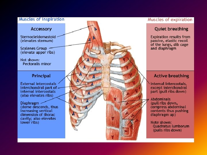
Intercostal Muscles Superficial Layer External Intercostals: (11 pairs) Run inferioranteriorly from rib above to rib below Action: Elevate Ribs Middle Layer: Internal Intercostals: (11 pairs) Run Inferiorposteriorly floors of costal grooves to superior borders of ribs inferior to them. Action: Elevate Ribs (Moore says depress) Deepest Layer Innermost Intercostals: similar to Internal Intercostals Innervation: Intercostal Nerve
Intercostal Muscles
Intercostal Vessels The thoracic wall is supplied by three sources of blood supply: Axillary Artery Supreme Thoracic (2) Lateral Thoracic (3) Subclavian Artery Internal Thoracic (or mammary) Artery (1) Anterior Intercostal branches Thoracic Aorta Intercostal Arteries (4)
Intercostal Vessels Azygos (right side, drains spaces 4 -11). Hemiazygos (left inferior side). Accessory hemiazygos (left superior side). Supreme intercostal vein. Internal thoracic vein.
NEUROVASCULAR SPACE Know the orientation of Nerve, Artery & Vein (NAV & VAN) Located between Internal & Innermost Intercostal Muscles
DEEP THORAX MUSCLES
Subcostal Muscle Lower Thorax Only Origin: Inner surface of near angle of rib. Insertion: 2 -3 Ribs below. Action: Lowers ribs when 12 th rib is fixed by quadratus lumborum. (First rib is locked and cannot be lowered so muscles of 1 st intercostal space can only pull 2 nd rib up. )
Transverse Thoracis Muscle On anterior chest wall Origin: Body and xiphoid of the sternum (ribs 4 -6). Insertion: Costal cartilages of ribs 2 -6. Action: Draw ribs down.
LUNGS
Pleura [of the Lung] A serous (watery) membrane – a “sack” – think of a deflated balloon but with a small amount of fluid between the walls in stead of powder. Parietal pleura – the side of the sack (deflated balloon) that contacts the thoracic wall. Visceral pleura – the side of the sack that contacts the lung. The space between the two sides is the pleural cavity. Note that the lung is NOT inside of the sack: Both surfaces of the deflated balloon curl around the lung.
Pleura [of the Lung] (cont. ) Parietal pleura blue Visceral pleura pink Areas of the parietal pleura are named for the structures they contact (medistinal, costal, diaphragmatic). Point of Reflection – where visceral side becomes the parietal side. (What happens at the sealed end of the balloon. ) Know the points of reflection.
Pleura [of the Lung] (cont. ) A “recess” is different than a point of reflection.
Pleura [of the Lung] (cont. ) Know the names of the recesses. Know that the lungs extend up above thoracic inlet. PLEURISY--FLUID ACCUMULATION IN THE PLEURAII. Drain from costodia-phargmatic recess along midaxillary line. inflammation of the pleura
Lungs Named points of interest: Apex & base. Surfaces – know the names. Lobes & fissures – right and left Impressions – relate to segments and surfaces. Know the lymph flow for the lungs.
Lung Segments
Orientation of Lobes in the Body RIGHT LUNG SUPERIOR LOBE (1) APICAL (2)ANTERIOR (3)POSTERIOR MIDDLE LOBE (1)LATERAL (2)MEDIAL INFERIOR LOBE (1)SUPERIOR (2)MEDIAL BASAL (3)ANTERIOR BASAL (4)LATERAL BASAL (5)POSTERIOR BASAL
Orientation of Lobes in the Body LEFT LUNG SUPERIOR (1)APICO-POSTERIOR (2)ANTERIOR (3)SUPERIOR (4)INFERIOR LOBE SUPERIOR INTERIOR BASAL MEADIAL BASAL LATERAL BASAL POSTERIOR BASAL
Bronchi Know the hierarchy by name and characteristic. (Should have covered this in histology / microanatomy. ) EACH LOBE RECEIVES A SECONDARY BRONCHUS 3 ON RIGHT (one branching off early) 2 ON LEFT
Bronchi 1. Trachea 2. Carina 3. Right main bronchus 4. Right superior lobe bronchus 5. Right middle lobe bronchus 6. Right lower lobe bronchus 7. Left main bronchus
Right superior lobe bronchus Apical segmental or lobular bronchus Anterior segmental or lobular bronchus Posterior segmental or lobular bronchus Terminal bronchioles
Respiration Pressure: intrathoracic < intrapleural < atmospheric Understand this. Know difference between inspiration & expiration; quiet & forced. What muscles are involved? When does gravity play a role? What happens when you stand on your head? What happens in zero-gravity?
A Little Physiology Dead air space – what is it? Collapsed lung – internal vs. external. Relate to pressure differentials.
SAMPLE QUIZ
1. The neurovascular space is found between the a. external intercostals and internal intercostals muscles b. external intercostals and the serratus anterior muscles c. internal intercostals and innermost intercostals muscles d. innermost intercostals muscles and the endothoracic fascia
2. Lymph from the medial half of the breast would first be drained to the a. lateral group of axillary lymph nodes b. subscapular group of axillary lymph nodes c. pectoral group of axillary lymph nodes d. parasternal lymph nodes
3. How many primary bronchi are there supplying the left lung? a. 1 b. 2 c. 3 d. 5
4. Blood from the Right 5 th intercostals space is drained by the intercostals vein which opens into the a. subclavian vein b. accessory hemiazygos vein c. hemiazygos vein d. azygos vein
5. What separates the superior lobe of the left lung from the inferior lobe of the left lung? horizontal fissure b. oblique fissure c. lingual d. pulmonary ligament a.
6. The tubercle of the rib articulates with which structure? a. facet on the transverse process of a thoracic vertebra b. sternum either directly or indirectly c. demifacets on the bodies of two adjacent vertebrae d. the superior articular process of a thoracic vertebra
7. The pulmonary artery enters the lung at the Lingual b. Hilus c. Apex d. Cardiac notch a.
8. Lymph from the right lung would first be drained into which group of lymph nodes? a. pulmonary lymph nodes b. parasternal lymph nodes c. paratrachael lymph nodes d. deep cervical lymph nodes
9. How many bronchopulmonary segments are there in the middle lobe of the right lung? a. 1 b. 2 c. 3 d. 4
10. Which of the following muscles acts to elevate the rib that lies inferior to it? a. external intercostal muscle b. internal intercostal muscle c. innermost intercostals muscle d. all of the above are correct
11. The sternal angle lies at which vertebral level? a. T 1 b. T 2 c. T 3 d. T 4
12. The lymph from the medial aspect of the breast will most probably be drained into which lymph nodes? a. pectoral group of axillary lymph nodes b. parasternal lymph nodes c. lateral group of axillary lymph nodes d. both a & c are correct
13. (3 pts) A patient presents with having difficulty gripping and holding objects. Physical examination is unremarkable except that there is a definite weakness in the grip of the right hand as compared to the left, and the right hand adducts when the wrist is flexed. Which nerve is probably damaged? Explain.
14. (2 pts) If a person aspirates something into their trachea, where will it most likely end up & why?
15. Describe the actions that take place to accomplish a normal, quiet inspiration and expiration.
PREVIEW OF WEEK 8
Heart & Pericardium One whole week on: Pericardium & surface features of heart Major vessels to & from the heart Coronary arteries & veins Chambers & valves of the heart Flow of blood through the heart Inside each chamber of the heart
Heart & Pericardium (cont. ) Heart sounds Skeleton of the heart Conduction system of the heart Innervation of the heart Misc. topics related to the heart