THORACOLUMBAR FASCIA LUMBAR It FASCIA deep fascia encloses

THORACO-LUMBAR FASCIA
- encloses deep muscles of the back. �")
� LUMBAR � It FASCIA (deep fascia)- encloses deep muscles of the back. � 3 layers-1)posterior-thickest. � 2)middle-thicker � 3)anterior-thinnest.
It has 3 strong layers-fills up the gap between the 12 th")
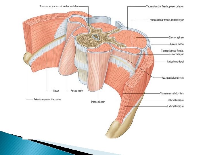
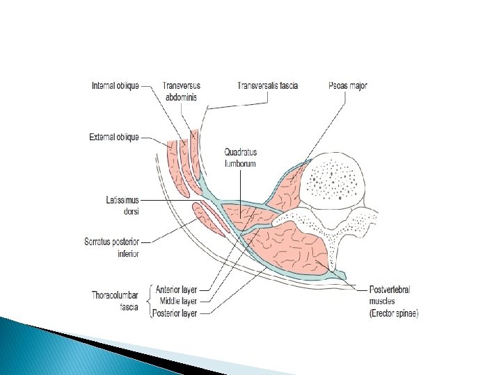
�Features� � 1)It has 3 strong layers-fills up the gap between the 12 th rib and the iliac crest. 2)Between posterior and middle layers-erector spinae and transversus spinalis muscles. 3)Between the middle and anterior layers-quadratus lumborum muscle. 4)The three layers form a dense aponeurotic sheet-origin to the internal oblique and transversus abdominis muscles.

� Extent- � Posterior layer-It covers the loin area, back of thorax and neck. � Middle region. and anterior layer-It covers the lumbar

� Anterior � � � layer- Medially- Anterior surface of the lumbar transverse processes. Laterally-It blends with the middle layer at the lateral border of the quadratus lumborum. Superiorly-lateral arcuate ligament-tip of Transverse process L 1 to 12 th rib. Inferiorly-inner lip of iliac crest and the ilio-lumbar ligament.

� � � Middle layer. Medially-Tips of the lumbar transverse processes and inter-transverse ligaments. Laterally-It blends with the anterior layer at the lateral border of the quadratus lumborum. Superiorly-lower border of the 12 th rib and lumbocostal ligament. Inferiorly-intermediate area of the iliac crest.

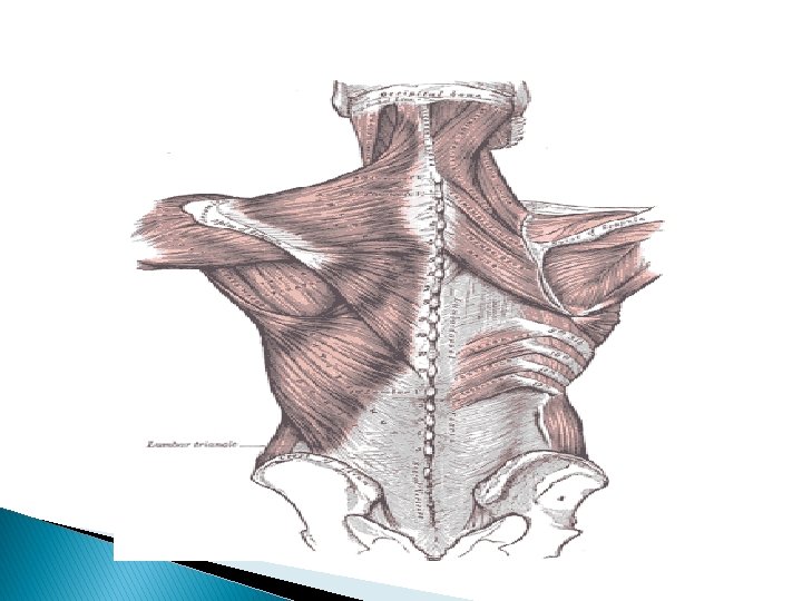
� Posterior layer- Medially-lumbar and sacral spines and interspinous ligaments. � Laterally-blends with middle layer at the lateral border of the erector spinae. � Superiorly-back of thorax - lumbar vertebral spines. � Inferiorly-outer lip of the iliac crest. �


� Thoracic part- � Medially-Spines of thoracic vertebrae. � Laterally-Angles of the ribs. � Above- It extends into the cervical region deep to the serratus posterior superior to fuse with the fascia of the neck.

� COELIAC GANGLIA� Largest ganglia in the body on either sides of coeliac trunk. � Irregular shape. � Larger upper part-greater splanchnic nerve. � Smaller lower part-aortico-renal-lesser splanchnic nerve.
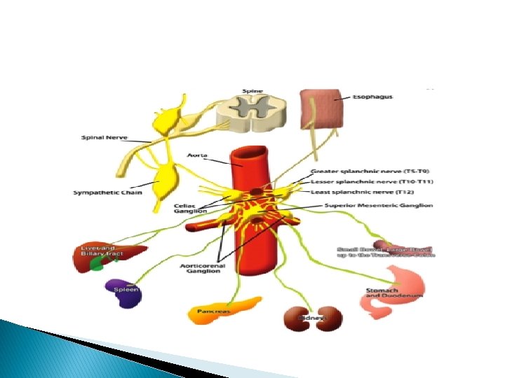

� COELIAC PLEXUS OR SOLAR PLEXUS-related to coeliac ganglion. � SITUATION-on aorta around the coeliac trunk, � and root of superior mesenteric artery.


� FIBERS FORMING THE PLEXUS� 1. Pre-ganglionic sympathetic fibres-through greater and lesser splanchnic nerves. � 2. Post-ganglionic sympathetic fibres-arising in the coeliac ganglion. � 3. Pre-ganglionic vagal fibres-posterior vagal trunk through the right and the left vagus nerves. � 4. sensory fibres from the diaphragm along the inferior phrenic arteries.

� BRANCHES� coeliac plexus forms a number of secondary branches which surround the aorta. � 1)Phrenic plexus� Along the inferior phrenic artery to the suprarenal gland. � 2)Hepatic plexus- around liver, gall bladder and bile ducts.
Left gastric plexus to stomach. � 4)Splenic plexus to spleen. � 5)Suprarenal plexus-pre-ganglionic")
� 3)Left gastric plexus to stomach. � 4)Splenic plexus to spleen. � 5)Suprarenal plexus-pre-ganglionic fibres end in the chromaffin cells of the suprarenal gland. � 6)Renal plexus-It is formed by the coeliac plexus, aorticorenal, lowest thoracic splanchnic nerve, first lumbar splanchnic nerve and aortic plexus. � It supplies kidney, upper part of ureter.
Testicular plexus� It supplies-testis, epididymis, and vas deferens. � 8)ovarian plexus� ovary and")
� 7)Testicular plexus� It supplies-testis, epididymis, and vas deferens. � 8)ovarian plexus� ovary and uterine tube. � 9)superior mesenteric plexus� It contains superior mesenteric ganglion. � It supplies the territory of superior mesenteric artery.
Inferior � It mesenteric plexus- is formed by the aortic plexus. is distributed")
� 10)Inferior � It mesenteric plexus- is formed by the aortic plexus. is distributed to the territory of the inferior mesenteric artery.

Thank u
- Slides: 22