Gastro Anatomy and Physiology Dr Nadia Farhat Ezzawi


 Esophageal Body (cervical & thoracic) Lower Esophageal Sphincter (LES)")
 is tonically closed and contracts during inspiration, preventing")


 lies in the upper abdomen behind the stomach.")

 that make and secrete insulin,")


- Slides: 34
Gastro Anatomy and Physiology Dr. Nadia Farhat Ezzawi Gastroenterologist BMC
Individual components of the gastrointestinal system. Oral cavity and Salivary glands Oesophagus Stomach Small intestine Large intestine Liver Gall bladder Pancreas
Salivary gland -Two submandibular. -Two Parotid. -Two sublingual. -> 400 minor salivary glands Distributed over lips, cheeks, palate, floor of mouth & retromolar area. .
Functions of salivary gland *Secretion of saliva 1500 ml of saliva / day -From the parotid gland: thin, watery fluid, -Sublingual and Submandibular glands: much thicker * facilitates swallowing * keeps the mouth moist & aids speech * serves as a solvent for molecules which stimulate the taste buds * cleans the mouth, gum, & teeth. * contains enzymes begins the chemical digestion of starches through the action of amylase, which breaks down polysaccharides into disaccharides.
oesophagus Upper Esophageal Sphincter (UES) Esophageal Body (cervical & thoracic) Lower Esophageal Sphincter (LES) length 18 to 24 cm
Oesophagus The upper esophageal sphincter (UES) is tonically closed and contracts during inspiration, preventing air from entering into the gastrointestinal tract, and reflux. It relaxes during swallow, belching and vomiting. The lower esophageal sphincter (LES) maintains a steady baseline tone (~20 mm Hg) to prevent gastric contents from entering the esophagus. The LES also contracts during periods of increased intra-abdominal pressure, preventing reflux due to the pressure build up in the abdomen
oesophagus. The esophagus has 2 muscle layers: the inner circular layer and the outer longitudinal layer. The longitudinal muscle shortens the esophagus, while the circular muscle forms lumen-occluding ring contractions. The combination of these localized contractions is responsible for peristalsis. The proximal esophagus contains striated muscle. the distal esophagus smooth muscle. Linning mucosa is stratified squamous epithelium changed to columnar epithelium at gastrooesophageal junction. The submucosa (SM) secretes mucus from mucous glands(MG) which aid the passage of food down the oesophagus.
physiology Normal phases of swallowing: Voluntary oropharyngeal phase – bolus is voluntarily moved into the pharynx Involuntary UES relaxation peristalsis (aboral movement) LES relaxation
Normal Phases of Swallowing Between swallows UES prevents air entering the esophagus during inspiration and prevents esophagopharyngeal reflux LES prevents gastroesophageal reflux peristaltic and non-peristaltic contractions in response to stimuli capacity for retrograde movement (belch, vomiting) and decompression
Structure of wall of the GIT
Stomach - It is J shaped organ in the upper left region of the abdominal cavity. - It has two openings- the oesophageal and the duodenal opening. - four regions. The cardia, fundus, body and pylorus. It is lined by simple columnar epithelium which contains mucous cells that secrete mucin. The muscularis layer is involuntary smooth muscle which physically breaks up the food particles -Inner oblique layer. -Middle circular layer -Outer longitudinal layer.
The stomach completes the mechnical and chemical digestion of the bolus. Producing chyme. Solid food remain in the stomach untill it has been broken down----- (chyme). Pass to duodenum. These muscles contract to churn the contents. This is whats called Mechanical digestion.
Chemical digestion Gastric enzymes: Hcl acid: secretes from the parietal cells. pepsin : from chief cells. Pepsin splits complex proteins into aminoacids Gastrin H : from the (g cells) in the antrum In response to protein meals. It stimulate the parietal cells to secrete gastric acids. This process is tightly controlled by a second hormone, somatostatin, which is a potent inhibitor of both gastrin and histamine release, and gastric acids. (turn off gastric secretions once digestion complete) secretion
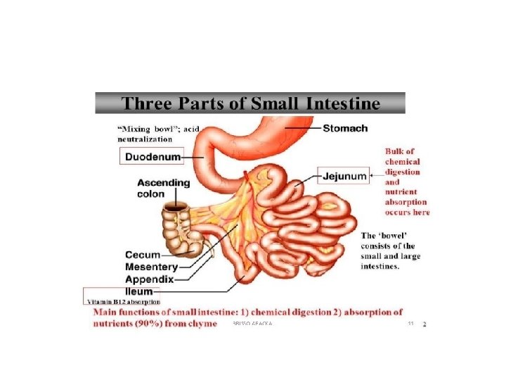
Small intestine Extends from the pylorus to the ileocaecal valve. It is about 6 meter long. It is approximately 2. 5 -3 cm in diameter. It receives bile juice and pancreatic juice through the pancreatic duct, controlled by the sphincter of oddi. Ileocecal sphincter: transition between small and large intestine.
is the site where most of the chemical and mechanical digestion is carried out, and where virtually all of the absorption of useful materials is carried out. three main sections to the small intestine: - The duodenum The jejunum The ileum -
duodenum: -C shaped organ, continous with stomach. -Surrounds the head of pancrease. -Receives gastric chyme , pancreatic juices and gall bladder (bile). -These digestive enzymes break down proteins and bile and emulsify fats into micelles. -It contains brunners gland ----- mucin rich alkaline---- neutralizees the stomach contents.
Jejunum: -Midsection of small intestine -Contains: plicae circulares, and villi that increases the surface area. -Products of digestion( sugar , aminoacids, and fatty acids) are absorbed into the blood stream here.
The ileum: -The final section of samll intestine. 3 meters long. -Contains villi similar to jejunum. -Absorbs -vit B 12. -bile acids -remaining nutrients Ends by ileocecal valve, ---- large intestine
Large intestine -Comes after the small intestine -Measures app. 1. 5 meters length. *Colon. *Rectum. *Anal canal.
The colon Is the last part of digestive system. Extracts water and salts forming solid wastes. It is the site in which flora aided fermintation of unabsorbed material occurs. Absorption is less (Na , fat soluble vitamines). Consists of the following sections : *Cecum *Ascending colon *Transverse colon *Descending colon *Sigmoid colon *Rectum
colon The Crypts- mucosal linning of the colon contains numerous straight tubular glands, The mucosa (M) is arranged into tightly-packed straight tubular glands (G)which consist of cells specialised for water absorption and mucus-secreting goblet cells to aid the passage of faeces. The large intestine also contains areas of called Peyer's patches), and they provide local immunological protection lymphoid tissue Tenia coli- longitudinal smooth muscle layer of the colon doesnot completely envelope the intestinal wall but forms three bands.
Rectum tube is a straight , muscular between: Sigmoid coln----- anal canal. Thick muscular tunic compared with the rest of gut. When fecal mass or flatus moves into rectum, it distends and defecation begins.
Anal canal -The last 2 -3 cm of the digestive tract. It can begins at the inferior end of the rectum and ends at the anus. - the smooth muscle is thicker than of the rectum. -forms the internal anal sphincter (superior end of the anal canal ). The external anal sphincter at the inferior end.
Defecation It is the final act of the digestion by which organisms eliminate solid, semisolid or liquid waste materials (feces) from the digestive tract via the anus. Waves of muscular contraction known as peristalsis in the rectum.
Normal functioning defecation reflex The act of defecation is preceded by a voluntary effort, which, in turn, probably gives rise to stimuli that magnify the visceral reflexes, although these originate primarily in the distension of the rectum. Centres that control defecation reflexes are found in the hypothalamus of the brain, in two regions of the spinal cord, and in the ganglionic plexus of the intestine. As the result of these reflexes, the internal anal sphincter relaxes. Time to move bowel- triggered by : -eating meal. -Drinking hot beverage. -45 min upon rising up from sleep. -
the defecation reflux will be disengaged after 15 minutes of being ignored. Prompt response to the defecation reflux prevents stool from drying out ---- difficult to pass. Dried stool stretched out the rectal sac -----requred more stool to stimulate thee reflux------- constipation.
pancrease The pancreas (meaning all flesh) lies in the upper abdomen behind the stomach. structurally, the pancreas has four sections; head, neck, body and tail; the tail stretches back to just in front of the spleen.
pancrease The pancreas consists mainly of exocrine glands that secrete digestive enzymes in the small intestine. the main enzymes produced are - lipases for fat - peptidases for proteins -amylases for carbohydrates. These are released into the duodenum via the duodenal ampulla, the same place that bile from the liver drains into. Pancreatic exocrine secretion is hormonally regulated, and the same hormone that encourages secretion (cholesystokinin) also encourages discharge of the gall bladder's store of bile. As bile is essentially an emulsifying agent, it makes fats water soluble and gives the pancreatic enzymes lots of surface area to work on.
Endocrine pancreas, the portions of the pancreas (the islets) that make and secrete insulin, glucagon, somatostatin and pancreatic polypeptide into the blood. Islets comprise 1 -2% of pancreatic mass.
liver The liver is the largest organ in the body, normally weighing about 1. 5 kg. located in the upper righthand portion of the abdominal cavity, beneath the diaphragm, and on top of the stomach, right kidney, and intestines.
4 Lobes • Major: left and right Minor: caudate and quadrate Ducts • Common hepatic Cystic From gallbladder Common bile - Choledochus Joins pancreatic duct at hepatopancreatic ampulla
The liver is the main organ of metabolism and energy production; its other main functions include: Bile production Storage of iron, vitamins and trace elements detoxification conversion of waste products for excretion by the kidneys The liver is functionally divided into two lobes, right and left. The external division is marked on the front of the liver by thefalciform ligament, which joins the coronary ligament at the superior margin of the liver
Thank you