Pancreatitis Dr nadia ezzawi Gastroenterology department BMC objectives

Pancreatitis Dr. nadia ezzawi Gastroenterology department BMC

objectives • Anatomical and physiological back ground. • Difinition and types of pancreatitis. • Clinical features and complications of disease and their management.

Pancreas: Anatomy and Physiology • Retroperitoneal organ. It is almost completely covered by the stomach and duodenum. • 15 -20 cm in length • Head, neck, body and tail • curves behind the superior mesenteric vessels
 Inferior pancreatoduodenal")
Pancreas: blood supply • HEAD: Superior pancreatoduodenal A. (from gastroduodenal A. ) Inferior pancreatoduodenal A. (from SMA) • BODY AND TAIL: superior pancreatic A. pancreatic magna A. transverse pancreatic A. • VEIN: to splenic vein , SMV and portal vein

Physiology • Exocrine: • the acinar cells produce digestive juices, which are secreted into the intestine and are essential in the breakdown and metabolism of proteins, fats, and carbohydrates. • Endocrine: – A cell:glucagon – B cell:insulin – D cell :somatostatin – G cell :gastrin

Pancreatitis • Difinition : • It is an inflammatory process of the pancrease with associated escape of pancreatic enzymes into surrounding tissues. • Classified into: • Acute Pancreatitis. • Chronic Pancreatitis

Acute pancreatitis • It is an acute inflammatory process of the pancreas, accompanied by abdominal pain and elevations of serum pancreatic enzymes. • It has Variable severity and duration. • Abrupt onset and unpredictable course.

Classification based on physiological findings, laboratory values, and radiological imaging – Mild Acute pancreatitis (MAP). – Mild disease is not associated with complications or organ dysfunction and recovery is uneventful. – Severe Acute pancreatitis (SAP). – severe pancreatitis is characterized by pancreatic dysfunction, local and systemic complications, and a complicated recovery.

Other classification: -- acute interstitial pancreatitis. the gland architecture is preserved but is edematous. Inflammatory cells and interstitial edema are prominent within the parenchyma -- acute hemorrhagic pancreatitis. characterized by marked necrosis, hemorrhage of the tissue, and fat necrosis. There is marked pancreatic necrosis along with vascular inflammation and thrombosis.

Etiology of acute pancreatitis. • • • Biliary tract disease. Abuse of ethanol. Trauma and operation. Ischemia of pancreas. Drugs … (azathioprine). Idiopathic pancreatitis. Hypercalcemia. Hyperlipidemia. Infections and Parasites. Procedure…. (Endoscopic retrograde cholangiopancreatography )

Pathogenesis • “Self digestion”, oedema and necrosis. • Reflux of bile or duodenal juice • Trypsinogen was activated • Release of the enzymes------ fat necrosis in both pancreatic and peritoneal cavity. • Collection of pancreatic enzymes form pancreatic pseudocyst.

Clinical manifestations • • Abdominal pain Nausea, vomiting abdominal Distension abdominal Tenderness, rebound tenderness, muscular regard • Fever,jaundice, • Gray-Turner sign: flank ecchymoses Cullen sign: periumbilical ecchymoses • MODS(multiple organ dysfunction syndrome )

laboratory test • • • High Amylase level in serum and in urine( increased renal clearance). High Lipase level ( more specific). Blood : impaired RFT, liver function, high BS, low Pa. CO 2 , Raised serum calcium, DIC manifestations. • Imaging studies • USS: especially if the cause is stones, abcess, hge. • CT : Important finding include pancreatic swelling, peripancreatic fluid collection, an area of non enhancement because of necrosis. • ERCP. • MRCP. • Abdomen plain film.

X ray

CT scan abdomin

Diagnosis Ransons criteria On admission Within 48 hrs Age > 55 years Hematocrit decrease by > 10% WBC >16, 000 mm Urea nitrogen increase > 5 mg/ dl LDH > 350 IU/l Serum calcium < 8 mg/dl Glucose > 200 mg/dl Arterial Po 2 <60 mmhg AST > 250 IU /L Base deficit > 4 m. Eq/L Estimated fluid sequestration > 6 L

Glasgow criteria Age >55 Glucose > 180 mg/dl Urea > 45 mg/dl WBC > 1500 mm Albumin < 3. 2 g/dl Arterial Po 2 < 60 mmhg LDH > 600 Calcium < 8 mg/dl
 • Pancreatic abscess")
Complications of acute pancreatitis • Pancreatic necrosis( relaese of pancrearic lipase) • Pancreatic abscess • Pancreatic pseudocyst • Acute pancreatic pseudocyst: Collection surrounded by fibrous tissue or granular tissue. Peripancreatic fluid Those persisting beyond the phase of acute inflammation become pancreatic pseudocysts.

Treatment of acute pancreatitis • Non Operative trearment: • ICU :to prevent MODS • fasting the patient, nasogastric suction • Minimizing pancreatic secretion – antacids • Fluid replacement and Nutritional support – maintenance of adequate hydration – TPN,glucose , lipid, amino acid, protein • Analgesia • Antibiotics • Abdominal lavage

operative • Indication of Operation: • Biliary obstruction • Secondary pancreatic infection • Undetermined diagnosis, needs laparotomy. • Surgery usually is drainage of fluids, abscess. • Methods – – Percutaneous drainage Operative drainage Cystgastrostomy, cystjejunostomy Resection of pancreatic body and tail

Chronic pancreatitis • It is benign inflammatory process and fibrosing disorder characterized by • - irreversible morphologic changes, • - progressive and • - permanent loss of exocrine and endocrine function.

• Clinically the disease is characterized by recurrent episodes of sever and uncontrollable upper abdominal pain and by a loss of exocrine function (diarrhea , steatorrhea) and endocrine function ( DM).

Clinical manifestations • abdominal pain: – may be continuous, intermittent or absent – Pattern is often atypical • RUQ or LUQ of the back • Diffuse throughout upper abdomen • May be referred to the anterior chest or flank – Typical form: • Persistent , deep-seated, • Unresponsive to antacids • Worsened by alcohol intake or a heavy meal (especially fatty foods) • Often need narcotics

• Pancreatic insufficiency – Weight loss – Fat malabsorption: • Steatorrhea: 15% of patients present with steatorrhea and no pain. – Pancreatic diabetes: • Like DM 1 needs insulin , but risk of hypoglycemia is more than DM (because alfa cells is also affected). – Fat-soluble vitamin deficiency rare

Etiology • • • - toxic metabolic Idiopathic Genetic / hereditory Autoimmune / immunologic Recurrent acute pancreatitis Obstructive /mechanical

Lab data • Amylase and lipase : usually normal • CBC , electrolytes, and liver function tests are typically normal • Bilirubin and ALP may be increased • Impaired glucose intolerance and elevated fasting blood glucose • Sudan staining of feces or quantitative test for steatorrhea • fecal elastase (Among pancreatic function tests, fecal elastase measurement is the most sensitive and specific, especially in the early phases of pancreatic insufficiency)

• Classic triad “ pancreatic calcification , steatorrhea , and diabetes mellitus “usually establishes chronic pancreatitis • Classic triad : found in fewer than one-third • It is often necessary to perform secretin stimulation test (abnormal when 60% or more of pancreatic exocrine function has been lost) • A decreased serum trypsinogen (<20 ng/ml) or a fecal elastase level of <100 ug/mg of stool strongly suggests severe pancreatic insufficiency

Imaging study • • • CT, MRI, US calcifications ductal dilatation enlargement of the pancreas fluid collections (eg, pseudocysts

USS of pancreatic pseudocyst

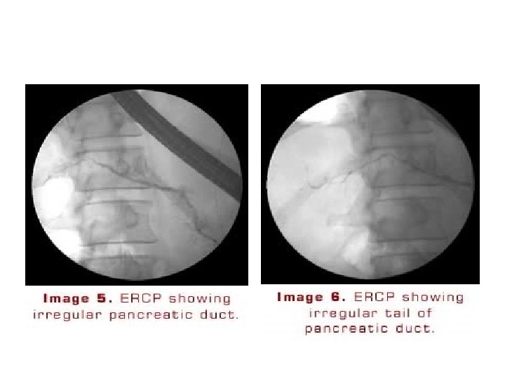
ERCP • May provide useful information on the status of the pancreatic ductal system • Abnormalities include : • 1)luminal narowing • 2)irregularities in the ductal system with stenosis, dilation, saculation, and ectasia • 3)blockage of the duct by calcium deposits Endoscopic ultrasonography • The most predictive endosonographic feature is the presence of stone

Complications of chronic pancreatitis • • pseudocyst formation bile duct or duodenal obstruction pancreatic ascites or pleural effusion splenic vein thrombosis Pseudoaneurysms pancreatic cancer acute attacks of pancreatitis( particularly alcoholics who continue drinking)

Treatment Establish a secure diagnosis. Cessation of alcohol intake. Small meals. Pancreatic enzyme supplements. Patients should also be treated with acid suppression (either with an H 2 receptor blocker or a proton pump inhibitor) to reduce inactivation of the enzymes from gastric acid. • Analgesics • • •

• pancreatitis can be suspected clinically, but requires, biochemical, radiologic, and sometimes histologic evidence to confirm the diagnosis. • measurement of amylase and lipase are useful for diagnosis of pancreatitis. • Imaging studies helps in diagnosis and comlication detection.

Thank you
- Slides: 35