FUNGAL INFECTION OF ORAL MUCOSA CANDIDIASIS Candidiasis is
")


 • • • Clinically seen as a white lesion")
 follow local use of antibiotic especially tetracycline which")
 It is seen in patient wearing ill-fitted upper")

 characterized by appearance of white plaque or patches that")
 stain hyphae are difficult to be seen but by")
 It occurs at the angles of the mouth which appears as")


 RLP is the most common & typical sharply defined lesion producing")


 is a chronic disseminated disease involving almost every organ of")


 shows the epithelium to be orthokeratinized or parakeratinized,")
 and complement (C 3) at")
- Slides: 68
FUNGAL INFECTION OF ORAL MUCOSA (CANDIDIASIS)
Candidiasis is a disease caused by infection with a yeast–like fungi. In about 50% of the population, Candida albicans can be shown in the normal oral flora without causing any evidence of infection. Predisposing factors of oral candidal infection: • Local Factors: – Mucosal trauma. - Denture (appliance) wearing. – Denture (appliance) hygiene - Tobacco smoking. – Carbohydrate-rich diet.
• Age : – Extremes of age: neonates, Infants, old age. • Drugs: – – – • • Broad-spectrum antibiotics. Steroids, local/systemic. Immunosuppressant / cytotoxic agent. Xerostomia: Drugs, Radiotherapy, Sjőgren syndrome. Systemic Disease: Decrease in Iron deficiency anemia Megaloblastic anemia Acute leukemia • Diabetes Mellitus HIV Infection and AIDS • Other Immunodeficiency states.
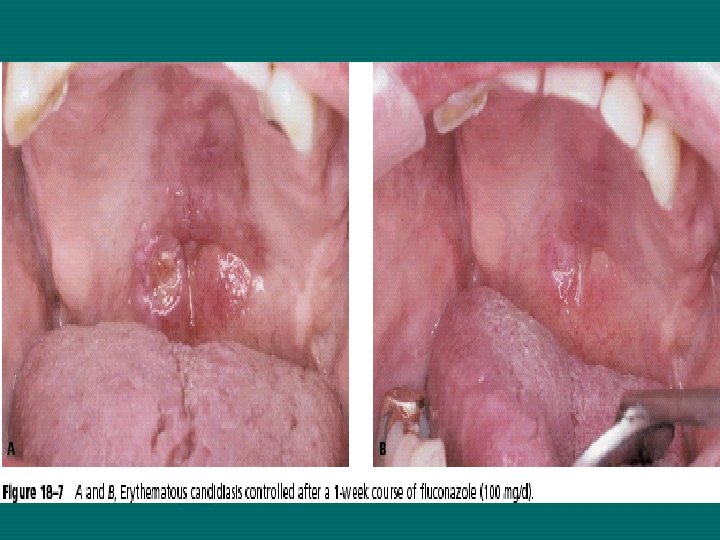
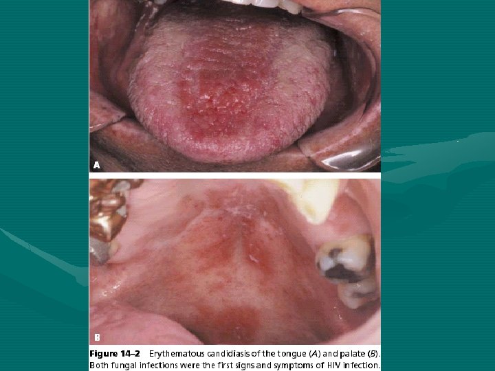
Types of candidal lesions • Acute candidiasis which include thrush and acute antibiotic stomatitis. • Chronic candidiasis: – Denture induced candidiasis. – Chronic hyperplastic candidiasis. – Chronic mucocutaneous candidiasis. – Erythematous candidiasis. • Angular stomatitis
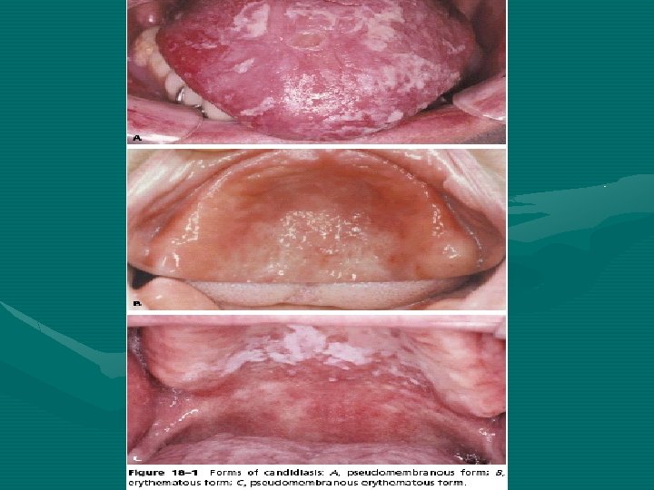
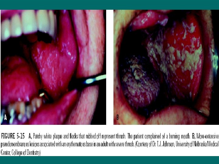
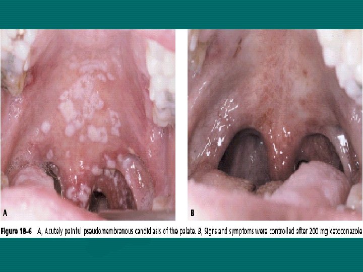
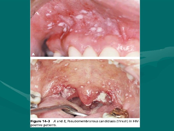
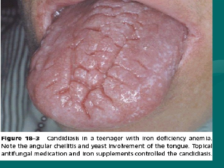
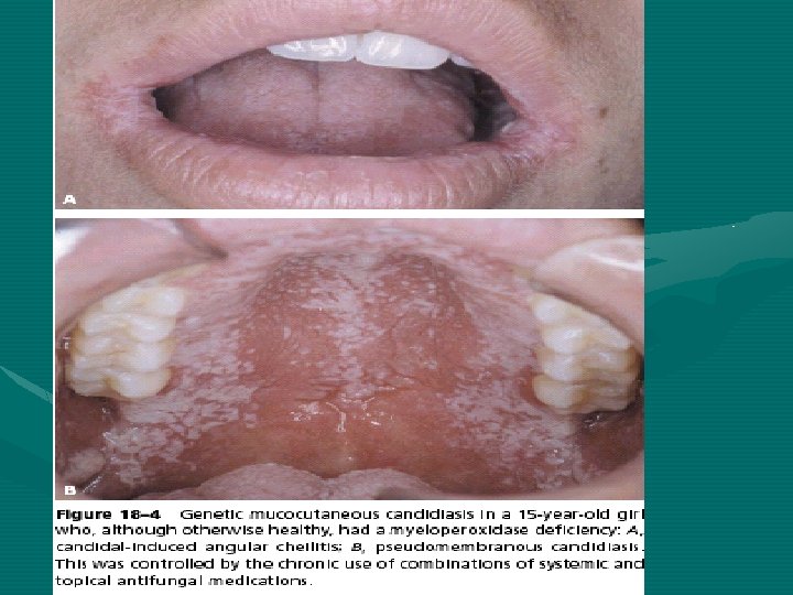
Acute candidiasis Thrush (pseudomembranous candidiasis) • • • Clinically seen as a white lesion or patch on the tongue, palate and cheek. resemble milk curds which can be removed by scraping them with wooden spatula or any gauze leaving the underlying mucosa appears normal or erythmatous. The white lesion contains Candida hyphae, with desquamated epithelial cells and food debris. Thrush may be initiated by exposure of the patient to broad spectrum antibiotics or impaired immune system.
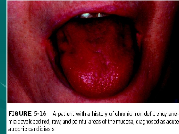
Acute antibiotic stomatitis (acute atrophic Candidiasis) follow local use of antibiotic especially tetracycline which cause suppression of normal oral flora. Clinically The whole oral mucosa is red and sore, spots of thrush may be present. The recovery will occur following removal of antibiotic.
Denture induced stomatitis (chronic atrophic candidiasis) It is seen in patient wearing ill-fitted upper denture which cut off the underlying mucosa from the protective action of saliva. This condition is characterized by varying degree of erythema, sometimes accompanied by peticheal hemorrhage localized to the denture bearing area of the upper dental prosthesis.
Whether this represent acute infection of Candida albicans or tissue response of the host to various micro-organism lying beneath the denture or this due to improper denture design or due to allergy to the denture base or inadequate curing of the denture acrylic.
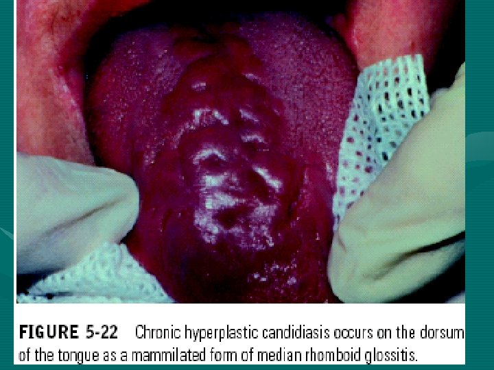
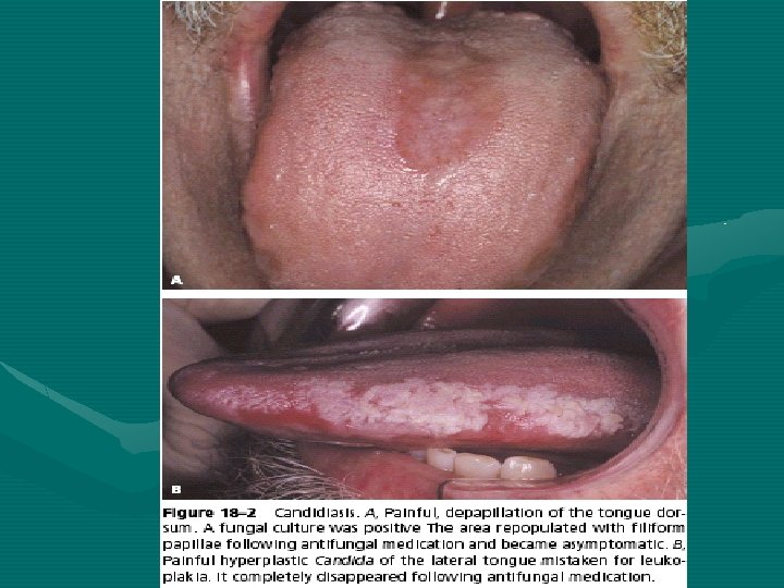
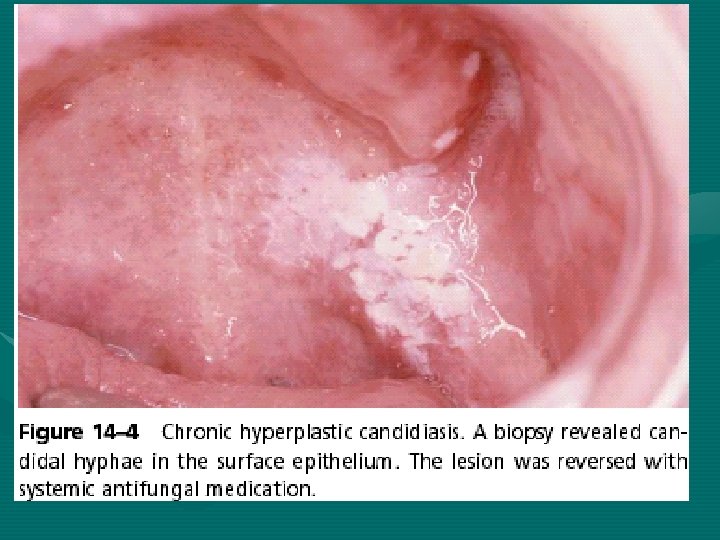
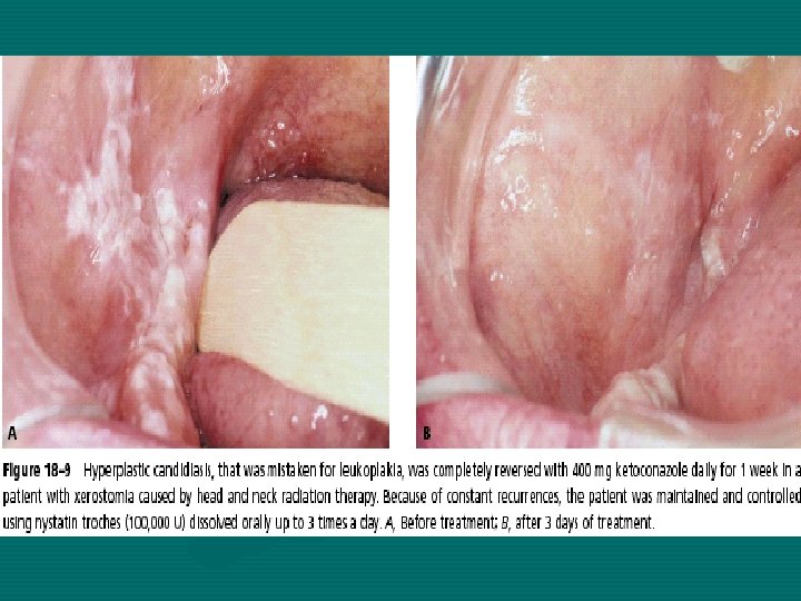
Chronic hyperplastic candidiasis (Candidal leukoplakia) characterized by appearance of white plaque or patches that can’t be removed by scarping. Such lesions are usually located on the anterior buccal mucosa and can’t clinically be distinguished from leukoplakia only by taking biopsy; it’s not clear whether this lesion caused by Candida or Candida is superimposed by the existed lesion.
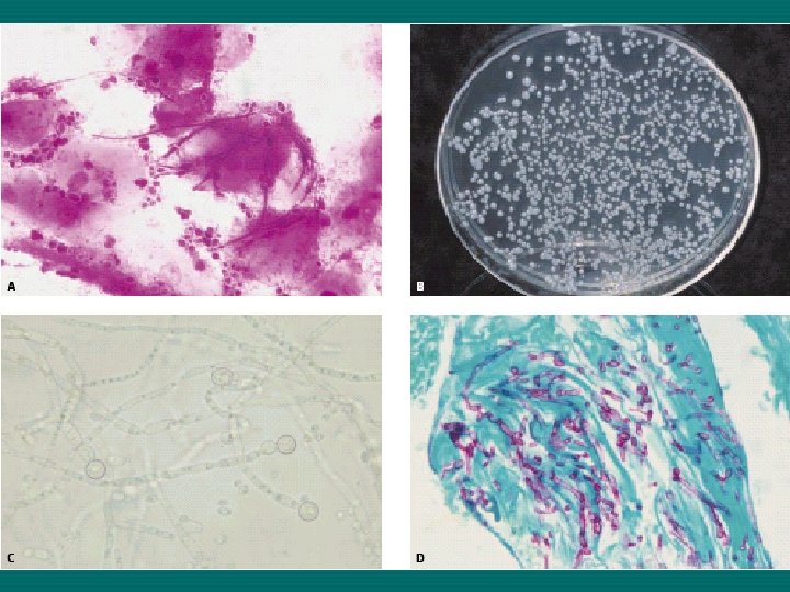
In hematoxylin and eosin (H&E) stain hyphae are difficult to be seen but by using PAS (periodic acid Schiff stain) clearly shows the hyphae growing through the full thickness of keratin to prickle cell layer.
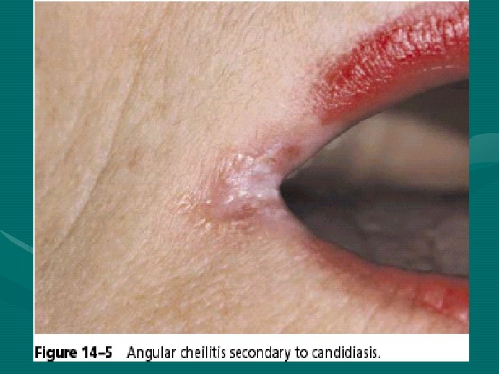
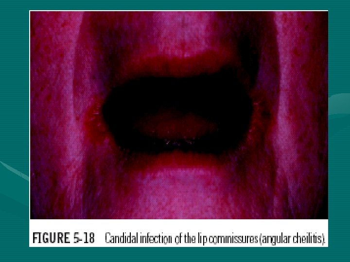
Angular stomatitis (perleche) It occurs at the angles of the mouth which appears as red-fissured scales. This is seen typically in older persons with reduced vertical dimensions and many folds at the corners of the mouth, so saliva tend to flow in these folds, keeping them moist and thus inducing a yeast infection, this may be caused by (C. ablicaus) or combined with other micro-organisms such as Staph. aureaus.
DERMATOLOGIC LESIONS OF THE ORAL MUCOSA
Lichen Planus is a common chronic inflammatory disease of skin and mucous membrane so called dermatological disease. The oral lesion has a characteristic feature in appearance and distribution.
Etiology of lichen planus It is unknown, but it appears to be Tlymphocytic mediated disorder, when cytotoxic T-Lymphocyte stimulated by antigen presenting cells (intraepithelial langerhans cells) emitting tumor necrosis factor (TNF) that contribute to degeneration of epithelium.
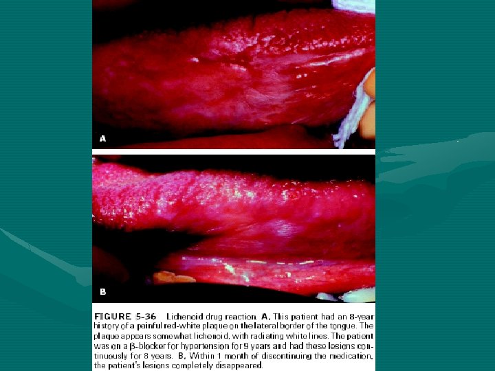
Predisposing factors of Lichen planus: • Genetic factor • Infective agents • Systemic diseases (diabetes mellitus, hypertension) • Graft-versus-host disease • Drug reaction (lichenoid reaction) e. g. : antimalarials, methyldopa. • Hypersensitivity to dental materials (mercury) • Tobacco smoking • Tobacco and/or betel chewing • Vitamin deficiency • Psychiatric disorders
Lichen Planus – Prevalence: 0. 5 -1. 9% of population. – Patients affected are 30 -50 years of age. – Oral lesions occur in about 50% of patients skin lesions, while skin lesion is about 1045% of Pts. (with oral lesion). Oral lichen planus may occur before, at the same time, or after skin lesion. – In oral lichen planus, skin lesion may or may not be present
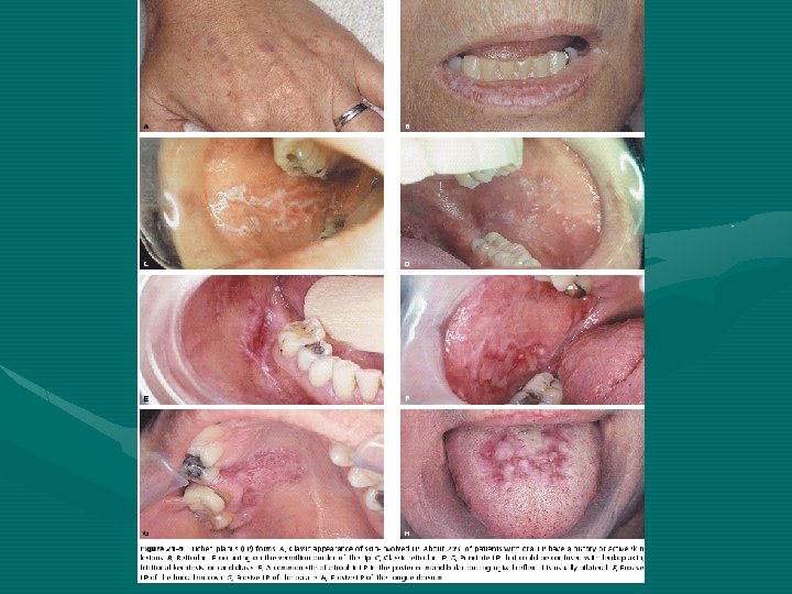
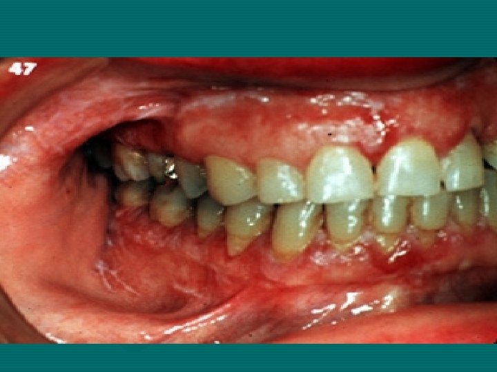
Clinically • Wide rang of clinical appearances that correlated with severity of disease process. Clinical presentations include: a-Reticular b-Erosive c-Plaque d. Atrophic e-Bullous f-Papular. • Lichen planus is common for patient to have a combination between reticular and erosive types in oral mucosa; plaque occurs a lone and resembles homogenous leukoplakia.
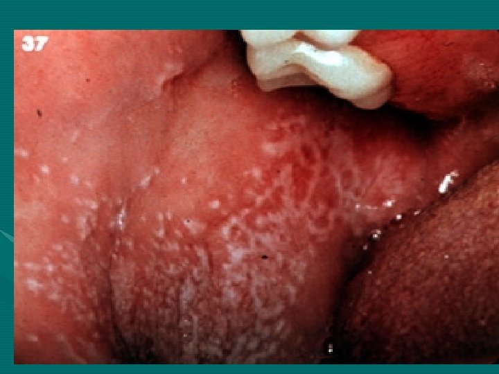
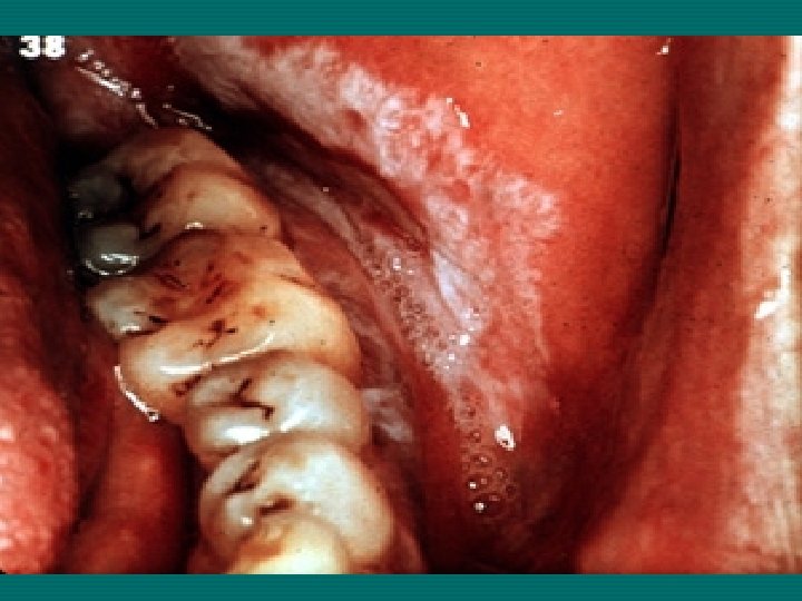
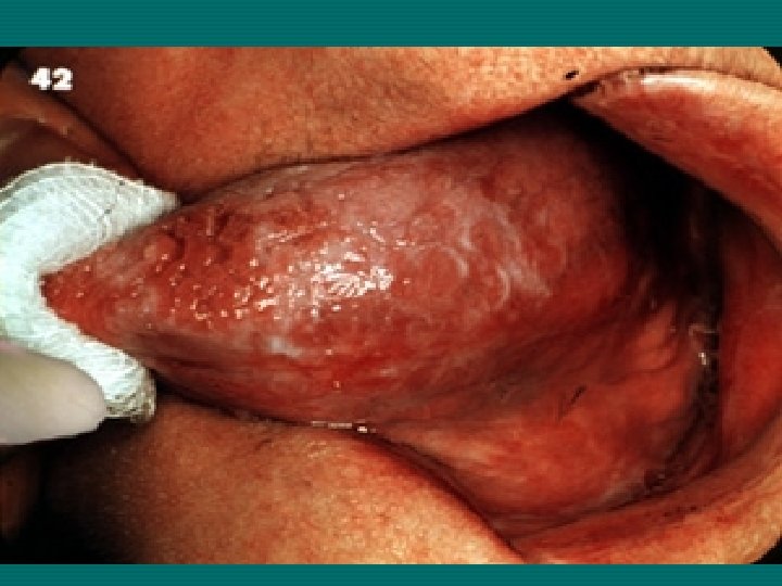
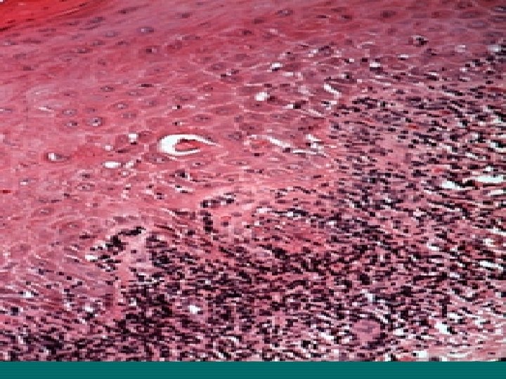
Reticular Lichen Planus(RLP) RLP is the most common & typical sharply defined lesion producing network of reticular appearance against erythematous background. Wickham striae referred to white lines. RLP occurs on the buccal mucosa and buccal vestibule usually bilateral and asymptomatic.
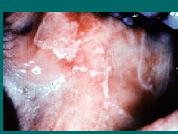
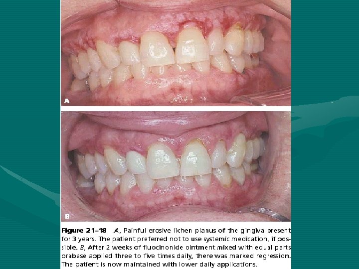
Erosive LP Appears as mixture of erythematous white pseudomembranous area, striae may radiate form the margin of these erosions, and Pts complain of sore mouth and mouth becomes sensitive to cold and hot and spicy food. It is difficult to be differentiated from candidiasis, pemphigus vulgaris, pemphigoid and discoid lupus erythematosus.
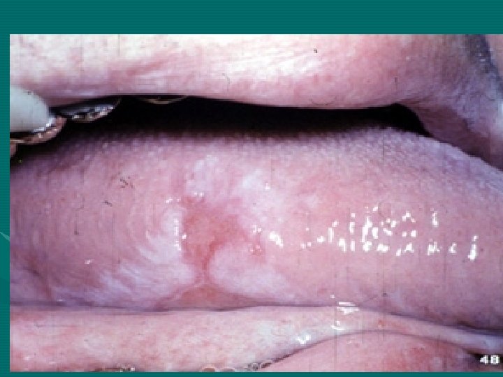
Plaque LP Appears as white raised or flatted area on the dorsum of the tongue where producing irregular white smooth area and raised plaque.
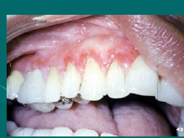
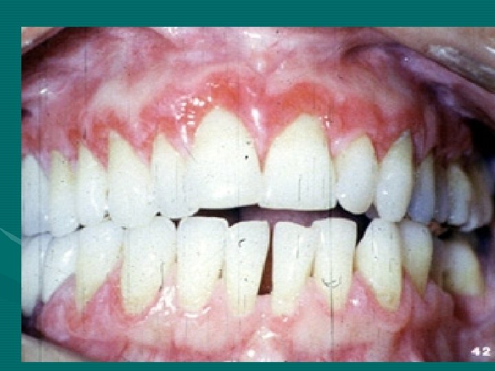
Atrophic LP It is identical to erythematous background of reticular form and may be a part of transitional stage from reticular to erosive most common on the gingival producing desquamated gingivitis.
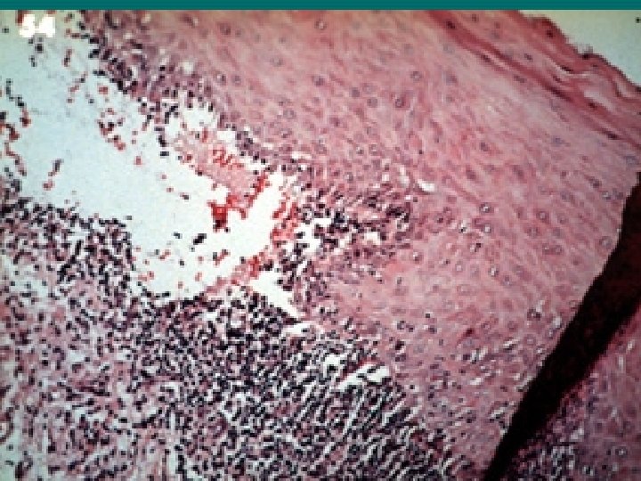
Bullous It is a form consisting of large bulla, when bulla is ruptured, erosion or ulcer will form, mostly occurs in the posterior buccal mucosa.
Papular Small white papules that may coalesce.
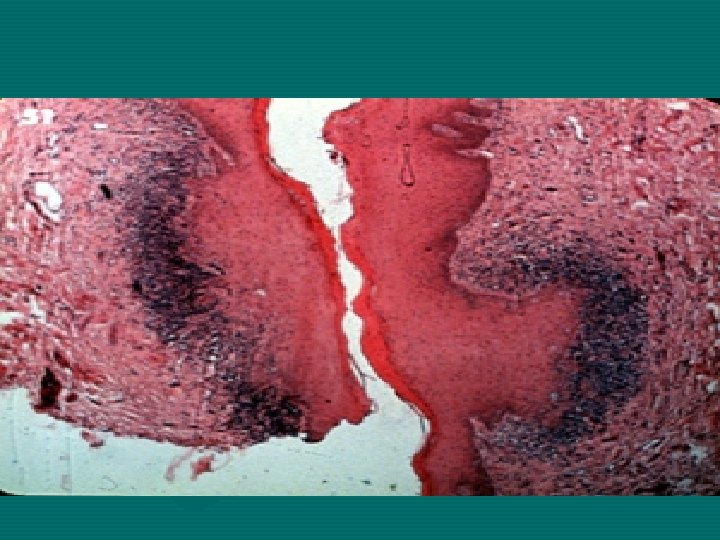
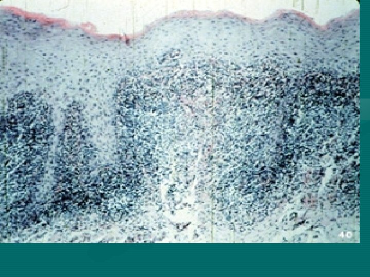
Histopathology LP is hyperorthokeratosis or hyperparakeratosis and acanthosis. The epithelium shows saw tooth profile of rete pegs, liquefactive degeneration of basal cell layer. The lymphoplasmocytic infiltrate mainly (T-lymphocyte just beneath epithelium (subepithelial inflammatory infiltrate).
Sometimes within the epithelium rounded or ovoid amorphous eosinophilic bodies, referred to as civatte bodies which represents dead keratinocyte or other necrotic epithelial component that are transported to connective tissue for phagocytosis. Immunofluorescent examination demonstrates deposit of fibrinogen along basement membrane (+ve PAS). .
Malignant changes estimated to occur 0. 4 -2% of patients when lesion persists for 5 or more years. Some cases thought to be LP proceeding to malignancy on histological bases, where real cases of epithelial dysplasia (lichenoid dysplasia) considered as premalignant condition
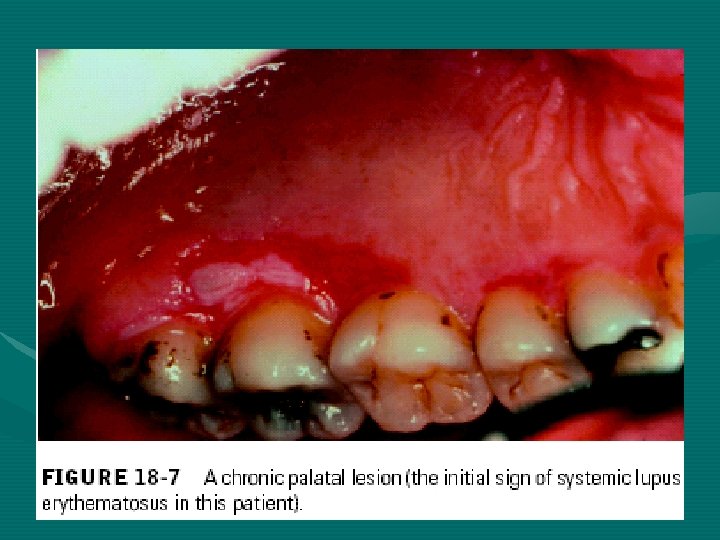
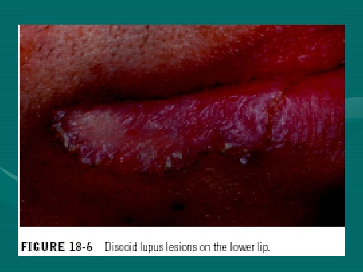
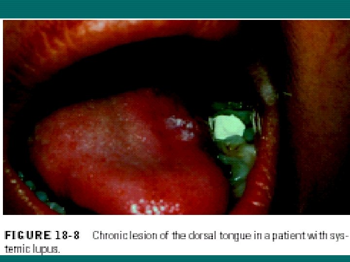
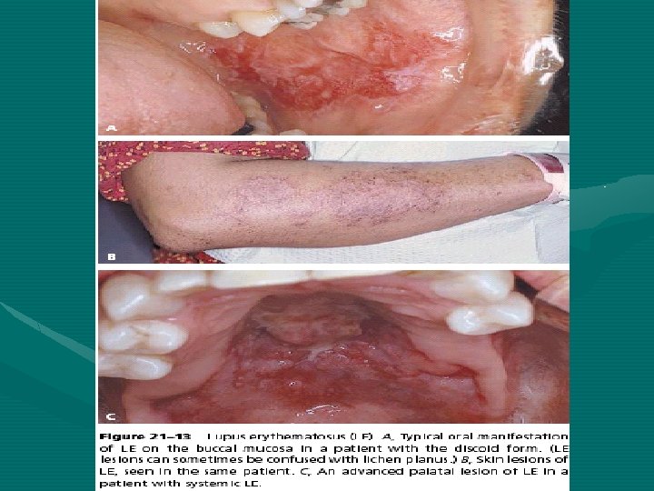
Lupus Erythematous Lupus erythematosus(LE) is a chronic disseminated disease involving almost every organ of the body. Females affected > males. Two main forms of the LE are recognized: • Chronic discoid LE • Systemic Lupus Erythematosus SLE
Etiology Is unknown, but genetic factor are important, and autoantibodies are present in all cases of the SLE. The most specific diagnostic test for SLE is the antinuclear antigen (ANA) and the LE cell tests. Theses tests are (-ve) for DLE.
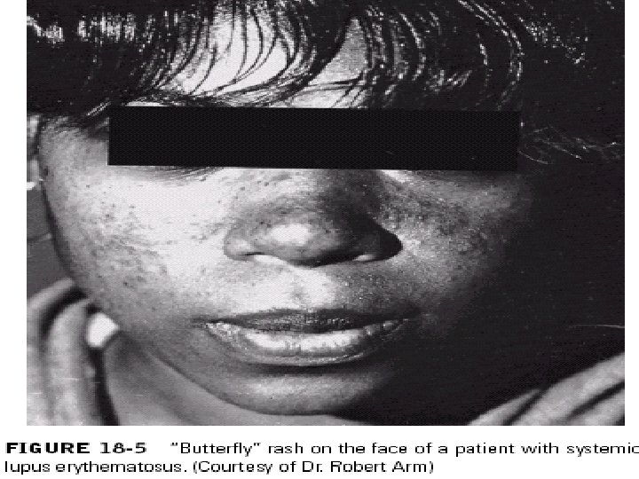
Chronic discoid LE Occurs in skin and usually in face, present as scaly red patches which later heal with scar formation. Symmetrical distribution of skin lesion over the nose and cheek is called butterfly pattern. Oral lesion has been represented in up to 50% of cases of discoid LE. The cheeks are most frequently affected. Oral lesion is composed from area of erythema or ulceration surrounded by a white keratotic
Systemic Lupus Erythematosus Skin rashes and lymphadenopathy. Skin rashes are maculopapular, photosensitive and typically occur on the cheeks. Kidney, liver and nervous system are also frequently involved in SLE. Oral lesion is composed of erythematous petechiae with superficial erosions on the buccal mucosa. Death may occur from the disease, but nowadays effective treatment give good prognosis.
Histopathology Oral lesion (epically of DLE) shows the epithelium to be orthokeratinized or parakeratinized, with area of hyperplasia and atrophy. Dome-shaped extensions of keratinized cells may extend from superficial layer down into the hyperplastic rete processes (keratin plugging). Subepithelial and deeply situated perivascular foci of lymphocytes are present in connective tissue and there may be liquefactive degeneration of basal cells.
-Immunofluorescent studies show abundant deposits of immunoglobulin (Ig. G) and complement (C 3) at the basement membrane zone (lupus band).