Candidiasis Candidiasis is infectious in Candida species generally


 Niger or bird seed agar melanin production : C. neoformans")
- Slides: 15
Candidiasis
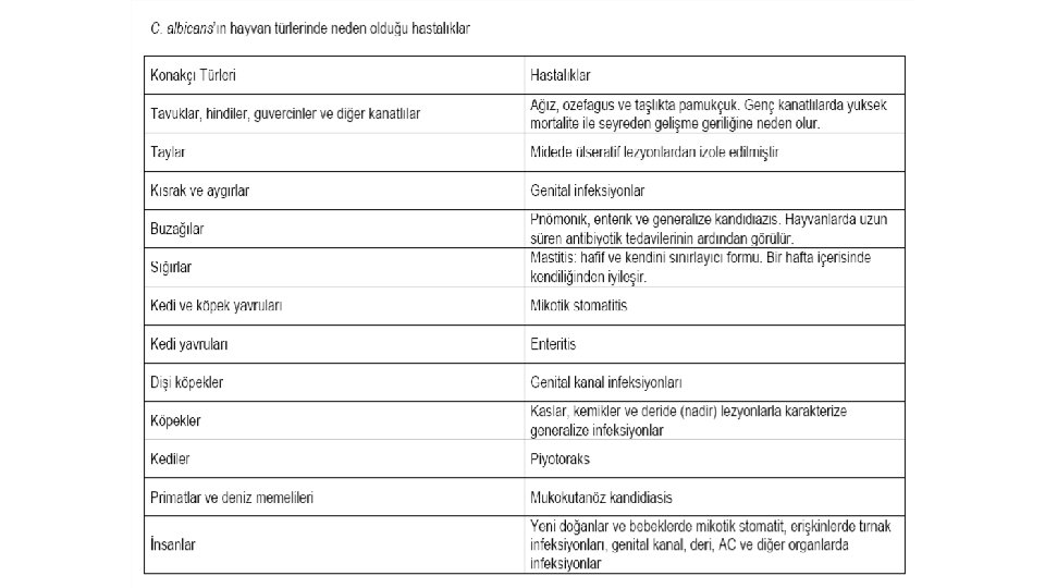
Candidiasis, is infectious in Candida species generally in the digestive tract and mucous membranes, predominantly in C. albicans in the vast majority of humans and animals (90%). • C. albicans, is located commensally in the mouth, esophagus, stomach, intestinal tract, subcutaneous tissues, lung, breast tissue and genital tract. affective lesions are encountered. • Most of the leading infections are of endogenous origin and predisposing causes such as immunosuppression, long-term antibiotic therapy and inadequate care-feeding are the reasons for the infection. • Infection, acute in young animals; is a chronic course in adult animals.
Clinical Symptoms • In poultry, clinical signs are hardly observable. • The cases are usually sporadic and rarely endemic. Upper respiratory tract and digestive system are affected the most. • In acute cases, yellow-white lesions are loose in the mucosa of the necropsy coats; In chronic cases, it is observed on the surface of the mucosa and a structure covered with necrotic membrane like towel. • Mycoses from C. albicans are rarely encountered in ruminants and pigs In cases, abortus, mastitis, mycotic stomatitis, pneumonia and rumenitis are usually observed. • In dogs and cats skin, otitis, intestinal candidiasis, genital candidiasis
Laboratory Diagnosis • • For the diagnosis of the disease, depending on the location of localization, mouth, esophagus, stomach, cow, milk, uterine flow, skin scraping etc. materials should be sent to the appropriate laboratory conditions. Direct Microscopy • Skin and mucosa excavations are treated with 10% KOH and examined under a microscope. Oval-budding yeast-like cells and short mycelial structures are observed. • Lactophenol is examined by cotton bluish or Gram stain on the frother where it is prepared from uterine fluids and milk. • Culture • Sabouraud Dextrose Agar is planted with both antibiotics and antibiotics from the laboratory materials. The petri dishes are allowed to incubate at 25 ° C and 37 ° C for 3 -5 days. • Some Candida species are inhibited by cycloheximide. • Plates are cultivated with a small inoculum volume as in the same bacteria.
Identification Colony morphology • • C. albicans colonies usually form within 3 -5 days. They form colonies of 4 -5 mm in diameter, with a sweet, fruity smell that is bright, high-convex in white or creamy color. Microscopic morphology • A small piece of Lactophenol Cotton Blue can be prepared from a single colon, or pre -fixed colonies can be stained with Gram Stain or Methylene Blue. • C. albicans bloody agar and Sabouraud Dextrose Agar to form thin-walled, budding cells. Demonstration of Germ Tubes • A small inoculum prepared from breeding colonies is inoculated into 0. 5 ml of sheep, cattle, rabbit or human serum and incubated for 2 -3 hours at 37 °C. • The prepared mixture is examined in a drop phase contrast microscope or light microscope. The appearance of small tubers that protrude out of some yeast cells is characteristic of C. albicans.
Treatment and Protection • Candidiasis is largely due to predisposing factors. For this reason, predisposing factors must be removed first. • Nystatin can be added to copper sulphate and baits for preservative drinking water. • Some studies have shown that formic acid applications may be beneficial to feeds. • Amphotericin is primarily used in animals to be treated. In addition, topical applications can be performed on lesioned areas.
Cryptococcosis
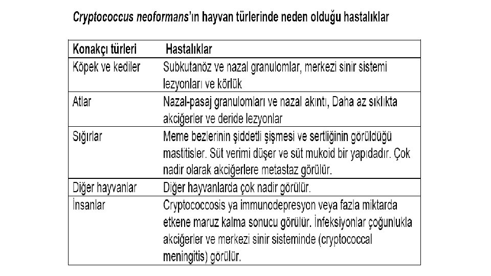
• Cryptococcosis, is a subacute and chronic disease characterized by Cryptococcus neoformans in humans and animals. Within 19 Cryptococcus species only Cryptococcus neoformans are pathogenic for humans and animals. • Thin-walled budding yeast that varies from spherical to oval and varies in diameter from 2. 5 to 20 μm. • • Cells are surrounded by a polysaccharide capsule in a mucoid structure with varying thickness, and this capsule is larger in animal tissues. The young cells are single and bud with a thin neck from the main cell. • Cryptococcus neoformans is a member of the Fungi Imperfecti class. • Cryptococcosis (European blastomycosis, torulosis) is a subacute or chronic infection involving the central nervous system, respiratory system and eye.
Epizotology • C. neoformans are very common on earth. Fruit juices, milk, soil, healthy animals were found in the skin, mucous membranes and intestinal tract. . • Due to the high content of creatine in the pigeon's ovary, it is present in excess in the stool and can survive for longer than 1 year in pigeon feces. • Creatinine can be used by C. neoformans, while inhibiting many other microorganisms.
Pathogenesis • The infectious infection is usually through respiration, firstly localization in the nasal cavity or paranasal sinuses, and then transmission to the brain and brain membranes. • The infection of the brain membranes is known as tuberculous meningitis. • Occasionally, subcutaneous granulomas occur in the disease, mostly in the cervical or pedal regions. • C. neoformans can affect any mammal, but cryptococcosis is more common in cats, dogs, cattle, horses, and humans. . • Antifagocytic and immunosuppressive capsules play a role in the active virus. • Cryptococcal lesions macroscopically resemble myxomatous neoplasms. These include capsular slime, yeast cells, some inflammatory cells, histiocytes, epitheloids and giant cells.
Laboratory Diagnosis • Cryptococcus should be studied very carefully when working with materials thought to contain C. neoformans (ideally in the biosafety cabinet) because it can cause serious illnesses in the causative organ. • Cerebrospinal fluid, lesions or exudates, milk taken from animal with mastitis, biopsy specimens and tissues Microscopy • The preparation can be prepared from cerebrospinal fluid or clean exudates and examined by India ink or nigrosin staining. With these dyes, the capsule can be shown characteristically. • Histological sections of tissue biopsies taken from the lesions can be stained with PAS-hematoxylin stain. With this staining, yeast cells will be dyed instead of capsules. The capsule will be observed as an empty area around the cell • In Mayer's mucicarmine stain, the yeast wall and capsules are painted red, which is determinant for C. neoformans. • LPCB or nigrosine staining displays spherical, capsule-encapsulated budding cells.
• Culture C. neoformans does not contain bloody agar and cycloheximide, but Sabouraud Dextrose is very good in agar. • Cultures are incubated aerobically for up to 2 weeks at 37 ° C. • Capsule breeding can be increased by incubation at 37 ° C in 5% CO 2 environment in agar. • While saprophytic cryptococcus species can not grow at 37 ° C, C. neoformans grows easily at temperatures up to 40 ° C. • The colony recurrence is not observed until about 2 weeks of incubation. • Colonies tend to mucoid as they become S-type, moist, bright and aging. It is initially white, and the latter forms a yellowish shadow. At 25 ° C and 37 ° C, mucoid yeast colonies are formed and are separated from the dimorphic fungi by this breeding shape.
Biochemical tests • b) Niger or bird seed agar melanin production : C. neoformans is one of the few Cryptococcus species that use creatinine in media containing diphenolic and polyphenolic compounds and that produce melanin pigmented (brown) colonies. The media are intensively cultivated and incubated at 37 ° C aerobically for at least 1 week. The dark brown pigment occurs around the breeding colon first and then on the entire medium. c) Biochemical profile : Biochemical profile of isolate API 20 C and Uni-Yeast-Tek commercial systems is determined for accurate diagnosis. Mouse inoculation Mice are inoculated intraperitoneally. If they do not die by themselves, euthanasia is administered 2 weeks later and there are gelatinous lesions in the abdominal cavity and lungs. C. neoformans is the only Cryptococcus species that is pathogenic to mice. Immunological tests Lam latex agglutination test kits have been developed for the determination of antigen in serum and cerebrospinal fluid. • Indirect FA tests are used to detect antibodies. Antibodies may not always be displayed as they may be combined with circulating antigen.