
Oral mucosa The surface of oral cavity is








- Slides: 31
Oral mucosa The surface of oral cavity is a mucous membrane it structure varies in adaptation to function in different regions of the oral cavity.
• Basic classification of OMM divided into 3 major Types : • 1 - Masticatory mucosa ( gingiva and hard palate ). • 2 - Lining mucosa (lip , cheek , vestibular fornix , alveolar mucosa , floor of the mouth and soft palate ). • 3 - Specialized mucosa ( dorsum of tongue and taste buds ).
• The structure of OMM resemble skin composed of : • 1 - Epithelium • 2 - Connective tissue (lamina properia )
1 Oral epithelium : Is stratified squamous epi. Either keratinized , parakeratinized or non keratinized. Keratinized oral epi. Consist of 4 layers : 1 - Stratum basale 2 - Stratum spinosum 3 - Stratum granulosum 4 - Stratum corneum Take their name from their morphological appearance
1 Basal cell layer : - is made up of cells that synthesize DNA cells and under go mitosis providing new cells. 2 straturm spinosum ( prickle cell layer} Cell are irregular, polyhedral about 20 - 25 layer and larger than b. c. l. cells
Stratum granuiosum: flatter and wider cells , these cells are larger than str. Sp. L. it's named for their basophilic keratohyalin granules Stratum cornium : - is made up of kertinized squamae with larger and flatter cells than str. Granuiosum , nuclei and organelles are disappeared
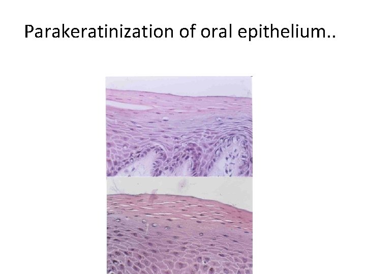
Parakeratinized oral epi: It is similar to the keratinized in it's layers except that the granular layer may be absent or not evident, the other differences in the str. Cornium ( cornified layer), the surface layer retain neucli that are pyknotic and condensed , and other partially lysed cell organelles until they desquamate.
Non keratinized epi. Have no cornified layer, therefore : 1 - Basal cell layer similar to those of keratinized epi 2 - Stratum intermedium 3 - Stratum superficial
The non keratiocyte cells 1. Melanocyte cells : - dendrities cells present in the basal cell layer store melanine in form of melanosomes which elaborate melanine pigment responsible for pigmentation of o. m. embryonic origin from n. c. c. (neural crest cell).
2. Langerhanes cell: - dendritic cell present in upper layer of SI and mucosal epi. In the zone of ortho keratinzation in str. Granulosum derived from haemopoitic tissues (i. e. from bone marrow ) involved in immune response 3. Merkels cell : - non - dendritic cell found in the basal layer, it's origin from n. c. c. have nerve tissue presum to be specialized neural pressure sensetive receptor cell means
4. Lymphocytes and polymorphonuclear leukocyte cells lymphocyte originate from hemopoietic stem cell present in every type of epi. And in any layer of epi. It is associated with inflammatory response
2 Lamina properia l. p. is a c. t. of variable thickness that supports the epi. It divided into 1 - papillary part 2 - reticular part
submucosa consist of c. t. of varying thickness and density it attaches mucosa membrane to the underlying structures weather attachment loose or firm depends on variety of submucosa. Glands, blood vessels , nerves and adipose tissue are present in submucosa
The following zones are seen in hard palate: 1 - gingival region adjacent to the teeth 2 - palatine raphe median area extend from incisive papilla posteriorly 3 - anterolatteral area or fatty zone between raphe and gingiva 4 - posterolatteral area or glandular zone between raphe and gingiva
Gingiva : - extends from dentogingival junction to the alveolar mucosa it subjected to the friction and pressure of mastication epi. Of gingiva is str. Sq. epi. Either orthokeratinzed ( 15%} most often parakeratinzed ( 75% ) and may be non keratinzed (10% ) of the population
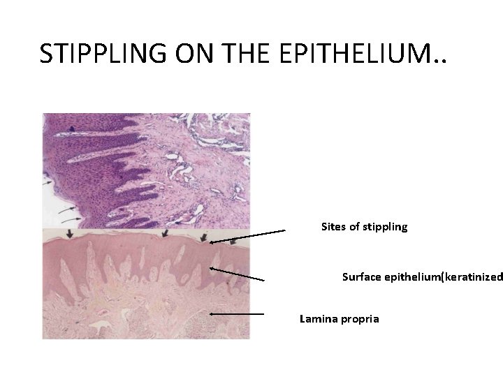
The surface of gingiva characterized by stippled appearance , portion of epi. appear elevated and between elevation there are shallow depressions the net result is stippling . The depression correspond to the center of heavier epi. ridges. These are functional adaptation to mechanical forces. dis appearance of stippling indicated odema and involvement of gingiva in gingivitis
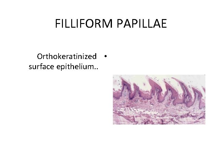
Specialized mucosa Dorsal surface of the tongue is rough and irregular divided into anterior 2/3 and posterior 1/3 by Vshaped lined called terminal sulcus. On anterior part Found numerous papilla Filliform papilla which are fine pointed epithelial structures containing core of c. t, the covering epith, is keratinized, these papilla do not contain taste buds
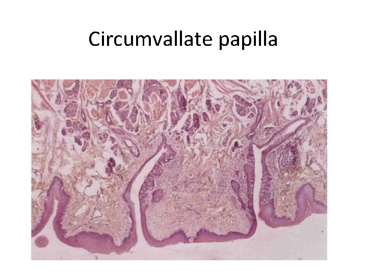
Fungiform papilla Usually found between filiform. p. isolated, round reddish prominences it contain few taste buds 13 found on their dorsal surface. Its red colour due to rich capillary network visible through its their epith. Circumvallate papilla 8 -10 vallate papilla in front of the V-shaped sulcus bet the body and the base of the tongue. The lateral surface of the vallate papillae, the epith contains a numerous taste buds.
Functions of OMM 1 - protection 2 - sensation 3 - secretion 4 - thermal regulation
Gingival sulcus and Dentogingival junction Gingival sulcus or crevice is the name given to the invagination made by the gingival as it joins with the tooth surface. The gingiva does not join the tooth at the gingival margin. It forms a small infolding Known as the sulcus. The sulcus extend from the free gingival to dentogingival junction. The sulcuar (crevicular) ep. is nonkeratinized
Junctional (attachment) epi thelium Two types 1. primary attachment epithelium derived from reduced E. epithelia 2. Secondary attachment epithelium derived from gingival epithelia
Periodontal pocket: -Pathological deepening of gingival sulcus with apical migration of junctional Epithelium along root Surface. -Periodontal pockets are caused by microorganisms (as spirochetes and motile rod). -Their products which produce pathologic tissue changes that lead to deepening of the gingival sulcus the cellular and fluid inflammatory exudates causes degeneration of the surrounding C. T. including gingival fibers.
Non-Keratinization in oral epithelium in human gingiva. . Superficial layer Intermediate layer Basal layer Prickle cell layer
Parakeratinization of oral epithelium. .
STIPPLING ON THE EPITHELIUM. . Sites of stippling Surface epithelium(keratinized Lamina propria
Circumvallate papilla
FILLIFORM PAPILLAE Orthokeratinized • surface epithelium. .
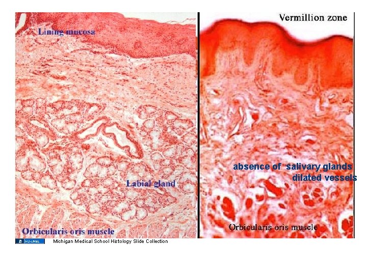
absence of salivary glands dilated vessels Michigan Medical School Histology Slide Collection
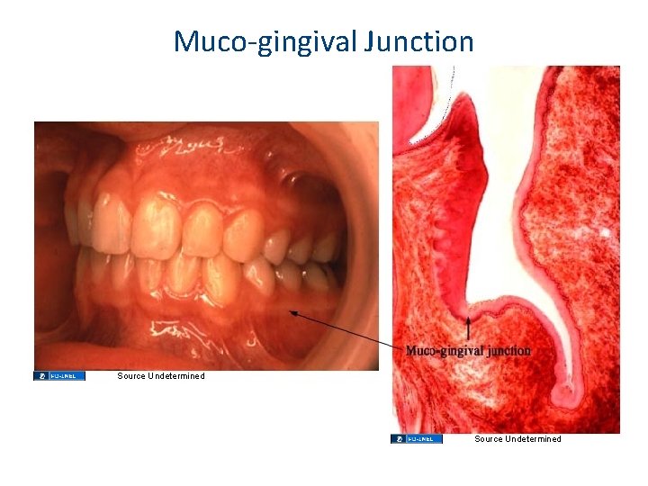
Muco-gingival Junction Source Undetermined
Orofacial Histology and Embryology, Moss-Salentijn, L. , et al. , F. A. Davis Co.
Filiform and Fungiform Papillae Keratinized epithelium, no taste buds Source Undetermined Non-keratinized epithelium with secondary papillae and scattered taste buds. Source Undetermined