INFECTIONS OF THE ORAL MUCOSA n VIRAL INFECTION


 A. Primary herpetic gingivostomatitis (type I H. S. V) In a")


 Occurs mostly on the firmly")
 It’s an acute infectious viral disease the")


 is transmitted by exchange of blood or body fluids")


- Slides: 34
INFECTIONS OF THE ORAL MUCOSA n VIRAL INFECTION n BACTERIAL INFECTION n FUNGAL INFECTION
Viral infectious diseases
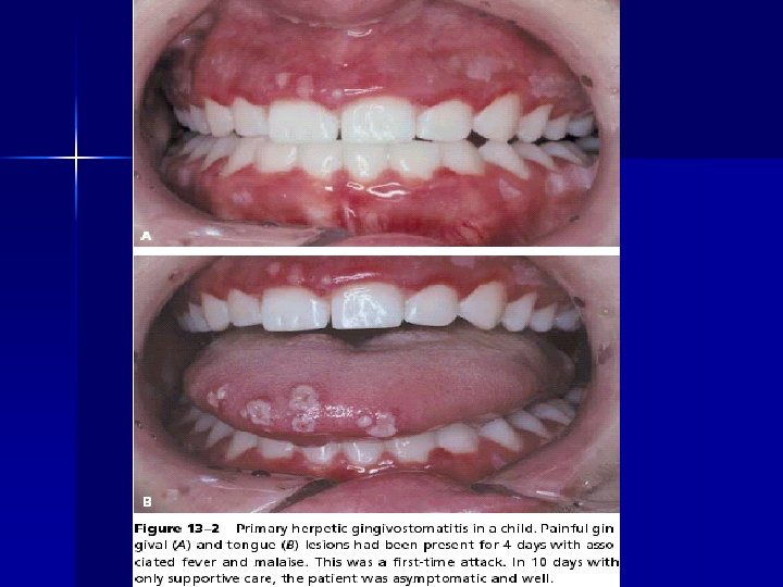
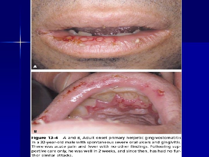
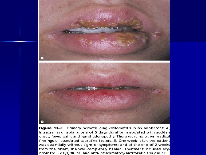
Herpes simplex virus(HSV) A. Primary herpetic gingivostomatitis (type I H. S. V) In a person without circulating antibodies. It’s developed in children and young adult, rarely in children under 6 months and mainly transmitted by saliva, incubation period is 5 days.
In the oral cavity is characterized by multiple vesicular eruptions located in the all attached oral mucosa, gingival and movable mucosa, chiefly lips. Buccal mucosa, pharynx and tonsils may be involved.
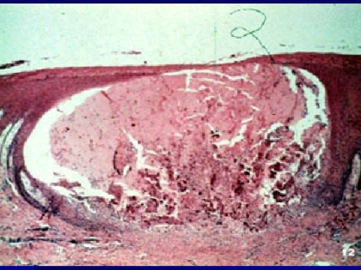
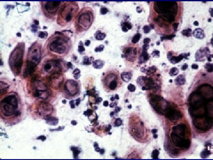
Histologically intraepithelial vesicles formations with necrotic exudates and neutrophil infiltrate accompanied by epithelial cells showing nuclear ballooning degeneration.
Treatment supported care consist of the prescribing a soft diet, analgesic to reduce pain and fever with antibiotic to prevent secondary infection, soothing mouth wash to wet the lesion.
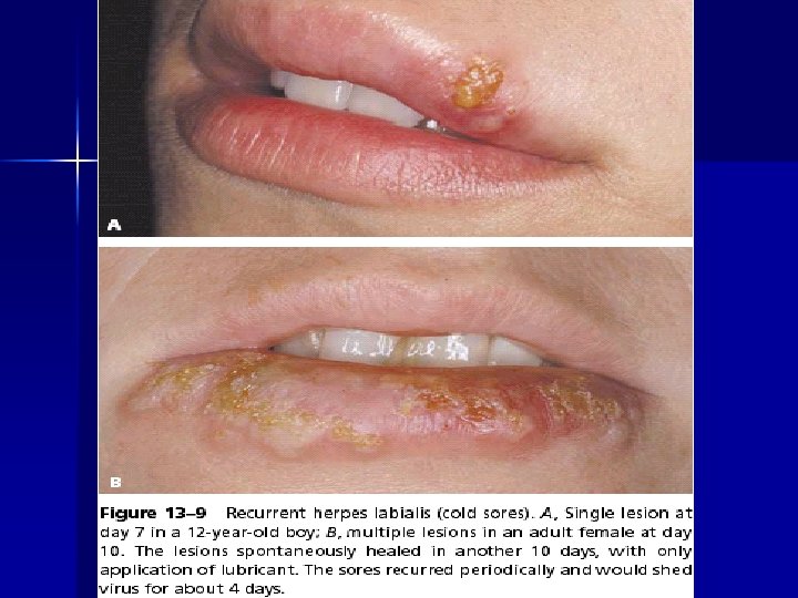
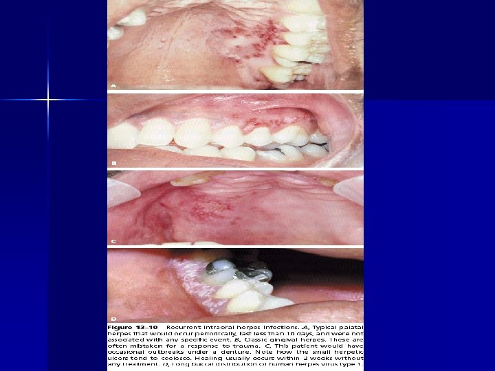
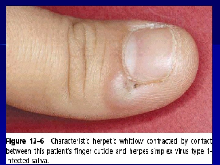
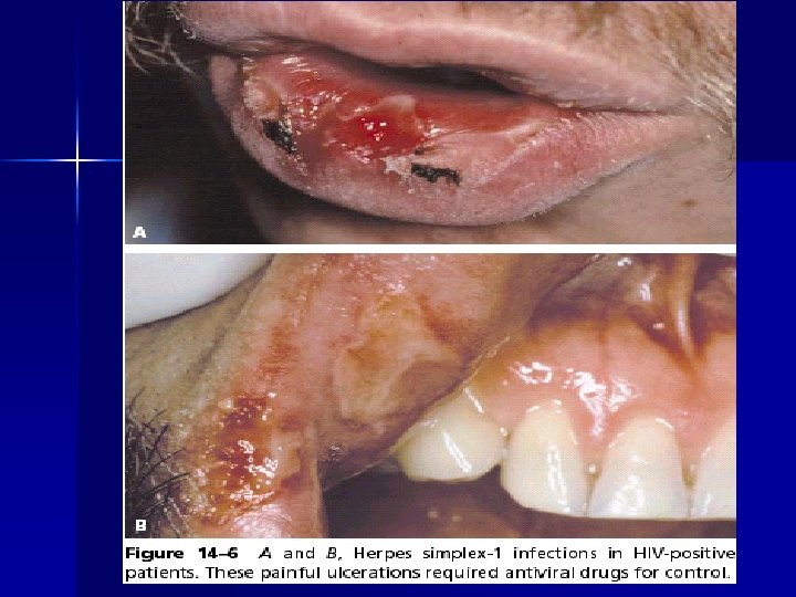
B. Secondary herpes simplex infection: 1. Recurrent (Herpes Labialis) Occurs mostly on the firmly attached mucosa such as hard palate and attached gingiva and not movable mucosa. Heals in 10 days and no treatment is applied, with reassurance of patient. 2. Herpetic whitlow infection: is viral dermatitis of finger and also called terminal pulp infection.
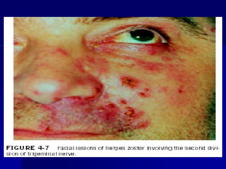
Varicella-zoster infection • Primary Varicella zoster(chicken pox) It’s an acute infectious viral disease the incubation period last for 2 -5 weeks, appears as vesicles with an erythematous area on boundary and are extremely pruritic, fever and mild generalized lymphadenopathy are also present. Resolved within 4 -8 days.
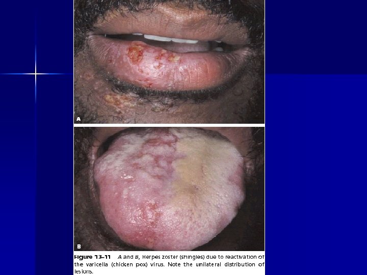
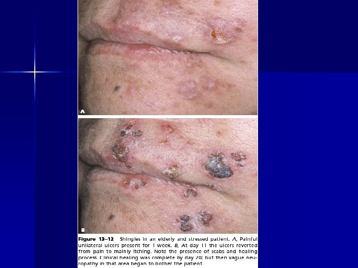
• Secondary varicella zoster infection along the peripheral nervous system commonly from dorsal root ganglia. It starts as painful vesicular eruption on skin and also may affect oral cavity when the trigeminal ganglia is infected with varicella. Patient gets malaise, fever and enlarged lymph node at area and runs in a course of about 2 weeks.
Herpangina Viral disease caused by “coxsackie group A virus” there will be fever and ill-feeling followed by vesicle rupture, lesion occurs mainly in soft palate and tonsilar area. The disease run a course of (7 -10) days.
Hand-foot &mouth disease is a highly contagious viral infection The virus is transferred from one individual to another through airborne spread or fecal-oral contamination.
Clinical Features: about 90% affects children younger than 5 years of age. After a short incubation period, the condition resolves spontaneously in 1 to 2 weeks. Signs and symptoms are usually mild to moderate in intensity and include low-grade fever, malaise, lymphadenopathy, and sore mouth. Pain from oral lesions is often the patient's chief complaint.
Measles a highly contagious viral infection caused by a member of the paramyxovirus family. Typically, oral eruptions consist of early pinpoint elevations over the soft palate that combines with ultimate involvement of the pharynx with bright erythema. German measles share some clinical features with measles, such as fever, respiratory symptoms
Clinical Featurs: After an incubation period of 7 to 10 days, prodromal symptoms of fever, malaise, conjunctivitis, photophobia, and cough develop. In 1 to 2 days small erythematous macules with white necrotic centers appear in the buccal mucosa, these lesion spots, known as Koplik's spots. precede the skin rash by 1 to 2 days.
HUMAN IMMUNODEFICIENCY VIRUS INFECTION (HIV) is transmitted by exchange of blood or body fluids principally through sexual contact. The injection of blood or from mother to child. Transmission of the virus may be followed by infection which is detected by the appearance of HIV antibodies in the blood.
This generally occurs within 3 months of exposure. A few patients have an acute HIV infection at this time, the clinical features of which include pyrexia, skin rash, headache, diarrhea, sore throat, and erythema of the buccal and palatal mucosa.
EXPOSURE TO INFECTION NO INFECTION
INFECTION HIV SEROPOSITIVE asymptomatic ACUTE HIV INFECTION Pyrexia, skin rash, headache , diarrhoea, sore throat &erythema of buccal&palatal mucosa
HIV SEROPOSITIVE asymptomatic ARC PGL AIDS l. Adenopathy p. pyrexia Diarr. , weight loss Fatique &malaise
Kaposi sarcoma
Kaposi Sarcoma. Classic Kaposi sarcoma in an older man presenting as multiple purple macules and plaques on the lower leg.