Esophageal Atresia And Tracheo esophageal Fistula Malformations Dr


- Slides: 31
Esophageal Atresia And Tracheo -esophageal Fistula Malformations Dr. Manish Kumar Gupta Assistant Professor Department of Paediatric Surgery AIIMS, Rishikesh
• • • Introduction Embryology Epidemiology Associated anomalies Classification DiagnosisØAntenatal diagnosis ØPostnatal diagnosis • Management- ØPre-operative management ØOperative management ØPost-operative management • Complications
Introduction • To anastomose the ends of an infant’s esophagus, the surgeon must be as delicate and precise as a skilled watchmaker. No other operation offers a greater opportunity for pure technical artistry. -Dr. Willis Potts(1950) • The first successful repair was done in 1940. • Most-neonatal centres are performing repair now days with success up to 90%.
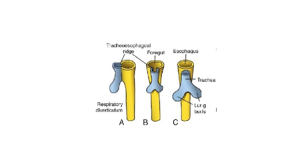
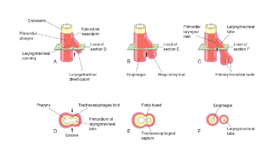
Embyrology • 4 th week of gestation: • the foregut ----->a ventral respiratory part ----->a dorsal esophageal part
Incomplete fusion of the folds defective trachea-esophageal septum abnormal connection between the trachea and esophagus
Epidemiology Incidence: 1 in 2500 to 3000 live births. Slight male preponderance- 1. 26: 1 • Chromosomal associations: • • Di. George syndrome Trisomy 21, 13 and 18 Opitz syndrome 13 q, 17 q and 16 q 24 deletions. • Single gene mutations: • Feingold syndrome • CHARGE syndrome • Fanconi anaemia • Environmental associations: • Methimazole in early pregnancy • Prolonged use of contraceptive pills • Progesterone and estrogen exposure • Maternal diabetes • Thalidomide exposure • Fetal alcohol syndrome • Maternal phenylketonuria
Associated anomalies • The most frequent associated malformations encountered in syndromic EA are: • • • Cardiac (13– 34%) Vertebral (6– 21%) Limb (5– 19%) Anorectal (10– 16%) Renal (5– 14%)
• Two syndromic associations: • VACTER-L: Vertebral, anorectal, cardiac, tracheo-esophageal, renal and limb • CHARGE: Coloboma, heart defects, atresia of the choanae, developmental retardation, genital hypoplasia and ear deformities.
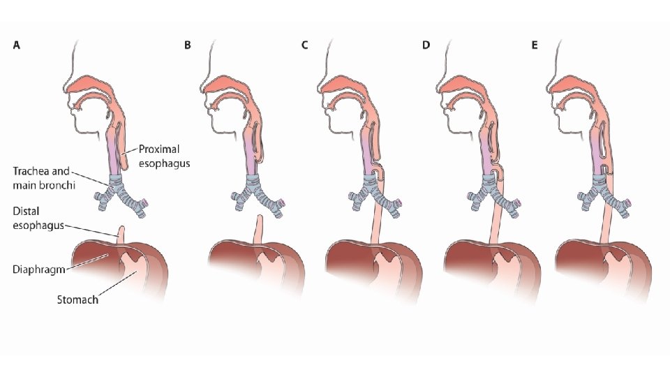
Classification • Many classification system has been proposed: • • Vogt Waterston Ladd Gross • Gross classification is most commonly used. • Based on anatomic variation
Diagnosis • Antenatal diagnosis: Ultrasonography • Polyhydramnios suggestive of EA • Absent or small stomach bubble • Fetal MRI.
Diagnosis • Postnatal: • Premature babies. • Symptomatic within first few hours of life. • Variants A, B, C and D- excessive drooling of saliva. • Choking, regurgitation and coughing on oral feeds. • Variants C and D- as the child cries, air goes into the stomach and causes abdominal distension and respiratory distress. • Variants C, D and E- gastric juice may regurgitate through the fistula causing chemical pneumonitis.
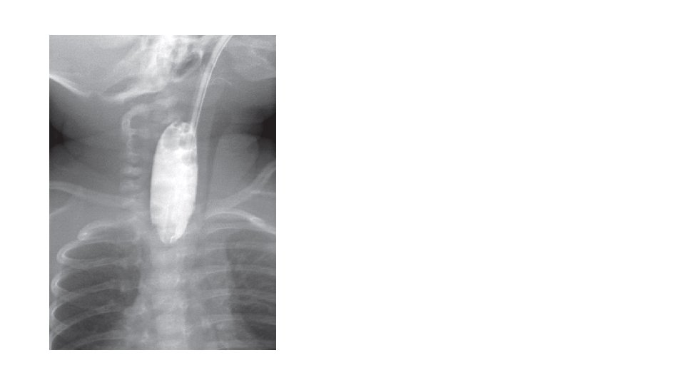
• Inability to pass an orogastric tube into the stomach. • X-ray: • Plain chest radiograph: dilated upper pouch. • If a soft feeding tube is used, the tube will coil in the upper pouch.
Gasless abdomen: Pure esophageal atresia and proximal fistula No route for gas to enter abdomen
• Plain chest radiographs help in assessment of the gap between the proximal and distal end. • Helps to plan for surgical management. • • Absence of air in abdomen is associated with long gap. Gap < 2 vertebrae Primary anastomosis Gap > 2 and < 6 vertebrae Delayed anastomosis Gap > 6 vertebrae Esophageal replacement
Bronchoscopy
Pre-operative management • Oral suctioning through 10 F double lumen Replogle tube. • Position of child: Propped up position- least gastric reflux. • Intravenous access and I. V. antibiotics. • I. V. fluids (10% dextrose and hypotonic saline)and Vitamin K administration. • If respiratory distress is present due to pneumonitis, mechanical ventilation may be done at low pressures. • High pressure ventilation may cause gastric dilatation and gastric perforation as well as diaphragmatic splinting.
• Investigations for associated anomalies: • Echocardiography- to look for cardiac defects • USG abdomen- to look for any renal abnormality. • X-rays spine- to look for any vertebral anomaly • X-ray upper limbs- to look for any radial anomaly.
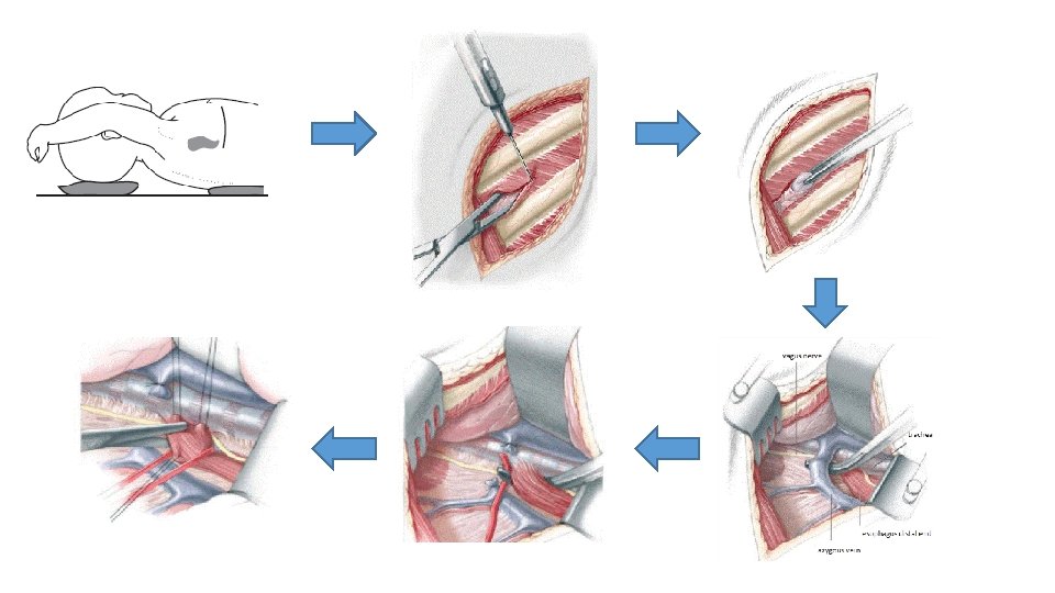
Operative management • Open thoracotomy: • Through 4 th intercostal space • Extrapleural approach • Posterior mediastinum is exposed. • Azygous vein is divided to reveal underlying TEF. • TEF is dissected circumferentially.
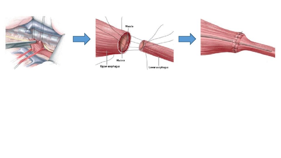
• Attachment of the fistula to the membranous portion of the trachea is taken down. • Tracheal opening is closed with non absorbable interrupted sutures. • Upper pouch of the esophagus is mobilised as much as possible. • The distal end of the upper pouch is opened and a nasogastric tube is passed through the nares to upper esophagus to the lower esophagus and then to the stomach. • Anastomosis of the ends of the esophagus are done over the nasograstric tube with absorbable sutures. • Wounds is closed after placement of chest drain.
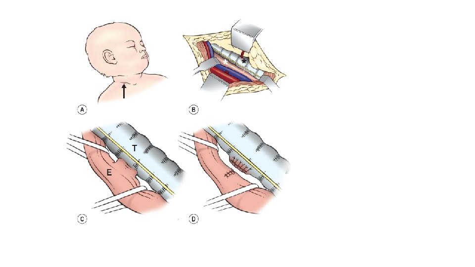
• Thoracoscopic approach is also being used. • Special cases: • H type fistula: division of the fistula done through the cervical approach.
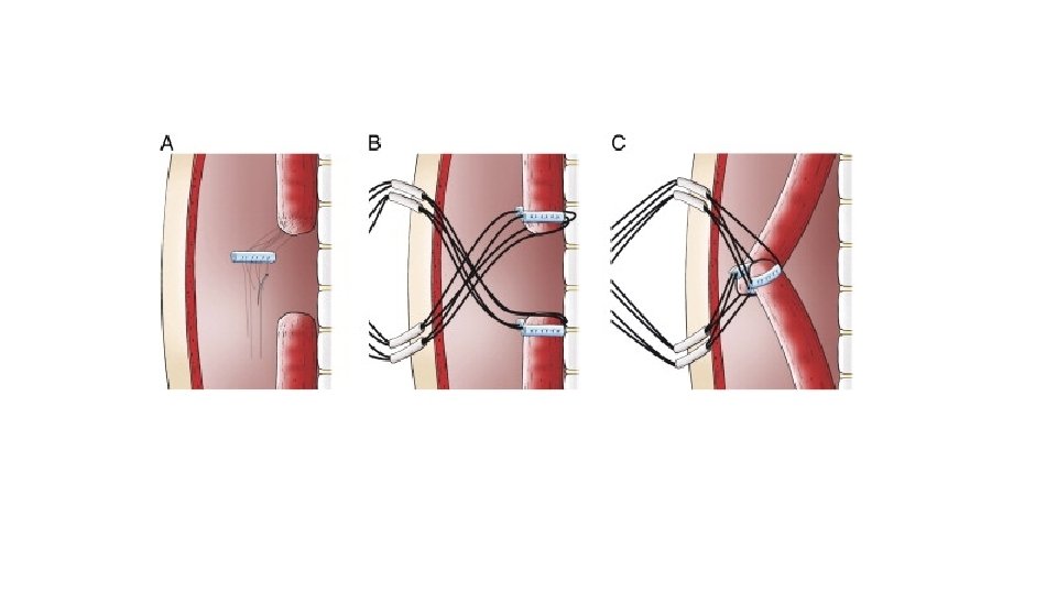
Long gap atresia • Delayed closure with lengthening procedure (at about 12 weeks of life) • • • Spontaneous growth Bougienage Upper pouch mobilisation Kimura’s extrathoracic elongation technique Foker traction suture technique Upper pouch myotomy and flaps • Esophageal replacement • • Stomach Colon Jejunum Ileum
Complications • Anastomotic leaks • Anastomotic stricture • Recurrent tracheoesophageal fistula • Tracheomalacia • Disordered peristalsis/ gastroesophageal reflux/ esophageal cancer • Vocal cord dysfunction • Respiratory morbidity