
Esophageal Atresia and Trachesophageal Fistulas MOHANNAD IBN HOMAID
Esophageal Atresia and Trachesophageal Fistulas MOHANNAD IBN HOMAID
Background Information �Definition of esophageal Artesia �The problem and its magnitude �The added problem of a TEF
Basic science �Embryology �Pathophysiology of different types �Associated anomalies
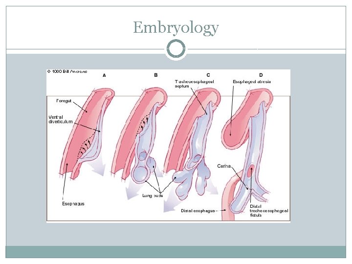
Embryology
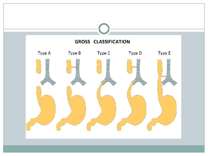
Pathophysiology �The problem of a blocked esophagus During Pregnancy In the neonatal Period �The problem of a Fistula If proximal vs distal �Associated anomalies � VACTERL and CAHRGE �The different Types
Back to the Clinical world �During pregnancy mother could develop Polydramnios �In the neonatal Period Excessive Drooling and secretion RDS and chocking upon feeding Aspiration Pneumonia Failure to pass an NG tube �Other Anomalies
Work Up �Routine Lab Work CBC and U/E, VBG and ABG BUN and Serum Cr �Imaging Studies Prenatal Ultrasonography Chest Radiography Echocardiography Renal Ultrasound Gap-o-Gram
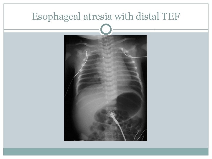
Esophageal atresia with distal TEF
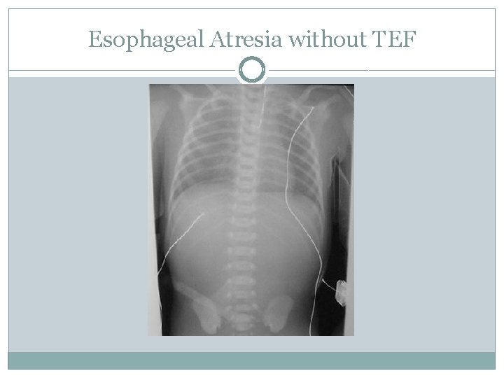
Esophageal Atresia without TEF
Gap-o-Gram �Why is it preformed �How is it preformed �Interpretation
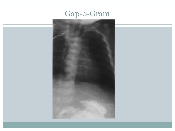
Gap-o-Gram
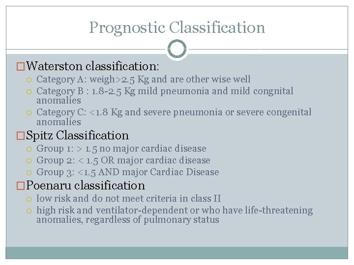
Prognostic Classification �Waterston classification: Category A: weigh>2. 5 Kg and are other wise well Category B : 1. 8 -2. 5 Kg mild pneumonia and mild congnital anomalies Category C: <1. 8 Kg and severe pneumonia or severe congenital anomalies �Spitz Classification Group 1: > 1. 5 no major cardiac disease Group 2: < 1. 5 OR major cardiac disease Group 3: <1. 5 AND major Cardiac Disease �Poenaru classification low risk and do not meet criteria in class II high risk and ventilator-dependent or who have life-threatening anomalies, regardless of pulmonary status
Treatment �Medical Therapy �Surgical Therapy �Preoperative Care �Postoperative Care �Complications �Follow up
Medical Therapy �Intravenous Fluid with adequate Glucose �Prophylactic Broad Spectrum Antibiotics �Replogle tube insertion
Surgical Therapy �In isolated Artesia Lengthening and anastomosis procedures Esophageal substitution Delayed vs immediate vs staged The gap-o-Gram �If TEF is present Divide and ligate the fistula
Pre operative care �In addition to medical therapy �Bronchoscopy for: Detection of an upper pouch fistula Localization of the distal fistula Assessment of post operative risk for tracheomalacia Assessment of specific vascular anomalies ( right sided aortic arch)
Post operative Care �Intubated and transferred to the NICU �Antibiotics are continued until chest drain is no longer needed �Suctioning the oral secretions
Complications �Early : Anastomotic leak Recurrent TEF Anastomotic Stricture �Late GERD Esophageal Dysmotiliy Tracheomalacia May appear early
Follow up �Appointment 1 -3 – 12 months after discharge Focusing on sings of respiratory distress and dysphagia �Radiologic assessment only if significant history of: Choking, cyanosis Regurgitation and dysphagia Failure to the thrive Coughing and wheezing
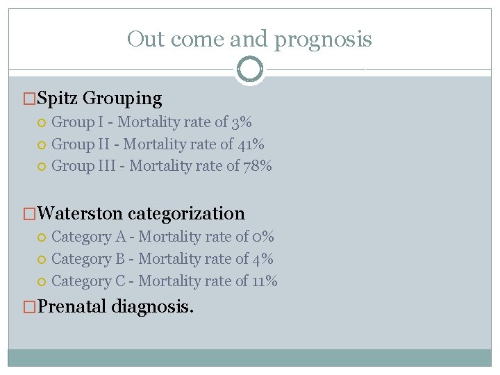
Out come and prognosis �Spitz Grouping Group I - Mortality rate of 3% Group II - Mortality rate of 41% Group III - Mortality rate of 78% �Waterston categorization Category A - Mortality rate of 0% Category B - Mortality rate of 4% Category C - Mortality rate of 11% �Prenatal diagnosis.

Questions
- Slides: 22