Development of the face and palate Dr Gallatz


 apparatus consist of: - pharyngeal arches - pharyngeal pouches - pharyngeal")


 from the mesenchyme covering the")


 project inferolaterally on each side of the tongue.")


 Cleft of the upper jaw (gnatoschisis)")


: Ø Due")


- Slides: 55
Development of the face and palate Dr Gallatz Katalin
Development of the face and pharyngeal arches 4 -5. week Pharyngeal arches begin to develop in the 4 th week as the neural crest cells migrate into the future head and neck region.
MIGRATION OF THE NEURAL CREST CELLS
The pharyngeal (branchial) apparatus consist of: - pharyngeal arches - pharyngeal pouches - pharyngeal grooves - pharyngeal mebranes
Each pharyngeal arch contain: - cartilage - nerve - artery - myoblast - mesenchyme
Stomodeum stomodeum: a wide shallow depression in the face, limited in its depth by the buccopharyngeal membrane
STOMODEUM SURROUNDED by the facial prominences Frontal prominence (frontonasal) from the mesenchyme covering the forebrain Maxillary prominence from mesenchyme of the I. pharyngeal arch Mandibular prominence From the mesenchyme of the I. pharyngeal arch
4. week On both sides of the frontonasal prominences thickenings of surface ectoderm form the NASAL PLACODS
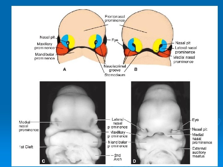
5. week 1. The nasal placods invaginate to form the nasal pits 2. Medial and lateral nasal processes are formed around the nasal pits 3. Maxillary and lateral nasal processes are separated by the nasolacrimal groove nasolacrimal duct
NASAL PITS MEDIAL AND LATERAL NASAL PROMINENCES
7 -10. week Maxillary prominences grow and press the medial nasal processes, they unite and fuse with the maxillary prominences. The inferior part of the fused medial nasal prominences form the INTERMAXILLARY SEGMENT
Intermaxillary segment is composed of 1. labial component philtrum of the lip 2. upper jaw component alveolar process of the incisor teeth 3. palatine component primary palate
- The ectodermal thickening betwen the lateral nasal and maxillary process invaginates into the underlying mesoderm, the nasolacrimal groove and cord is formed, later it canalicularises and it becomes NASOLACRIMAL DUCT. - The maxillary prominences unite with lateral nasal processes and also with the mandibular prominences.
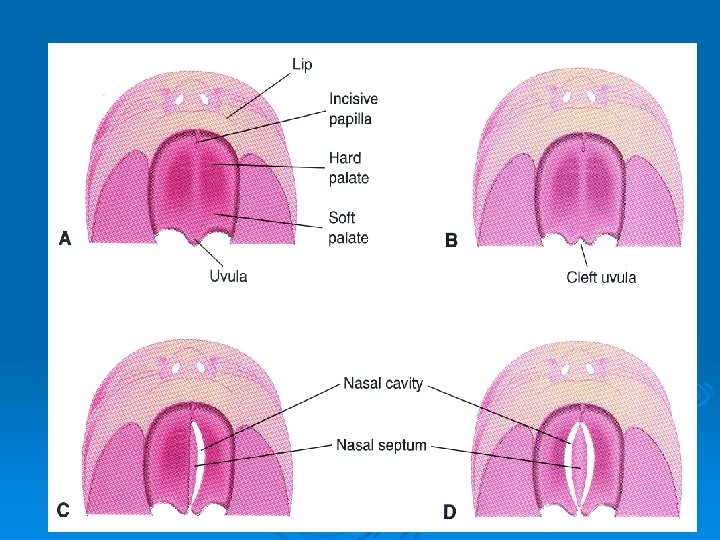
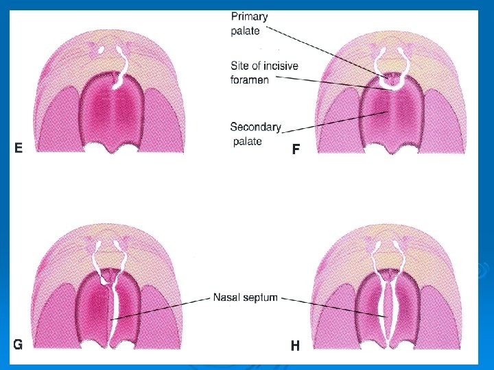
Palatogenesis 5 -12 th week Critical period 6 -9 th week The palate develops from two primordia: - the primary palate - the secondary palate
Development of the palate Primary palate develops from the palatine portion of the intermaxillary segment Secondary palate develops from the palatine process of the maxillary prominence
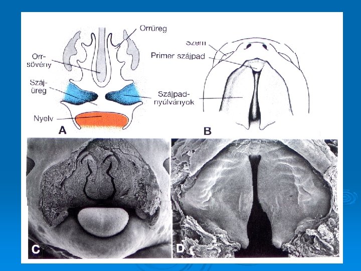
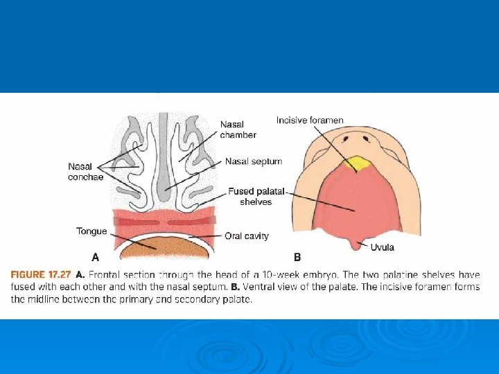
- Palatine processes (or palatal shelves) project inferolaterally on each side of the tongue. - As the mandible develop the tongue moves inferiorly, the palaine processes elongate and ascend to the horizontal position. They fuse in the median plane and also fuse with the nasal septum and with the primary palate Nasal septum develops as a downgrowth from the internal part of the merged medial nasal prominences
Derivates of the facial prominences Frontonasal prominence frontal part: forehead, root of the nose medial nasal process: - dorsum and tip of the nose, nasal septum intermaxillary segmentum: philtrum, alveolar process of the incisor teeth, primary palateral nasal process: alae (wings) of the nose Maxillary prominences: maxilla, lateral part of the upper lip, upper part of the cheek secondary palate Mandibular prominence: mandible, lower lip, inferior part of the cheek
Development of the nasal cavity - Nasal placods - nasal pits - oronasal membrane - primitive choana - primary and secundary palate - nasal conchas - olfactory region - paranasal sinuses
1. Development of the stomodeum and the facial prominences, 2. appearence of the nasal placods, nasal pits 3. development of the medial and lateral nasal prominences 5. week 4. growth of the maxillary prominences, 5. fusion of the medial nasal processes, 6. formation of the intermaxillary segment, 7. development ot the primary palate 6 -7. week, 8. formation of the nasolacrimal sulcus and duct, 9. fusion of the maxillary prominence with the and medial and lateral nasal prominences, 10. secondary palate, separation of the nasal and oral cavity Development of the Face
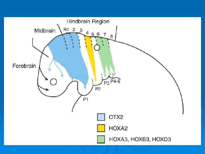
GENES IN DEVELOPMENT OF THE FACE
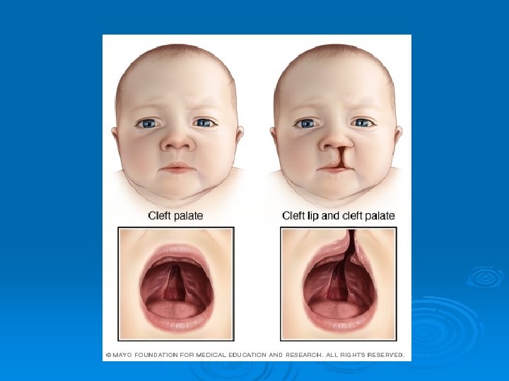
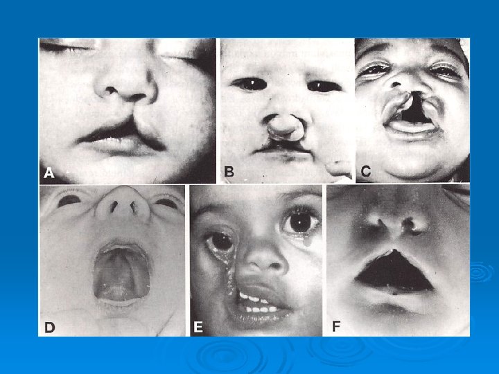
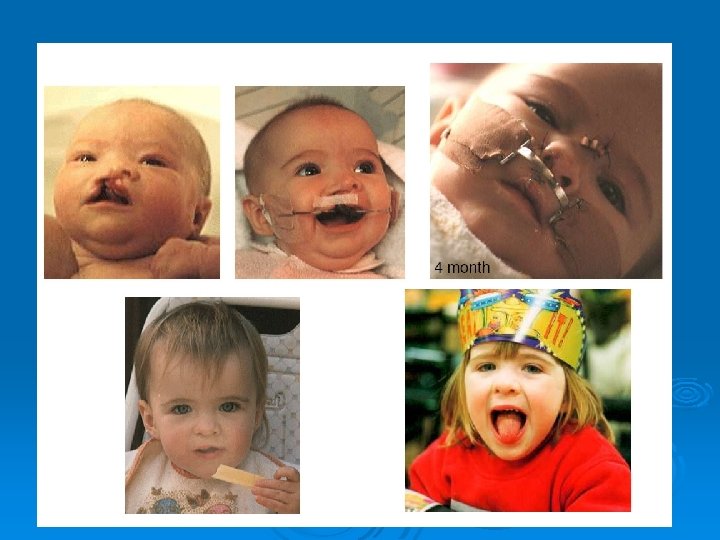
Malformations Median cleft lip Lateral cleft lip (cheiloschisis) Cleft of the upper jaw (gnatoschisis) Cleft palate (palatoschisis) Oblique facial cleft Transverse facial cleft Macrostomy, microstomy Cleft uvula
Malformations
Malformations Median cleft Macrostomia Cleft of inferior lip Microstomia Obligue facial cleft Bifid nose and incomplete median cleft
Congenital anomalies of the face 1. Lateral cleft lip ( hare lip): Ø Due to failure of fusion between the maxillary process with the intermaxillary segment, unilateral or bilateral.
Bilateral cleft lip
Congenital anomalies of the face 2. Median cleft lip : Ø Due to incomplete union of the 2 medial nasal process in the midline.
3. Oblique facial cleft : Ø Due to failure of union of the maxillary processes with lateral nasal processes.
Thank you for your attention! Development of the Face and Palate www. indiana. edu/~anat 550/hnanim/face. html Face And Palate - You. Tube ► 4: 07 www. youtube. com/watch? v=Dg. Z_tqucd. I 4
Hypobranchial eminence
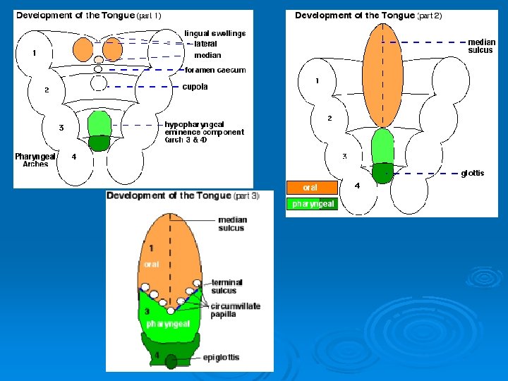
Development of the tongue ANTERIOR 2/3 At the end of the 4 th week on the base of the primordiali pharynx 3 tubercule develops from the mesenchyme of the Ist pharyngeal arch, The paired TUBERCULUM LATERALE and between and behind them the TUBERCULUM IMPAR. The lateral tubercules develop quickly, overgrow the tuberculum impar and unite in the midline ( median lingual sulcus)
Development of the tongue POSTERIOR 1/3 From the II-III. pharyngeal arch mesenchyme. COPULA from the ventromedial part of II. pharyngeal arch HYPOBRANCHIAL EMINENCE from the ventromedial part of III-IV. pharyngeal arch
Development of the tongue EPITHELIUM Anterior 2/3 ectoderm Posterior 1/3 endoderma MUSCLES From the myotomes of the occipital somites
Development of the tongue
DEVELOPMENTAL MALFORMATIONS BIFID TONGUE
ANKYLOGLOSSIA
MACROGLOSSIA
MICROGLOSSIA
LEUKOPLAKIA
HYPOGLOSSAL NERVE PARALYSIS
DEVELOPMENT OF THE THYROID GLAND
DEVELOPMENTAL MALFORMATIONS 1. - Thyreoglossal duct - a duct penetrates the mesenchyme from the foramen cecum, it bifurcates and forms the 2 lobes of the thyroid gland. - In case the duct does not disappear (ductus thyreoglossus persistens) it may contribute to the formation of median cervical cysts. They often form fistules too.
THYROGLOSSAL CYST
Thank you for your attention! Development of the Face and Palate www. indiana. edu/~anat 550/hnanim/face. html Face And Palate - You. Tube ► 4: 07 www. youtube. com/watch? v=Dg. Z_tqucd. I 4