Palate Hard palate anterior portion of mouths roof


")


")


 and sides. Projections of lamina propria Some contain")


 – pointed; tearing")
")
 dentition and times of eruptions")
 dentition and times of eruptions")
 Breaks into pieces Mixes with")


- Slides: 30
Palate Hard palate – anterior portion of mouth’s roof; formed by maxillae and palatine bones Soft palate – posterior portion of mouth's roof; formed by two muscular arches: Palatoglossal arch - extends from palate to tongue anterior to palatine tonsils Palatopharyngeal arch - extends from palate to pharyngeal wall posterior to palatine tonsil
Figure 24. 5 Structures of the mouth (oral cavity)
Salivary Glands General Saliva Functions: Keeps the oral and pharyngeal mucous membranes moist. Lubricates and dissolves food. Starts carbohydrate chemical digestion. Most saliva is secreted by salivary glands. Some saliva comes from buccal glands in the mucous membrane that lines the mouth. Minor salivary glands include labial, buccal, lingual and palatal glands which make a small contribution to saliva.
Salivary Glands Outside the mouth ducts oral cavity 1. Parotid glands - below ear, over masseter; parotid ducts open into vestibule beside 2 nd maxillary molar. 2. Submandibular glands - under mandible; submandibular ducts open lateral to lingual frenulum 3. Sublingual glands - floor of mouth deep to tongue; lesser sublingual ducts open into floor of mouth
Figure 24. 6 The three major salivary glands – parotid, sublingual, and submandibular
Salivary Glands Salivary gland cells are organized into acini. Acini: small saclike cavities (clusters) in a gland surrounded by secretory cells. Serous acini - secrete a watery fluid Mucous acini - secrete a slimy, mucus secretion Parotid glands – serous acini only Submandibular glands – mostly serous acini and a few mucous acini Sublingual glands - mostly mucous acini and a few serous acini
Figure 24. 6 b Histology of the submandibular gland
Composition and Functions of Saliva Composition: 1. 2. 99. 5% water 0. 5% solutes (salts, gases, mucous, ions, enzymes, lysozyme, Ig. A) Functions: 1. 2. 3. 4. 5. 6. 7. Lubricate food for easier swallowing Dissolve food for tasting Moistens mucous membranes Bicarbonate ions buffer acidic foods Salivary amylase begins chemical digestion of starch (maltose) Lysozyme antibacterial Ig. A 1 st line of defense
Salivation The secretion of saliva. Salivation is under ANS control Moistens mucous membranes Assists speech Almost all salivary components are reabsorbed Dehydration – conserve water by not producing saliva
Salivation 1. Increase salivation Sight, smell, sounds, thoughts/memory. Tongue stimulation taste buds Salivatory nuclei in brainstem increase salivation Parasympathetic Facial 2. nerve impulses stimulate saliva (VII) and glossopharyngeal (IX) nerves. Stop salivation Sympathetic nerves inhibit salivation
Tongue Composed of skeletal muscle covered with mucous membrane. Forms oral cavity’s floor. Participates in chewing, swallowing, and speech. Glands on tongue’s dorsum secrete lingual lipase
Tongue Lingual frenulum - attaches tongue to mouth’s floor Tongue-tied Ankyloglossia Abnormally short frenulum Speech impediment Can be surgically repaired
Tongue Papillae cover upper surface (dorsum) and sides. Projections of lamina propria Some contain taste buds
Figure 11. 7 Muscles that Move the Tongue - FYI
Teeth Project into the mouth Alveolar processes of mandible and maxilla Covered by gingivae
Teeth Adapted for mechanical digestion. A typical tooth consists of crown, neck, and root. Composed primarily of dentin, which encloses the crown’s pulp cavity the roots’ root canals. Root’s dentin is covered by cementum Cementum attaches root to periodontal ligament Fibrous connective tissue Anchor Shock absorber
Figure 24. 7 A typical tooth and surrounding structures
Tooth Types 1. Incisors – chisel shaped; cutting 2. Cuspids (canines) – pointed; tearing 3. Premolars – bicuspid; crushing and grinding 4. Molars – four cusps; crushing and grinding
Dentition There are two sets of teeth in an individual’s lifetime: 1. Deciduous (primary) teeth 20 teeth that start erupting at 6 months 2. Permanent (secondary) teeth 32 teeth that erupt between 6 and 12 years of age
Figure 24. 8 Deciduous (primary) dentition and times of eruptions
Figure 24. 8 b Permanent (secondary) dentition and times of eruptions
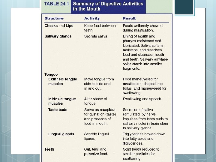
Digestion in the Mouth Mechanical digestion (mastication or chewing) Breaks into pieces Mixes with saliva bolus Chemical digestion Amylase Begins starch digestion at p. H of 6. 5 or 7. 0 found in mouth When bolus & enzymes hit the p. H of 2. 5 (gastric juices), hydrolysis ceases Lingual lipase Secreted by glands in tongue Begins breakdown of triglycerides into fatty acids and glycerol once it reaches the acidic p. H in the stomach
PHARYNX
Figures 23. 2 / 24. 10 Pharynx “Throat” – but don’t call it that… Funnel-shaped tube extending from internal nares to the esophagus and larynx Skeletal muscle lined by mucous membrane. 3 parts: Nasopharynx – only respiratory fnc Oropharynx – digestive and respiratory fnc Laryngopharynx – digestive and respiratory fnc
Figure 23. 2 The pharynx
Pharynx Deglutition mucus or swallowing is facilitated by saliva and Starts when bolus is pushed into the oropharynx Sensory nerves send signals to deglutition center in brainstem Soft palate is lifted, epiglottis is bent Ingested food mouth oropharynx laryngopharynx muscular contractions esophagus stomach
ESOPHAGUS
Figure 24. 1 Esophagus Secrete mucus and transport food to the stomach. Collapsible muscular tube = flexible In front of vertebrae Posterior to trachea Posterior to the heart Connects pharynx to stomach Pierces diaphragm at hiatus