
HISTOLOGY OF DIGESTIVE TRACT By DR SOBIA IBRAHIM
HISTOLOGY OF DIGESTIVE TRACT By DR SOBIA IBRAHIM Assistant professor ANATOMY KEMU
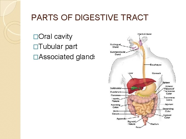
PARTS OF DIGESTIVE TRACT �Oral cavity �Tubular part �Associated glands
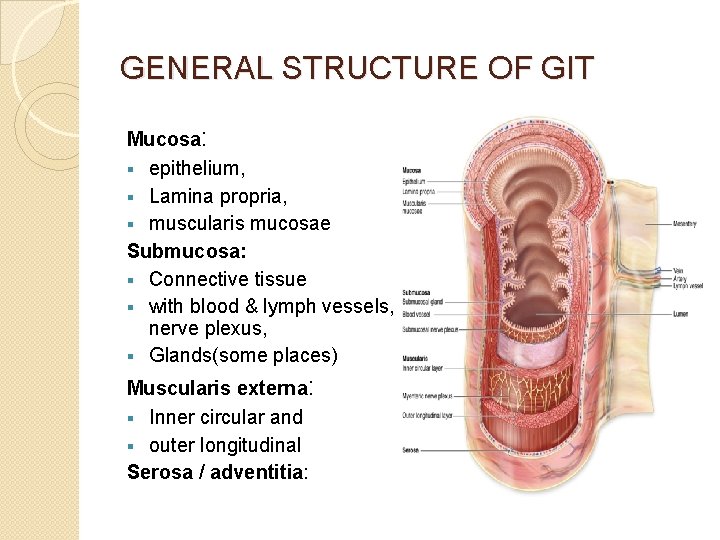
GENERAL STRUCTURE OF GIT Mucosa: § epithelium, § Lamina propria, § muscularis mucosae Submucosa: § Connective tissue § with blood & lymph vessels, nerve plexus, § Glands(some places) Muscularis externa: § Inner circular and § outer longitudinal Serosa / adventitia:
NERVE PLEXUSES � Autonomic plexuses � Submucosal /Meissner’s � Myenteric /Auerbach’s � Parasympathetic pre&post ganglionic fibres � Sympathetic post ganglionic fibres � Sensory nerve fibres � Parasympathetic ganglia
MAIN FUNCTIONS OF MUCOSA EPITHELIAL LINING Ø Selectively permeable memb. Transport & digestion of food Ø Promote absorption of products of digestion Ø Produce hormones affecting activity of GIT Ø Produce mucus for lubrication & protection LAMINA PROPRIA Ø Macrophages & lymphoid cells rich zone Ø Immunoglobulin A (Ig. A) production MUSCULARIS MUCOSAE Ø Promotes movement of mucosa, increasing its contact with food Ø
MEDICAL APPLICATION � HIRSCHSPRUNG DISEASE (congenital megacolon) � CHAGAS DISEASE (Trypanosoma cruzi infection) � Nerve plexuses severely damaged& neurons destroyed � Disturbance of GIT motility & frequent dilatations of intestines � Effects of emotional stress on GIT
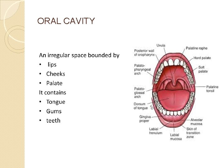
ORAL CAVITY
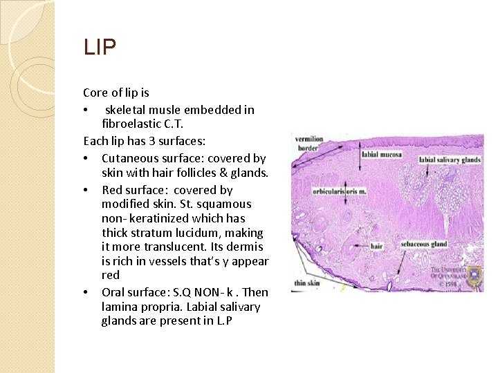
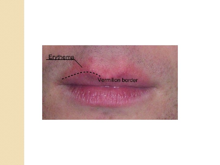
LIP Core of lip is • skeletal musle embedded in fibroelastic C. T. Each lip has 3 surfaces: • Cutaneous surface: covered by skin with hair follicles & glands. • Red surface: covered by modified skin. St. squamous non- keratinized which has thick stratum lucidum, making it more translucent. Its dermis is rich in vessels that’s y appear red • Oral surface: S. Q NON- k. Then lamina propria. Labial salivary glands are present in L. P
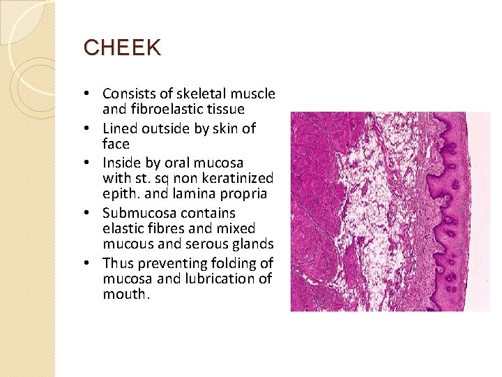
CHEEK • Consists of skeletal muscle and fibroelastic tissue • Lined outside by skin of face • Inside by oral mucosa with st. sq non keratinized epith. and lamina propria • Submucosa contains elastic fibres and mixed mucous and serous glands • Thus preventing folding of mucosa and lubrication of mouth.
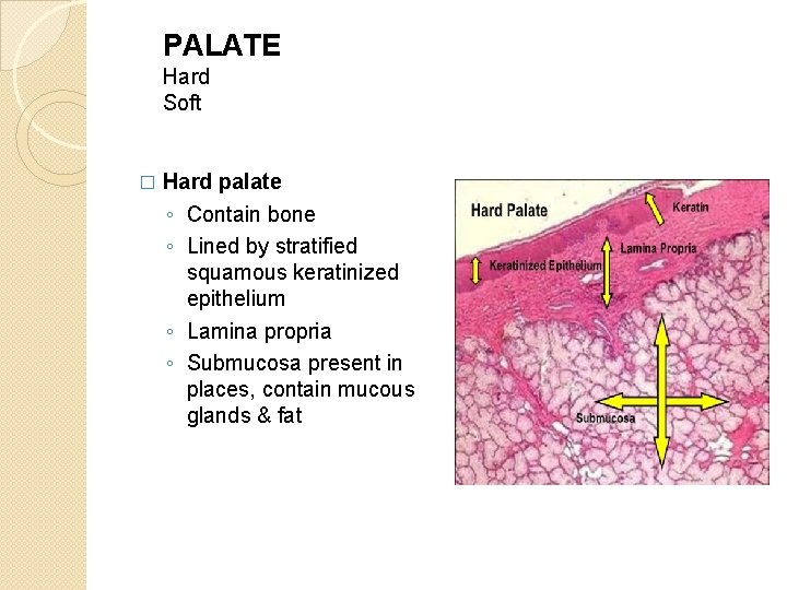
PALATE Hard Soft � Hard palate ◦ Contain bone ◦ Lined by stratified squamous keratinized epithelium ◦ Lamina propria ◦ Submucosa present in places, contain mucous glands & fat
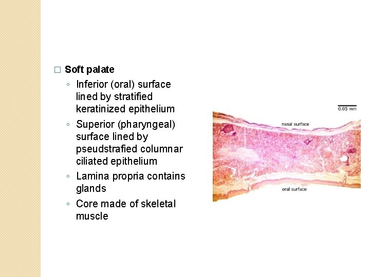
� Soft palate ◦ Inferior (oral) surface lined by stratified keratinized epithelium ◦ Superior (pharyngeal) surface lined by pseudstrafied columnar ciliated epithelium ◦ Lamina propria contains glands ◦ Core made of skeletal muscle
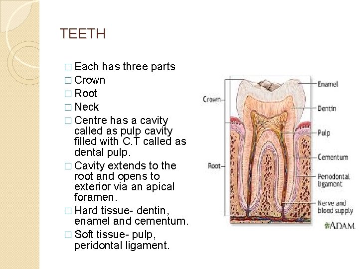
TEETH � Each has three parts � Crown � Root � Neck � Centre has a cavity called as pulp cavity filled with C. T called as dental pulp. � Cavity extends to the root and opens to exterior via an apical foramen. � Hard tissue- dentin, enamel and cementum. � Soft tissue- pulp, peridontal ligament.
- Slides: 13