PALATE INTRODUCTION Palate roof of the oral cavity


 • Presence of supernumerary,")
 of the cortical bone Usually – midline of")


- Slides: 31
PALATE
INTRODUCTION: Palate : roof of the oral cavity. It has two parts –an anterior hard palate –a posterior soft palate
HARD PALATE Separates the oral cavity from the nasal cavities Consists of a bony plate covered above and below by mucosa Above: covered by respiratory mucosa and forms floor of nasal cavity Below: covered by oral and forms much of of oral cavity mucosa the roof
POSITION The anteriolateral margins of the palate are continuous with the alveolar arches and gums. The posterior margin gives attachment to the soft palate. The superior surface forms the floor of the nose. The inferior surface forms the roof of the oral cavity
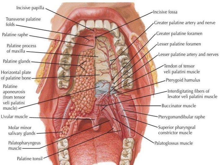
OSTEOLOGY: • Palatine processes of the maxillae form the anterior 3/4 of the hard palate • Horizontal plates of the palatine bones form the posterior 1/4
SUTURE: • INTERMAXILLARY SUTURE • INTERPALATINE SUTURE • PALATOMAXILLARY SUTURE INCISIVE CANAL: CONTENTS: Greater palatine vessels Nasopalatine nerve (terminal part) GREATER PALATINE FORAMEN: CONTENTS: Greater palatine vessels Anterior palatine nerve LESSER PALATINE FORAMEN: CONTENTS: Middle and Posterior palatine nerves
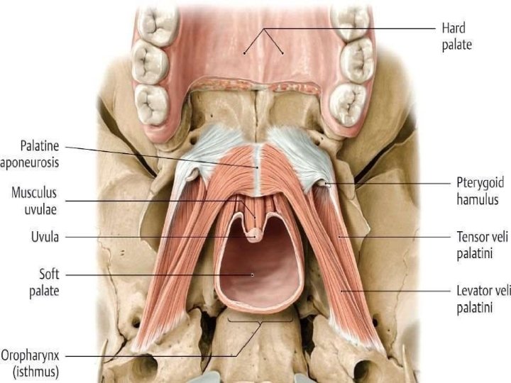
SOFT PALATE Movable, muscular fold, suspended from the posterior border of the hard palate. It separates the nasopharynx from the oropharynx. Acts as a valve that can be: depressed to help close the oropharyngeal isthmus; elevated to separate the nasopharynx from the oropharynx.
MUSCLES OF SOFT PALATE • Tensor velipalatini • Levator velipalatini • Musculus uvulae • Palato pharyngeus • Palato glossus
TENSOR VELI PALATINI ORIGIN: Lateral side of auditary tube Scaphoid fossa of sphenoid bone INSERTION: Palatine aponeurosis NERVE SUPPLY: Mandibular nerve via br to medial pterygoid muscle ACTION: Tightens the soft palate Opens the auditory tube
LEVATOR VELI ORIGIN: PALATINI Petrous temporal bone Inferior aspect of auditory tube INSERTION: Upper surface of palatine aponeurosis NERVE SUPPLY: Vagus N via pharyngeal plexus ACTION: Elevates the soft palate
ORIGIN: MUSCULUS UVULAE Posterior nasal spine of hard palate INSERTION: Connective tissue of uvula NERVE SUPPLY: Vagus N via pharyngeal plexus ACTION: Elevates and retracts uvula thickens central region of soft palate
PALATOGLOSSUS ORIGIN: Inferior surface of palatine aponeurosis INSERTION: Lateral margin of tongue NERVE SUPPLY: Vagus N via pharyngeal plexus ACTION: Depresses palate Moves palatoglossal arch toward midline elevates back of the tongue
PALATOPHARYNGEUS ORIGIN: Superior surface of palatine aponeurosis INSERTION: Pharyngeal wall NERVE SUPPLY: Vagus N via pharyngeal plexus ACTION: Depresses soft palate moves palatopharyngeal arch toward midline elevates pharynx
PASSAVANT’S RIDGE • Some of the upper fibres of the palatopharyngeus passes circularly deep to mucous membrane of the pharynx • Forms a sphincter internal to the superior constrictor • This constitute the passavant’s muscle • On contraction raises a ridge called passavant’s ridge • Best developed in cleft palate cases
BLOOD SUPPLY • Greater palatine branch of the maxillary artery • Ascending palatine branch of the facial artery • Palatine branch of the Ascending pharyngeal artery
VEINS: Pterygoid plexuses tonsillar plexuses of veins. LYMPHATICS: Upper deep cervical retropharyngeal lymph nodes.
NERVE SUPPLY • Supplied by the greater and lesser palatine nerves and the nasopalatine nerve • General sensory fibers carried in all these nerves originate in the pterygopalatine fossa from the maxillary nerve • Special sensory and scretomotor nerves are contained in lesser palatine nerves.
DEVELOPMENT OF PALATE develops as two parts • The Primary Palate • The Secondary Palate Development Of The Primary Palate : Fusion of the two medial nasal processes with the fronto nasal process results in the formation of primary palate.
Development of Secondary Palate: The formation of secondary palate commences between 7 and 8 weeks and completes around the 3 rd month of the gestation. Three outgrowth appear in the oral cavity • The two palatal process • The nasal septum
• Each palatal process grows downwards first then upwards after the withdrawal of tongue(7 th week) • septum and the two shelves converges and fuse in the midline • The closure of the secondary palate proceeds gradually form the primary palate in a posterior direction.
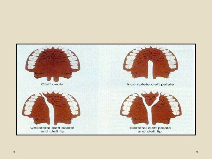
CLINICAL CONSIDERATIONS CLEFT PALATE: Congenital birth defect Defective fusion of the various components of palate gives rise to cleft in palate
Cleft of palate occurs in number of ways: • Defective growth of palatal shelves • Delayed or total failure of shelves to elevate and attain a horizontal position • Lack of contact between shelves • Post fusion rupture of shelves
Problems associated with cleft palate • Dental problems • Aesthetic problems • Hearing and speech problems • Psychological problems
Dental problems • Congenitally missing teeth( mostly upper lateral incisors) • Presence of supernumerary, neonatal and natal teeth • Ectopically erupted tooth • Enamel hypoplasia • Microdontia, macrodontia • Fused teeth • Gemination, dilaceration • Tendency towards class III skeletal pattern • Posterior and anterior cross bite • Deep bite • Spacing/ crowding • Protruding premaxilla
TORUS PALATINUS Localized nodular enlargement (exostosis) of the cortical bone Usually – midline of the palate Pose a mechanical problem in the construction of denture
INFLAMMATORY PAPILLARY HYPERPLASIA • Common lesion that develops on the central hard palate • in response to chronic denture irritation
SMOKER’S PALATE • Nicotine stomatitis • An erythematous irritation is initially seen, followed by a whitish palatal mucosa reflecting a hyperkeratosis • Red dots representing orifices of accessory salivary glands seen
HIGH ARCHED PALATE Develomental feature that may occur in isolation or in association with a number of conditions Acquired condition caused by chronic thumb sucking High arched palate may cause narrowed airway and sleep disordered breathing Pose difficulty in the construction of denture