
12 5 2015 Development of Face Nose Palate






- Slides: 44
12. 5. 2015 Development of Face, Nose & Palate Dr. Archana Rani Associate Professor Department of Anatomy KGMU UP, Lucknow
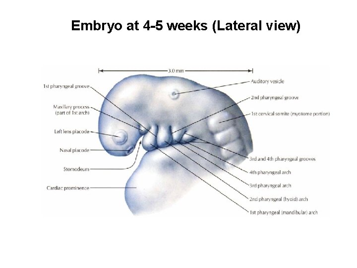
Embryo at 4 -5 weeks (Lateral view)
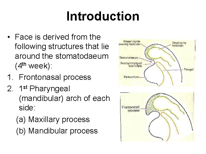
Introduction • Face is derived from the following structures that lie around the stomatodaeum (4 th week): 1. Frontonasal process 2. 1 st Pharyngeal (mandibular) arch of each side: (a) Maxillary process (b) Mandibular process
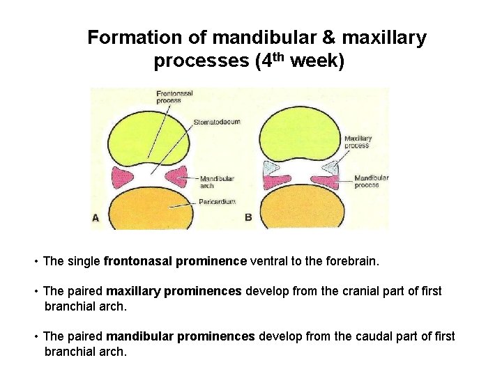
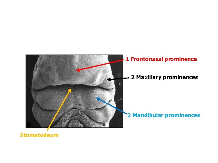
Formation of mandibular & maxillary processes (4 th week) • The single frontonasal prominence ventral to the forebrain. • The paired maxillary prominences develop from the cranial part of first branchial arch. • The paired mandibular prominences develop from the caudal part of first branchial arch.
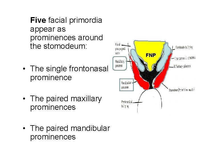
Five facial primordia appear as prominences around the stomodeum: FNP • The single frontonasal prominence • The paired maxillary prominences • The paired mandibular prominences
1 Frontonasal prominence 2 Maxillary prominences 2 Mandibular prominences Stomatodeum
Further development of face • Formation of nasal placodes and lens placodes (4 th week). • Nasal placodes sinks below to form nasal pits (5 th week). • Elevations of the nasal pits form the medial and lateral nasal processes. • Nasal placodes are primordia of the nose and nasal cavities.
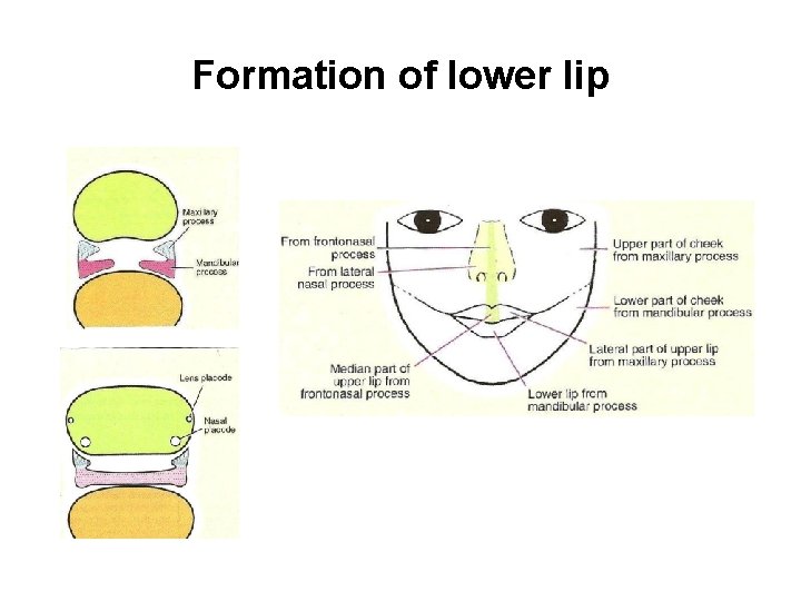
Formation of lower lip
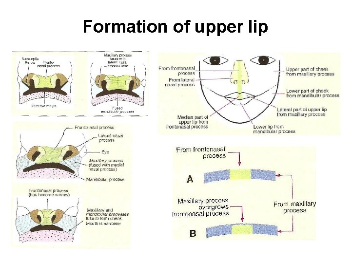
Formation of upper lip
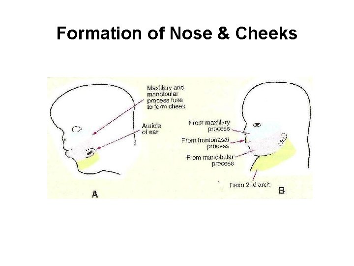
Formation of Nose & Cheeks
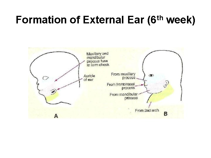
Formation of External Ear (6 th week)
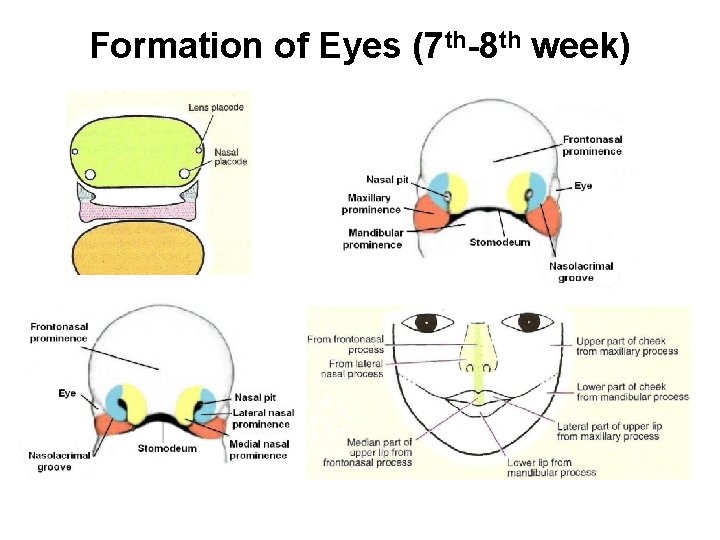
Formation of Eyes (7 th-8 th week)
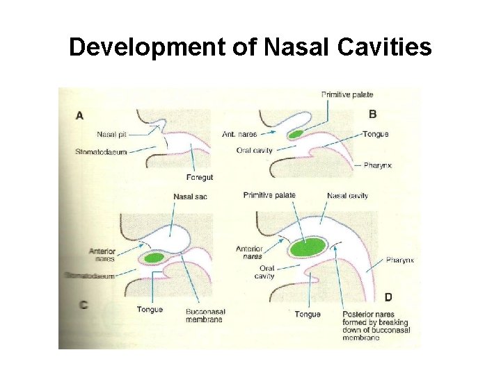
Development of Nasal Cavities
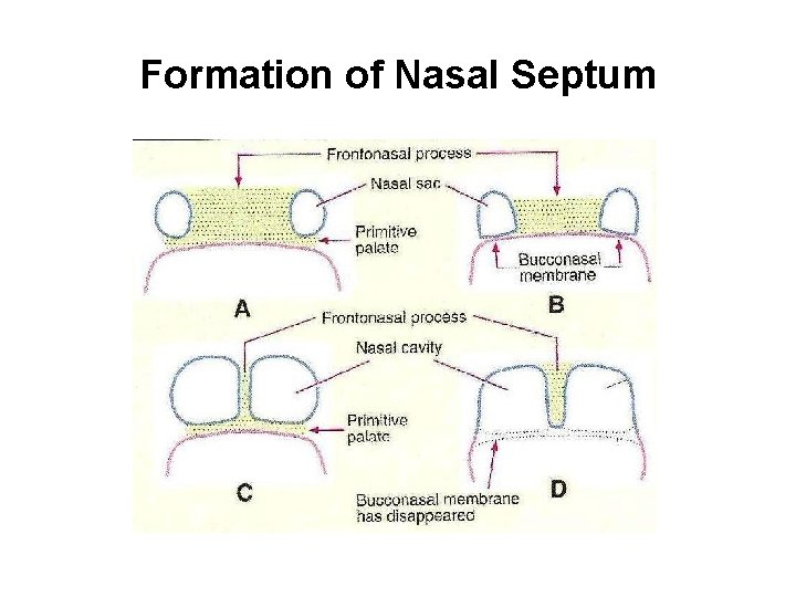
Formation of Nasal Septum
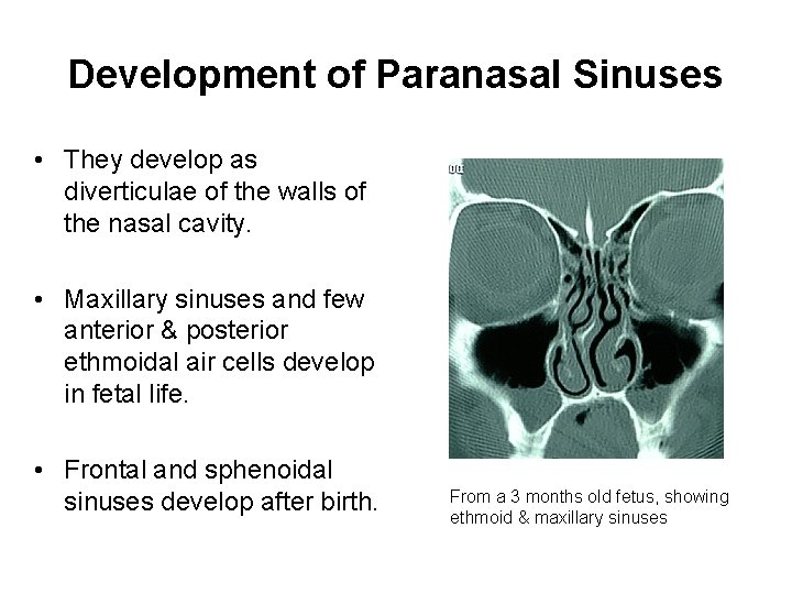
Development of Paranasal Sinuses • They develop as diverticulae of the walls of the nasal cavity. • Maxillary sinuses and few anterior & posterior ethmoidal air cells develop in fetal life. • Frontal and sphenoidal sinuses develop after birth. From a 3 months old fetus, showing ethmoid & maxillary sinuses
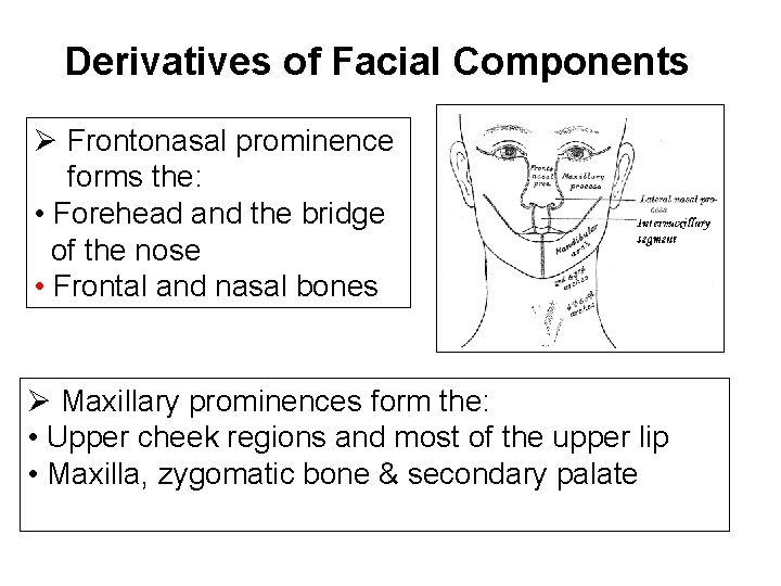
Derivatives of Facial Components Ø Frontonasal prominence forms the: • Forehead and the bridge of the nose • Frontal and nasal bones Ø Maxillary prominences form the: • Upper cheek regions and most of the upper lip • Maxilla, zygomatic bone & secondary palate
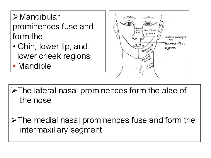
ØMandibular prominences fuse and form the: • Chin, lower lip, and lower cheek regions • Mandible ØThe lateral nasal prominences form the alae of the nose ØThe medial nasal prominences fuse and form the intermaxillary segment
Development of Palate (Palatogenesis)
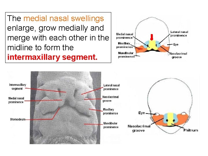
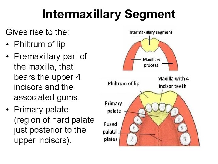
The medial nasal swellings enlarge, grow medially and merge with each other in the midline to form the intermaxillary segment. Human embryo: 7 weeks
Intermaxillary Segment Gives rise to the: • Philtrum of lip • Premaxillary part of the maxilla, that bears the upper 4 incisors and the associated gums. • Primary palate (region of hard palate just posterior to the upper incisors).
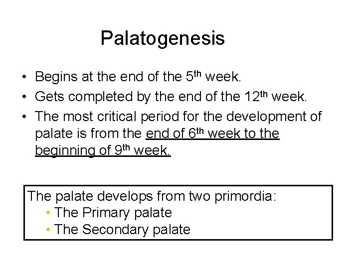
Palatogenesis • Begins at the end of the 5 th week. • Gets completed by the end of the 12 th week. • The most critical period for the development of palate is from the end of 6 th week to the beginning of 9 th week. The palate develops from two primordia: • The Primary palate • The Secondary palate
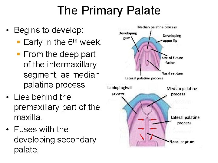
The Primary Palate • Begins to develop: § Early in the 6 th week. § From the deep part of the intermaxillary segment, as median palatine process. • Lies behind the premaxillary part of the maxilla. • Fuses with the developing secondary palate.
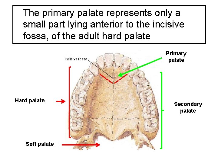
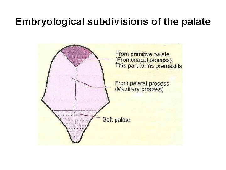
The primary palate represents only a small part lying anterior to the incisive fossa, of the adult hard palate Primary palate Hard palate Soft palate Secondary palate
The Secondary Palate • Is the primordia of hard and soft palate posterior to the incisive fossa. • Begins to develop: § Early in the 6 th week. § From the internal aspect of the maxillary processes, as lateral palatine process.
• In the beginning, the lateral palatine processes project inferomedially on each side of the tongue. • With the development of the jaws, the tongue moves inferiorly. • During 7 th & 8 th weeks, the lateral palatine processes elongate and ascend to a horizontal position above the tongue. Tongue
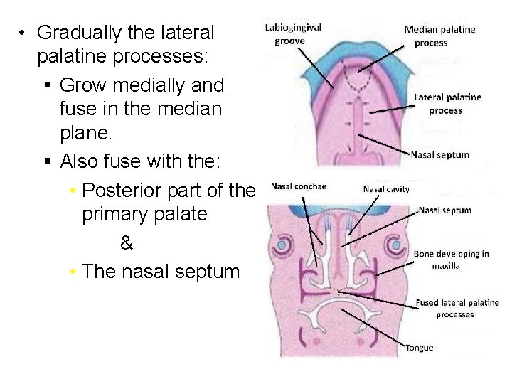
• Gradually the lateral palatine processes: § Grow medially and fuse in the median plane. § Also fuse with the: • Posterior part of the primary palate & • The nasal septum
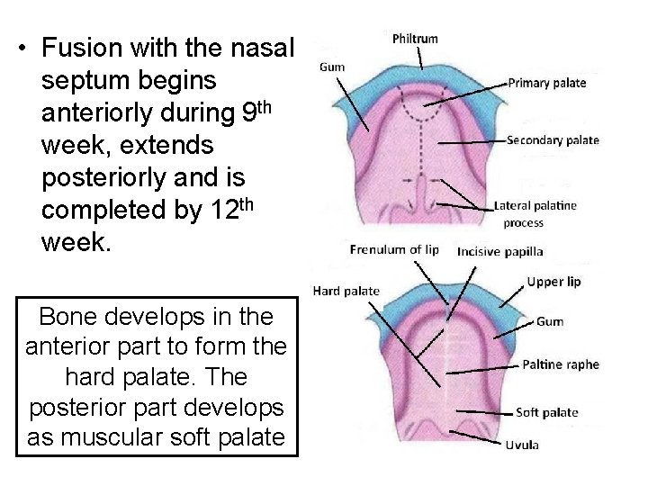
• Fusion with the nasal septum begins anteriorly during 9 th week, extends posteriorly and is completed by 12 th week. Bone develops in the anterior part to form the hard palate. The posterior part develops as muscular soft palate
Embryological subdivisions of the palate
Anomalies related to Face, Nose & Palate
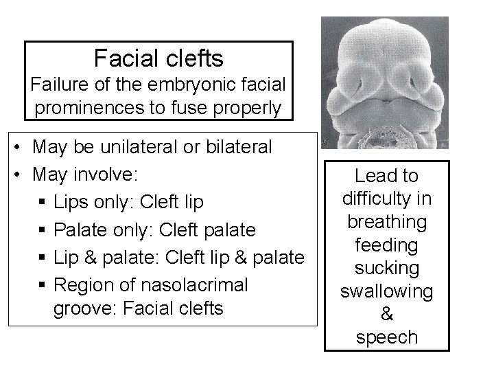
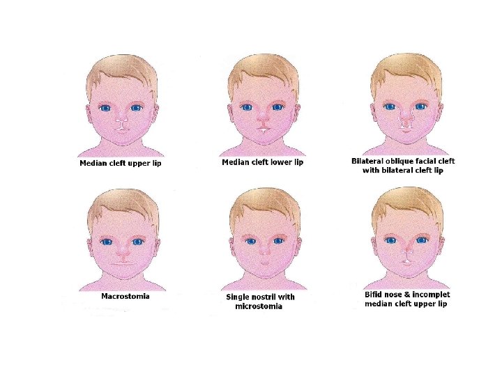
Facial clefts Failure of the embryonic facial prominences to fuse properly • May be unilateral or bilateral • May involve: § Lips only: Cleft lip § Palate only: Cleft palate § Lip & palate: Cleft lip & palate § Region of nasolacrimal groove: Facial clefts Lead to difficulty in breathing feeding sucking swallowing & speech
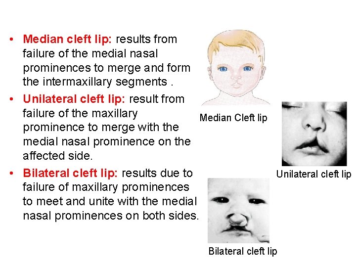
• Median cleft lip: results from failure of the medial nasal prominences to merge and form the intermaxillary segments. • Unilateral cleft lip: result from failure of the maxillary Median Cleft lip prominence to merge with the medial nasal prominence on the affected side. • Bilateral cleft lip: results due to Unilateral cleft lip failure of maxillary prominences to meet and unite with the medial nasal prominences on both sides. Bilateral cleft lip
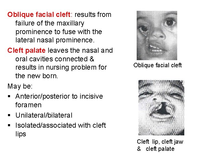
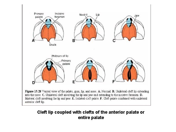
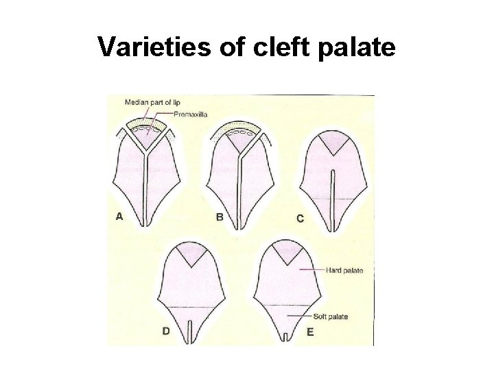
Oblique facial cleft: results from failure of the maxillary prominence to fuse with the lateral nasal prominence. Cleft palate leaves the nasal and oral cavities connected & results in nursing problem for the new born. May be: § Anterior/posterior to incisive foramen § Unilateral/bilateral § Isolated/associated with cleft lips Oblique facial cleft Cleft lip, cleft jaw & cleft palate
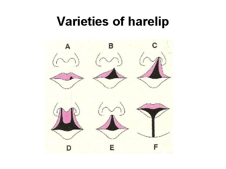
Varieties of harelip
Developmental Anomalies of Face Oblique facial cleft Cyclops & Proboscis
Cleft lip coupled with clefts of the anterior palate or entire palate
Varieties of cleft palate
Other Anomalies • Mandibulofacial dysostosis (Treacher Collins syndrome) • Retrognathia & agnathia • Hypertelorism
REFERENCES 1. Langman’s Medical Embryology, 11 th Edition. 3. I. B. Singh. Human Embryology, 10 th Edition.
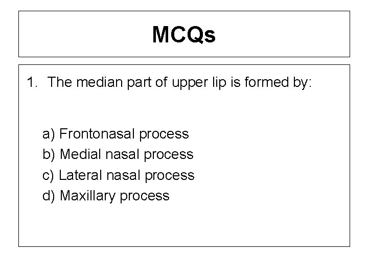
MCQs 1. The median part of upper lip is formed by: a) Frontonasal process b) Medial nasal process c) Lateral nasal process d) Maxillary process
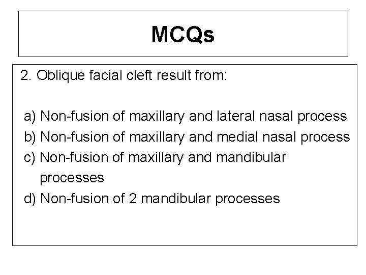
MCQs 2. Oblique facial cleft result from: a) Non-fusion of maxillary and lateral nasal process b) Non-fusion of maxillary and medial nasal process c) Non-fusion of maxillary and mandibular processes d) Non-fusion of 2 mandibular processes
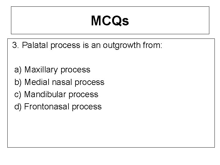
MCQs 3. Palatal process is an outgrowth from: a) Maxillary process b) Medial nasal process c) Mandibular process d) Frontonasal process
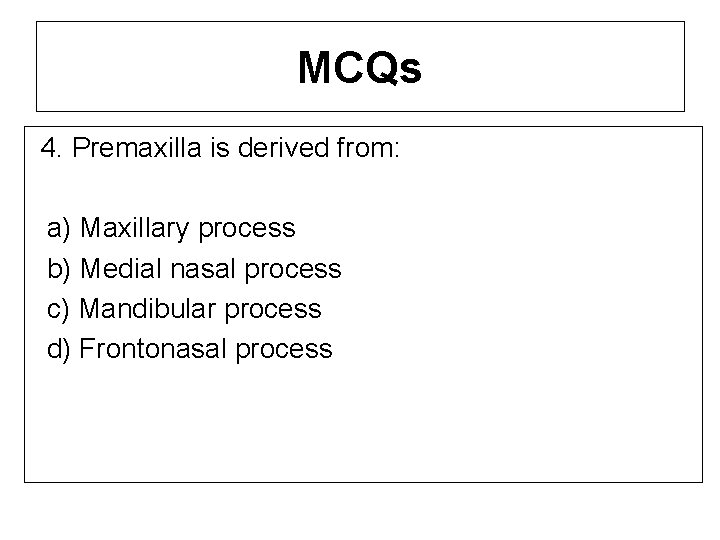
MCQs 4. Premaxilla is derived from: a) Maxillary process b) Medial nasal process c) Mandibular process d) Frontonasal process
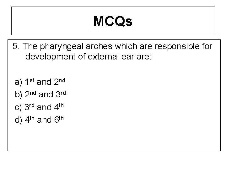
MCQs 5. The pharyngeal arches which are responsible for development of external ear are: a) 1 st and 2 nd b) 2 nd and 3 rd c) 3 rd and 4 th d) 4 th and 6 th